|
Bouchard's Nodes
Bouchard's nodes are hard, bony outgrowths or gelatinous cysts on the proximal interphalangeal joints (the middle joints of fingers or toes). They are seen in osteoarthritis, where they are caused by formation of calcific spurs of the articular (joint) cartilage. Much less commonly, they may be seen in rheumatoid arthritis, where nodes are caused by antibody deposition to the synovium. Bouchard's nodes are comparable in presentation to Heberden's nodes, which are similar osteoarthritic growths on the distal interphalangeal joints, but are significantly less common. Eponym Bouchard's nodes are named after French pathologist Pathology is the study of the causes and effects of disease or injury. The word ''pathology'' also refers to the study of disease in general, incorporating a wide range of biology research fields and medical practices. However, when used in t ... Charles Jacques Bouchard (1837–1915). See also * Heberden's node References External links A ... [...More Info...] [...Related Items...] OR: [Wikipedia] [Google] [Baidu] |
Cyst
A cyst is a closed sac, having a distinct envelope and division compared with the nearby tissue. Hence, it is a cluster of cells that have grouped together to form a sac (like the manner in which water molecules group together to form a bubble); however, the distinguishing aspect of a cyst is that the cells forming the "shell" of such a sac are distinctly abnormal (in both appearance and behaviour) when compared with all surrounding cells for that given location. A cyst may contain air, fluids, or semi-solid material. A collection of pus is called an abscess, not a cyst. Once formed, a cyst may resolve on its own. When a cyst fails to resolve, it may need to be removed surgically, but that would depend upon its type and location. Cancer-related cysts are formed as a defense mechanism for the body following the development of mutations that lead to an uncontrolled cellular division. Once that mutation has occurred, the affected cells divide incessantly and become cancerous, ... [...More Info...] [...Related Items...] OR: [Wikipedia] [Google] [Baidu] |
Proximal Interphalangeal Joint
The interphalangeal joints of the hand are the hinge joints between the phalanges of the fingers that provide flexion towards the palm of the hand. There are two sets in each finger (except in the thumb, which has only one joint): * "proximal interphalangeal joints" (PIJ or PIP), those between the first (also called proximal) and second (intermediate) phalanges * "distal interphalangeal joints" (DIJ or DIP), those between the second (intermediate) and third (distal) phalanges Anatomically, the proximal and distal interphalangeal joints are very similar. There are some minor differences in how the palmar plates are attached proximally and in the segmentation of the flexor tendon sheath, but the major differences are the smaller dimension and reduced mobility of the distal joint. Joint structure The PIP joint exhibits great lateral stability. Its transverse diameter is greater than its antero-posterior diameter and its thick collateral ligaments are tight in all positions duri ... [...More Info...] [...Related Items...] OR: [Wikipedia] [Google] [Baidu] |
Osteoarthritis
Osteoarthritis (OA) is a type of degenerative joint disease that results from breakdown of joint cartilage and underlying bone which affects 1 in 7 adults in the United States. It is believed to be the fourth leading cause of disability in the world. The most common symptoms are joint pain and stiffness. Usually the symptoms progress slowly over years. Initially they may occur only after exercise but can become constant over time. Other symptoms may include joint swelling, decreased range of motion, and, when the back is affected, weakness or numbness of the arms and legs. The most commonly involved joints are the two near the ends of the fingers and the joint at the base of the thumbs; the knee and hip joints; and the joints of the neck and lower back. Joints on one side of the body are often more affected than those on the other. The symptoms can interfere with work and normal daily activities. Unlike some other types of arthritis, only the joints, not internal organs, are ... [...More Info...] [...Related Items...] OR: [Wikipedia] [Google] [Baidu] |
Calcification
Calcification is the accumulation of calcium salts in a body tissue. It normally occurs in the formation of bone, but calcium can be deposited abnormally in soft tissue,Miller, J. D. Cardiovascular calcification: Orbicular origins. ''Nature Materials'' 12, 476-478 (2013). causing it to harden. Calcifications may be classified on whether there is mineral balance or not, and the location of the calcification. Calcification may also refer to the processes of normal mineral deposition in biological systems, such as the formation of stromatolites or mollusc shells (see Biomineralization). Signs and symptoms Calcification can manifest itself in many ways in the body depending on the location. In the pulpal structure of a tooth, calcification often presents asymptomatically, and is diagnosed as an incidental finding during radiographic interpretation. Individual teeth with calcified pulp will typically respond negatively to vitality testing; teeth with calcified pulp often lack ... [...More Info...] [...Related Items...] OR: [Wikipedia] [Google] [Baidu] |
Bone Spur
An exostosis, also known as bone spur, is the formation of new bone on the surface of a bone. Exostoses can cause chronic pain ranging from mild to debilitatingly severe, depending on the shape, size, and location of the lesion. It is most commonly found in places like the ribs, where small bone growths form, but sometimes larger growths can grow on places like the ankles, knees, shoulders, elbows and hips. Very rarely are they on the skull. Exostoses are sometimes shaped like spurs, such as calcaneal spurs. Osteomyelitis, a bone infection, may leave the adjacent bone with exostosis formation. Charcot foot, the neuropathic breakdown of the feet seen primarily in diabetics, can also leave bone spurs that may then become symptomatic. They normally form on the bones of joints, and can grow upwards. For example, if an extra bone formed on the ankle, it might grow up to the shin. When used in the phrases "cartilaginous exostosis" or "osteocartilaginous exostosis", the term is consid ... [...More Info...] [...Related Items...] OR: [Wikipedia] [Google] [Baidu] |
Cartilage
Cartilage is a resilient and smooth type of connective tissue. In tetrapods, it covers and protects the ends of long bones at the joints as articular cartilage, and is a structural component of many body parts including the rib cage, the neck and the bronchial tubes, and the intervertebral discs. In other taxa, such as chondrichthyans, but also in cyclostomes, it may constitute a much greater proportion of the skeleton. It is not as hard and rigid as bone, but it is much stiffer and much less flexible than muscle. The matrix of cartilage is made up of glycosaminoglycans, proteoglycans, collagen fibers and, sometimes, elastin. Because of its rigidity, cartilage often serves the purpose of holding tubes open in the body. Examples include the rings of the trachea, such as the cricoid cartilage and carina. Cartilage is composed of specialized cells called chondrocytes that produce a large amount of collagenous extracellular matrix, abundant ground substance that is rich in p ... [...More Info...] [...Related Items...] OR: [Wikipedia] [Google] [Baidu] |
Rheumatoid Arthritis
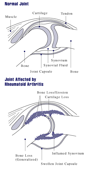
Rheumatoid arthritis (RA) is a long-term autoimmune disorder that primarily affects synovial joint, joints. It typically results in warm, swollen, and painful joints. Pain and stiffness often worsen following rest. Most commonly, the wrist and hands are involved, with the same joints typically involved on both sides of the body. The disease may also affect other parts of the body, including skin, eyes, lungs, heart, nerves and blood. This may result in a anemia, low red blood cell count, pleurisy, inflammation around the lungs, and pericarditis, inflammation around the heart. Fever and low energy may also be present. Often, symptoms come on gradually over weeks to months. While the cause of rheumatoid arthritis is not clear, it is believed to involve a combination of genetics, genetic and environmental factors. The underlying mechanism involves the body's immune system attacking the joints. This results in inflammation and thickening of the synovium, joint capsule. It also affec ... [...More Info...] [...Related Items...] OR: [Wikipedia] [Google] [Baidu] |
Synovium
The synovial membrane (also known as the synovial stratum, synovium or stratum synoviale) is a specialized connective tissue that lines the inner surface of capsules of synovial joints and tendon sheath. It makes direct contact with the fibrous membrane on the outside surface and with the synovial fluid lubricant on the inside surface. In contact with the synovial fluid at the tissue surface are many rounded macrophage-like synovial cells (type A) and also type B cells, which are also known as fibroblast-like synoviocytes (FLS). Type A cells maintain the synovial fluid by removing wear-and-tear debris. As for the FLS, they produce hyaluronan, as well as other extracellular components in the synovial fluid. Structure The synovial membrane is variable but often has two layers: * The outer layer, or subintima, can be of almost any type of connective tissue – fibrous (dense collagenous type), adipose (fatty; e.g. in intra-articular fat pads) or areolar (loose collagenous t ... [...More Info...] [...Related Items...] OR: [Wikipedia] [Google] [Baidu] |
Heberden's Node
Heberden's nodes are hard or bony swellings that can develop in the distal interphalangeal joints (DIP) (the joints closest to the end of the fingers and toes). They are a sign of osteoarthritis and are caused by formation of osteophytes (calcific spurs) of the articular (joint) cartilage in response to repeated trauma at the joint. Heberden's nodes typically develop in middle age, beginning either with a chronic swelling of the affected joints or the sudden painful onset of redness, numbness, and loss of manual dexterity. This initial inflammation and pain eventually subsides, and the patient is left with a permanent bony outgrowth that often skews the fingertip sideways. Bouchard's nodes may also be present; these are similar bony growths in the proximal interphalangeal (PIP) joints (middle joints of the fingers), and are also associated with osteoarthritis. Heberden's nodes are more common in women than in men, and there seems to be a genetic component involved in predisposi ... [...More Info...] [...Related Items...] OR: [Wikipedia] [Google] [Baidu] |
Distal Interphalangeal Joint
Distal interphalangeal joints are the articulations between the phalanges of the hand or foot. This term therefore includes: * Interphalangeal joints of the hand The interphalangeal joints of the hand are the hinge joints between the phalanges of the fingers that provide flexion towards the palm of the hand. There are two sets in each finger (except in the thumb, which has only one joint): * "proximal in ... * Interphalangeal joints of the foot Joints {{Short pages monitor ... [...More Info...] [...Related Items...] OR: [Wikipedia] [Google] [Baidu] |
Pathologist
Pathology is the study of the causes and effects of disease or injury. The word ''pathology'' also refers to the study of disease in general, incorporating a wide range of biology research fields and medical practices. However, when used in the context of modern medical treatment, the term is often used in a narrower fashion to refer to processes and tests that fall within the contemporary medical field of "general pathology", an area which includes a number of distinct but inter-related medical specialties that diagnose disease, mostly through analysis of tissue, cell, and body fluid samples. Idiomatically, "a pathology" may also refer to the predicted or actual progression of particular diseases (as in the statement "the many different forms of cancer have diverse pathologies", in which case a more proper choice of word would be " pathophysiologies"), and the affix ''pathy'' is sometimes used to indicate a state of disease in cases of both physical ailment (as in cardiomy ... [...More Info...] [...Related Items...] OR: [Wikipedia] [Google] [Baidu] |
Charles Jacques Bouchard
Charles Jacques Bouchard (6 September 1837 – 28 October 1915) was a French pathologist and an esperantist born in Montier-en-Der, a commune the department of Haute-Marne. Biography He studied medicine in Lyon and Paris, where he obtained his doctorate in 1866. In 1874 he became a physician at Bicêtre Hospital, and in 1879 was appointed chair of general pathology. In 1886, he became a member of the Academie de Médecine. Bouchard is remembered for his work with infectious and nutritional diseases. He was a student of Jean Charcot at the Pitié-Salpêtrière Hospital, and with Charcot described a disorder that would later be known as a " Charcot-Bouchard aneurysm". It is described as a small aneurysm on cerebral perforated vessels that could be the cause of intracranial hemorrhages. Bouchard wrote about the aneurysm in his doctorate thesis ''Étude sur quelques points de la pathogénie des hémorrhagies cérébrales''. Bouchard promoted a theory of intestinal autointoxicat ... [...More Info...] [...Related Items...] OR: [Wikipedia] [Google] [Baidu] |


.jpg)
