|
Binucleated Cell Overlay
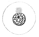
Binucleated cells are cells that contain two nuclei. This type of cell is most commonly found in cancer cells and may arise from a variety of causes. Binucleation can be easily visualized through staining and microscopy. In general, binucleation has negative effects on cell viability and subsequent mitosis. They also occur physiologically in hepatocytes, chondrocytes and in fungi (dikaryon). Causes * Cleavage furrow regression: Cells divide and almost complete division but then the cleavage furrow begins to regress and the cells merge. This is thought to be caused by nondisjunction in chromosomes but the mechanism by which it occurs is not well understood. * Failed cytokinesis: The cell can fail to form a cleavage furrow, leading to both nuclei remaining in one cell. * Multipolar spindles: Cells contain three or more centrioles, resulting in multiple poles. This leads to the cells pulling chromosomes in many directions that end in multiple nuclei found in one cell. * Mergin ... [...More Info...] [...Related Items...] OR: [Wikipedia] [Google] [Baidu] |
Pathology
Pathology is the study of the causes and effects of disease or injury. The word ''pathology'' also refers to the study of disease in general, incorporating a wide range of biology research fields and medical practices. However, when used in the context of modern medical treatment, the term is often used in a narrower fashion to refer to processes and tests that fall within the contemporary medical field of "general pathology", an area which includes a number of distinct but inter-related medical specialties that diagnose disease, mostly through analysis of tissue, cell, and body fluid samples. Idiomatically, "a pathology" may also refer to the predicted or actual progression of particular diseases (as in the statement "the many different forms of cancer have diverse pathologies", in which case a more proper choice of word would be " pathophysiologies"), and the affix ''pathy'' is sometimes used to indicate a state of disease in cases of both physical ailment (as in cardiomy ... [...More Info...] [...Related Items...] OR: [Wikipedia] [Google] [Baidu] |
Multipolar Spindles
Multipolar spindles are spindle formations characteristic of cancer cells. Spindle formation is mostly conducted by the aster of the centrosome which it forms around itself. In a mitotic cell wherever two asters convene the formation of a spindle occurs. Mitosis consists of two independent processes: the intra-chromosomal and the extra-chromosomal (formation of spindle) changes both of these being in total coordination of each other. In cancer cells, it has been observed that the formation of the spindles comes before when compared to the chromosomes. Because the prophase stage is brief, metaphase begins earlier than in normal cells. Chromosomes unable to reach the metaphase plate are stranded behind. These chromosomes still have asters attached to them and when met with other asters, form multiple spindles. Characteristics Cells with multipolar spindles are characterized by more than two centrosomes, usually four, and sometimes have a second metaphase plate. The multiple centro ... [...More Info...] [...Related Items...] OR: [Wikipedia] [Google] [Baidu] |
Mutations
In biology, a mutation is an alteration in the nucleic acid sequence of the genome of an organism, virus, or extrachromosomal DNA. Viral genomes contain either DNA or RNA. Mutations result from errors during DNA or viral replication, mitosis, or meiosis or other types of damage to DNA (such as pyrimidine dimers caused by exposure to ultraviolet radiation), which then may undergo error-prone repair (especially microhomology-mediated end joining), cause an error during other forms of repair, or cause an error during replication (translesion synthesis). Mutations may also result from insertion or deletion of segments of DNA due to mobile genetic elements. Mutations may or may not produce detectable changes in the observable characteristics (phenotype) of an organism. Mutations play a part in both normal and abnormal biological processes including: evolution, cancer, and the development of the immune system, including junctional diversity. Mutation is the ultimate source o ... [...More Info...] [...Related Items...] OR: [Wikipedia] [Google] [Baidu] |
Interphase
Interphase is the portion of the cell cycle that is not accompanied by visible changes under the microscope, and includes the G1, S and G2 phases. During interphase, the cell grows (G1), replicates its DNA (S) and prepares for mitosis (G2). A cell in interphase is not simply quiescent. The term quiescent (i.e. dormant) would be misleading since a cell in interphase is very busy synthesizing proteins, copying DNA into RNA, engulfing extracellular material, processing signals, to name just a few activities. The cell is quiescent only in the sense of cell division (i.e. the cell is out of the cell cycle, G0). Interphase is the phase of the cell cycle in which a typical cell spends most of its life. Interphase is the 'daily living' or metabolic phase of the cell, in which the cell obtains nutrients and metabolizes them, grows, replicates its DNA in preparation for mitosis, and conducts other "normal" cell functions. Interphase was formerly called the resting phase. However, interp ... [...More Info...] [...Related Items...] OR: [Wikipedia] [Google] [Baidu] |
Chromatin Bridge
Chromatin bridge is a mitotic occurrence that forms when telomeres of sister chromatids fuse together and fail to completely segregate into their respective daughter cells. Because this event is most prevalent during anaphase, the term anaphase bridge is often used as a substitute. After the formation of individual daughter cells, the DNA bridge connecting homologous chromosomes remains fixed. As the daughter cells exit mitosis and re-enter interphase, the chromatin bridge becomes known as an interphase bridge. These phenomena are usually visualized using the laboratory techniques of staining and fluorescence microscopy. Background The faithful inheritance of genetic information from one cellular generation to the next heavily relies on the duplication of deoxyribonucleic acid (DNA), as well as the formation of two identical daughter cells. This complicated cellular process, known as mitosis, depends on a multitude of cellular checkpoints, signals, interactions and signal cascades ... [...More Info...] [...Related Items...] OR: [Wikipedia] [Google] [Baidu] |
Micronuclei
Micronucleus is the name given to the small nucleus that forms whenever a chromosome or a fragment of a chromosome is not incorporated into one of the daughter nuclei during cell division. It usually is a sign of genotoxic events and chromosomal instability. Micronuclei are commonly seen in cancerous cells and may indicate genomic damage events that can increase the risk of developmental or degenerative diseases. Micronuclei form during anaphase from lagging acentric chromosome or chromatid fragments caused by incorrectly repaired or unrepaired DNA breaks or by nondisjunction of chromosomes. This incorrect segregation of chromosomes may result from hypomethylation of repeat sequences present in pericentromeric DNA, irregularities in kinetochore proteins or their assembly, dysfunctional spindle apparatus, or flawed anaphase checkpoint genes. Micronuclei can contribute to genome instability by promoting a catastrophic mutational event called chromothripsis. Many micronucleus assays ha ... [...More Info...] [...Related Items...] OR: [Wikipedia] [Google] [Baidu] |
Multipolar Spindles
Multipolar spindles are spindle formations characteristic of cancer cells. Spindle formation is mostly conducted by the aster of the centrosome which it forms around itself. In a mitotic cell wherever two asters convene the formation of a spindle occurs. Mitosis consists of two independent processes: the intra-chromosomal and the extra-chromosomal (formation of spindle) changes both of these being in total coordination of each other. In cancer cells, it has been observed that the formation of the spindles comes before when compared to the chromosomes. Because the prophase stage is brief, metaphase begins earlier than in normal cells. Chromosomes unable to reach the metaphase plate are stranded behind. These chromosomes still have asters attached to them and when met with other asters, form multiple spindles. Characteristics Cells with multipolar spindles are characterized by more than two centrosomes, usually four, and sometimes have a second metaphase plate. The multiple centro ... [...More Info...] [...Related Items...] OR: [Wikipedia] [Google] [Baidu] |
Binucleated Cell
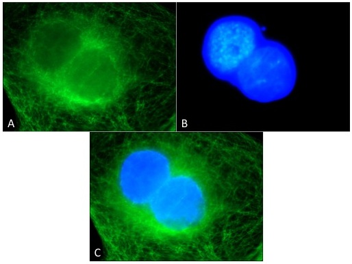
Binucleated cells are cells that contain two nuclei. This type of cell is most commonly found in cancer cells and may arise from a variety of causes. Binucleation can be easily visualized through staining and microscopy. In general, binucleation has negative effects on cell viability and subsequent mitosis. They also occur physiologically in hepatocytes, chondrocytes and in fungi (dikaryon). Causes * Cleavage furrow regression: Cells divide and almost complete division but then the cleavage furrow begins to regress and the cells merge. This is thought to be caused by nondisjunction in chromosomes but the mechanism by which it occurs is not well understood. * Failed cytokinesis: The cell can fail to form a cleavage furrow, leading to both nuclei remaining in one cell. * Multipolar spindles: Cells contain three or more centrioles, resulting in multiple poles. This leads to the cells pulling chromosomes in many directions that end in multiple nuclei found in one cell. * Merging ... [...More Info...] [...Related Items...] OR: [Wikipedia] [Google] [Baidu] |
Immunofluorescence
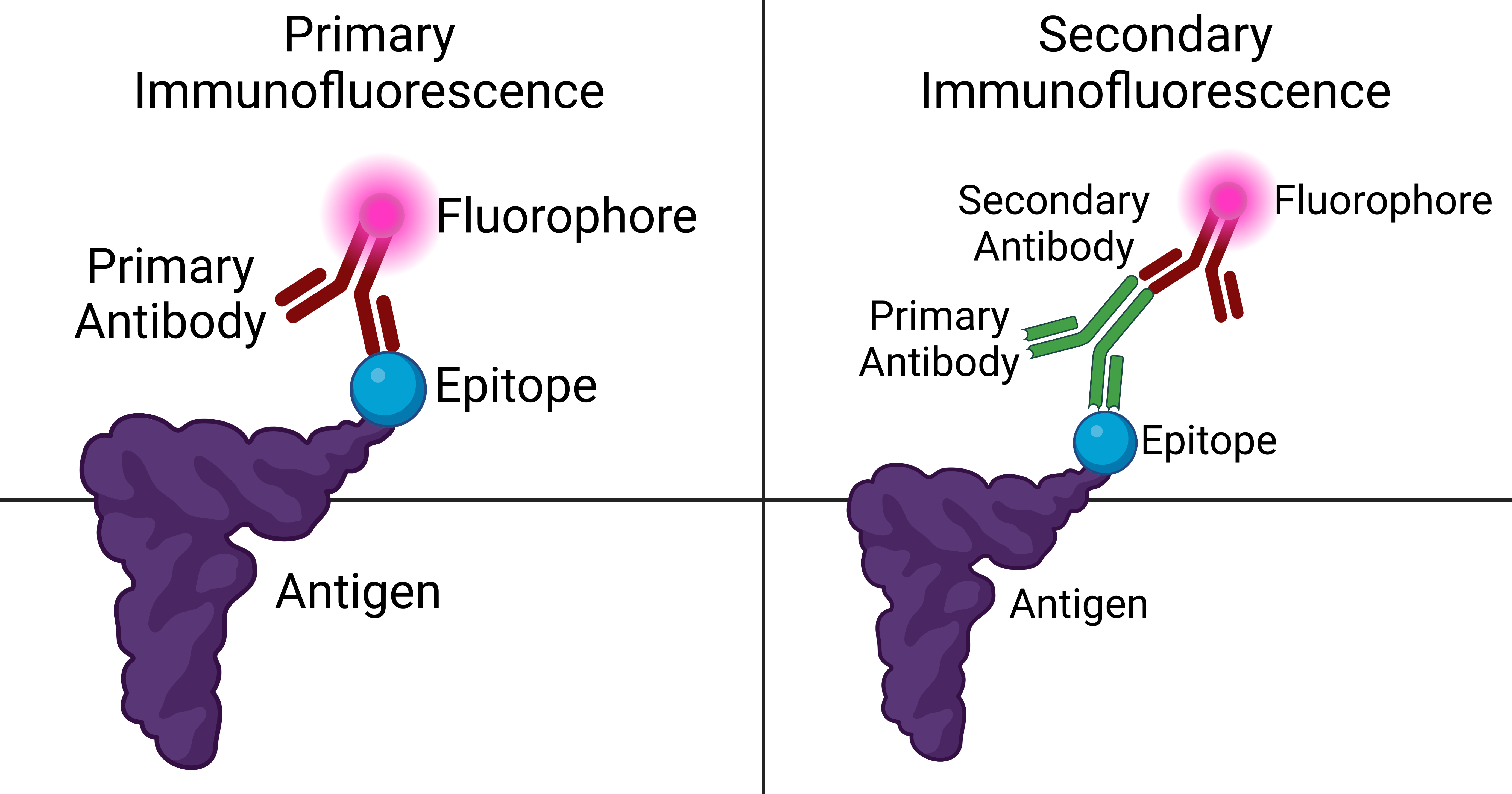
Immunofluorescence is a technique used for light microscopy with a fluorescence microscope and is used primarily on microbiological samples. This technique uses the specificity of antibodies to their antigen to target fluorescent dyes to specific biomolecule targets within a cell, and therefore allows visualization of the distribution of the target molecule through the sample. The specific region an antibody recognizes on an antigen is called an epitope. There have been efforts in epitope mapping since many antibodies can bind the same epitope and levels of binding between antibodies that recognize the same epitope can vary. Additionally, the binding of the fluorophore to the antibody itself cannot interfere with the immunological specificity of the antibody or the binding capacity of its antigen. Immunofluorescence is a widely used example of immunostaining (using antibodies to stain proteins) and is a specific example of immunohistochemistry (the use of the antibody-antigen rel ... [...More Info...] [...Related Items...] OR: [Wikipedia] [Google] [Baidu] |
Antibody
An antibody (Ab), also known as an immunoglobulin (Ig), is a large, Y-shaped protein used by the immune system to identify and neutralize foreign objects such as pathogenic bacteria and viruses. The antibody recognizes a unique molecule of the pathogen, called an antigen. Each tip of the "Y" of an antibody contains a paratope (analogous to a lock) that is specific for one particular epitope (analogous to a key) on an antigen, allowing these two structures to bind together with precision. Using this binding mechanism, an antibody can ''tag'' a microbe or an infected cell for attack by other parts of the immune system, or can neutralize it directly (for example, by blocking a part of a virus that is essential for its invasion). To allow the immune system to recognize millions of different antigens, the antigen-binding sites at both tips of the antibody come in an equally wide variety. In contrast, the remainder of the antibody is relatively constant. It only occurs in a few varia ... [...More Info...] [...Related Items...] OR: [Wikipedia] [Google] [Baidu] |
DAPI
DAPI (pronounced 'DAPPY', /ˈdæpiː/), or 4′,6-diamidino-2-phenylindole, is a fluorescent stain that binds strongly to adenine–thymine-rich regions in DNA. It is used extensively in fluorescence microscopy. As DAPI can pass through an intact cell membrane, it can be used to stain both live and fixed cells, though it passes through the membrane less efficiently in live cells and therefore provides a marker for membrane viability. History DAPI was first synthesised in 1971 in the laboratory of Otto Dann as part of a search for drugs to treat trypanosomiasis. Although it was unsuccessful as a drug, further investigation indicated it bound strongly to DNA and became more fluorescent when bound. This led to its use in identifying mitochondrial DNA in ultracentrifugation in 1975, the first recorded use of DAPI as a fluorescent DNA stain. Strong fluorescence when bound to DNA led to the rapid adoption of DAPI for fluorescent staining of DNA for fluorescence microscopy. Its use f ... [...More Info...] [...Related Items...] OR: [Wikipedia] [Google] [Baidu] |
Tubulin
Tubulin in molecular biology can refer either to the tubulin protein superfamily of globular proteins, or one of the member proteins of that superfamily. α- and β-tubulins polymerize into microtubules, a major component of the eukaryotic cytoskeleton. Microtubules function in many essential cellular processes, including mitosis. Tubulin-binding drugs kill cancerous cells by inhibiting microtubule dynamics, which are required for DNA segregation and therefore cell division. In eukaryotes, there are six members of the tubulin superfamily, although not all are present in all species.Turk E, Wills AA, Kwon T, Sedzinski J, Wallingford JB, Stearns "Zeta-Tubulin Is a Member of a Conserved Tubulin Module and Is a Component of the Centriolar Basal Foot in Multiciliated Cells"Current Biology (2015) 25:2177-2183. Both α and β tubulins have a mass of around 50 kDa and are thus in a similar range compared to actin (with a mass of ~42 kDa). In contrast, tubulin polymers (microtubules) te ... [...More Info...] [...Related Items...] OR: [Wikipedia] [Google] [Baidu] |
.jpg)


