|
Bent-over Row
A bent-over row (or barbell row) is a weight training exercise that targets a variety of back muscles. Which ones are targeted varies on form. The bent over row is often used for both bodybuilding and powerlifting. Implements There are several variants of this exercise, depending on whether dumbbells or a barbell is used and whether both arms are exercised at the same time: Two arm rows: * ''Two arm barbell bent-over-row:'' This version uses both arms to lift a barbell to the stomach in a bent-forward position. The hands are kept pronated and the back straight. * ''Two arm dumbbell bent-over-row'': The barbell is replaced by two dumbbells, one for each hand. * ''Pendlay row'': named after Glenn Pendlay; the back is parallel to the ground and the weight lifted from the floor. * '' Yates row'': named after Dorian Yates; a row done with underhand grip and a slightly more upright torso than a regular row. One arm rows: * ''One arm dumbbell bent-over-row'': This exercise is freque ... [...More Info...] [...Related Items...] OR: [Wikipedia] [Google] [Baidu] |
Weight Training
Weight training is a common type of strength training for developing the strength, size of skeletal muscles and maintenance of strength.Keogh, Justin W, and Paul W Winwood. “Report for: The Epidemiology of Injuries Across the Weight-Training Sports.” ''Altmetric – Vitamin C Antagonizes the Cytotoxic Effects of Antineoplastic Drugs'', Mar. 2017, summon.altmetric.com/details/8964732. It uses the force of gravity in the form of weighted bars, dumbbells or weight stacks in order to oppose the force generated by muscle through concentric or eccentric contraction. Weight training uses a variety of specialized equipment to target specific muscle groups and types of movement. Sports in which weight training is used include bodybuilding, weightlifting, powerlifting, strongman, highland games, hammer throw, shot put, discus throw, and javelin throw. Many other sports use strength training as part of their training regimen, notably: American football, baseball, bas ... [...More Info...] [...Related Items...] OR: [Wikipedia] [Google] [Baidu] |
Latissimus Dorsi Muscle
The latissimus dorsi () is a large, flat muscle on the back that stretches to the sides, behind the arm, and is partly covered by the trapezius on the back near the midline. The word latissimus dorsi (plural: ''latissimi dorsorum'') comes from Latin and means "broadest uscleof the back", from "latissimus" ( la, broadest)' and "dorsum" ( la, back). The pair of muscles are commonly known as "lats", especially among bodybuilders. The latissimus dorsi is the largest muscle in the upper body. The latissimus dorsi is responsible for extension, adduction, transverse extension also known as horizontal abduction (or horizontal extension), flexion from an extended position, and (medial) internal rotation of the shoulder joint. It also has a synergistic role in extension and lateral flexion of the lumbar spine. Due to bypassing the scapulothoracic joints and attaching directly to the spine, the actions the latissimi dorsi have on moving the arms can also influence the movement of the ... [...More Info...] [...Related Items...] OR: [Wikipedia] [Google] [Baidu] |
Rectus Abdominis
The rectus abdominis muscle, ( la, straight abdominal) also known as the "abdominal muscle" or simply the "abs", is a paired straight muscle. It is a paired muscle, separated by a midline band of connective tissue called the linea alba. It extends from the pubic symphysis, pubic crest and pubic tubercle inferiorly, to the xiphoid process and costal cartilages of ribs V to VII superiorly. The proximal attachments are the pubic crest and the pubic symphysis. It attaches distally at the costal cartilages of ribs 5-7 and the xiphoid process of the sternum. The rectus abdominis muscle is contained in the rectus sheath, which consists of the aponeuroses of the lateral abdominal muscles. The outer, most lateral line, defining the rectus is the linea semilunaris. Bands of connective tissue traverse the rectus abdominis, separating it into distinct muscle bellies. In the abdomens of people with low body fat, these muscle bellies can be viewed externally. They can appear in sets ... [...More Info...] [...Related Items...] OR: [Wikipedia] [Google] [Baidu] |
Transversus Abdominis
The transverse abdominal muscle (TVA), also known as the transverse abdominis, transversalis muscle and transversus abdominis muscle, is a muscle layer of the anterior and lateral (front and side) abdominal wall which is deep to (layered below) the internal oblique muscle. It is thought by most fitness instructors to be a significant component of the core. Structure The transverse abdominal, so called for the direction of its fibers, is the innermost of the flat muscles of the abdomen. It is positioned immediately inside of the internal oblique muscle. The transverse abdominal arises as fleshy fibers, from the lateral third of the inguinal ligament, from the anterior three-fourths of the inner lip of the iliac crest, from the inner surfaces of the cartilages of the lower six ribs, interdigitating with the diaphragm, and from the thoracolumbar fascia. It ends anteriorly in a broad aponeurosis (the Spigelian fascia), the lower fibers of which curve inferomedially (medially an ... [...More Info...] [...Related Items...] OR: [Wikipedia] [Google] [Baidu] |
Human Abdomen
The abdomen (colloquially called the belly, tummy, midriff, tucky or stomach) is the part of the body between the thorax (chest) and pelvis, in humans and in other vertebrates. The abdomen is the front part of the abdominal segment of the torso. The area occupied by the abdomen is called the abdominal cavity. In arthropods it is the posterior tagma of the body; it follows the thorax or cephalothorax. In humans, the abdomen stretches from the thorax at the thoracic diaphragm to the pelvis at the pelvic brim. The pelvic brim stretches from the lumbosacral joint (the intervertebral disc between L5 and S1) to the pubic symphysis and is the edge of the pelvic inlet. The space above this inlet and under the thoracic diaphragm is termed the abdominal cavity. The boundary of the abdominal cavity is the abdominal wall in the front and the peritoneal surface at the rear. In vertebrates, the abdomen is a large body cavity enclosed by the abdominal muscles, at front and to the side ... [...More Info...] [...Related Items...] OR: [Wikipedia] [Google] [Baidu] |
Fly (exercise)
A fly or flye is a strength training exercise in which the hand and arm move through an arc while the elbow is kept at a constant angle. Flies are used to work the muscles of the upper body. Because these exercises use the arms as levers at their longest possible length, the amount of weight that can be moved is significantly less than equivalent press exercises for the same muscles (the military press and bench press for the shoulder and chest respectively). Due to this leverage, fly exercises of all types have a large potential to damage the shoulder joint and its associated ligaments and the tendons of the muscles connecting to it. They should be done with caution and their effects first tested while using very light weights; which are gradually incremented after more strength is gained. Equipment Flies can be performed using any weight that can be held in the hand. The simplest equipment to use is a dumbbell, though the exercise can also be performed using a cable machin ... [...More Info...] [...Related Items...] OR: [Wikipedia] [Google] [Baidu] |
Lateral Deltoid
The deltoid muscle is the muscle forming the rounded contour of the human shoulder. It is also known as the 'common shoulder muscle', particularly in other animals such as the domestic cat. Anatomically, the deltoid muscle appears to be made up of three distinct sets of muscle fibers, namely the # anterior or clavicular part (pars clavicularis) # posterior or scapular part (pars scapularis) # intermediate or acromial part (pars acromialis) However, electromyography suggests that it consists of at least seven groups that can be independently coordinated by the nervous system. It was previously called the deltoideus (plural ''deltoidei'') and the name is still used by some anatomists. It is called so because it is in the shape of the Greek capital letter delta (Δ). Deltoid is also further shortened in slang as "delt". A study of 30 shoulders revealed an average mass of in humans, ranging from to . Structure Previous studies showed that the insertions of the tendons of the delt ... [...More Info...] [...Related Items...] OR: [Wikipedia] [Google] [Baidu] |
Rhomboid Muscles
The rhomboid muscles (), often simply called the rhomboids, are rhombus-shaped muscles associated with the scapula. There are two rhomboid muscles on each side of the upper back: * Rhomboid major muscle * Rhomboid minor muscle The large rhombus-shaped muscle, located under the trapezius muscle in the upper part of the thoracic region of the back, and the small muscle, in the same way, participate in the movement of the scapula. Anatomy The rhomboid major muscle originates from the spinous processes of vertebrae T2–T5, while it inserts to the medial border of the scapula. The rhomboid minor muscle originates from the nuchal ligament and spinous processes of vertebrae C7–T1. Both muscles are innervated by the dorsal scapular nerve, a branch of the brachial plexus. Their functions are the following: * Drawing scapula superomedially * Supporting scapula * Rotating glenoid cavity inferiorly Additional images File:Rhomboid muscles animation small.gif, Rhomboid muscles. ... [...More Info...] [...Related Items...] OR: [Wikipedia] [Google] [Baidu] |
Rotator Cuff
The rotator cuff is a group of muscles and their tendons that act to stabilize the human shoulder and allow for its extensive range of motion. Of the seven scapulohumeral muscles, four make up the rotator cuff. The four muscles are the supraspinatus muscle, the infraspinatus muscle, teres minor muscle, and the subscapularis muscle. Structure Muscles composing rotator cuff The supraspinatus muscle spreads out in a horizontal band to insert on the superior facet of the greater tubercle of the humerus. The greater tubercle projects as the most lateral structure of the humeral head. Medial to this, in turn, is the lesser tubercle of the humeral head. The subscapularis muscle origin is divided from the remainder of the rotator cuff origins as it is deep to the scapula. The four tendons of these muscles converge to form the rotator cuff tendon. These tendinous insertions along with the articular capsule, the coracohumeral ligament, and the glenohumeral ligament complex, b ... [...More Info...] [...Related Items...] OR: [Wikipedia] [Google] [Baidu] |
Teres Minor
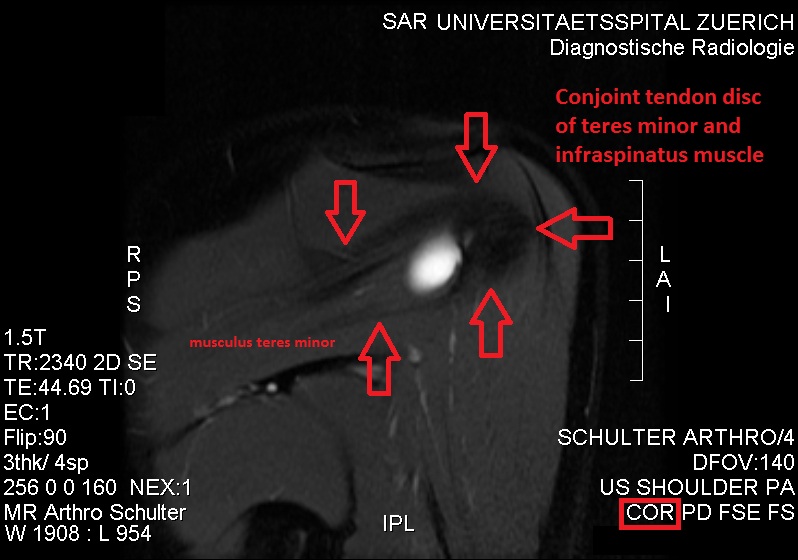
The teres minor (Latin ''teres'' meaning 'rounded') is a narrow, elongated muscle of the rotator cuff. The muscle originates from the lateral border and adjacent posterior surface of the corresponding right or left scapula and inserts at both the greater tubercle of the humerus and the posterior surface of the joint capsule. The primary function of the teres minor is to modulate the action of the deltoid, preventing the humeral head from sliding upward as the arm is abducted. It also functions to rotate the humerus laterally. The teres minor is innervated by the axillary nerve. Structure It arises from the dorsal surface of the axillary border of the scapula for the upper two-thirds of its extent, and from two aponeurotic laminae, one of which separates it from the infraspinatus muscle, the other from the teres major muscle. Its fibers run obliquely upwards and laterally; the upper ones end in a tendon which is inserted into the lowest of the three impressions on the greater tu ... [...More Info...] [...Related Items...] OR: [Wikipedia] [Google] [Baidu] |
Infraspinatus
In human anatomy, the infraspinatus muscle is a thick triangular muscle, which occupies the chief part of the infraspinatous fossa.'' Gray's Anatomy'', see infobox. As one of the four muscles of the rotator cuff, the main function of the infraspinatus is to externally rotate the humerus and stabilize the shoulder joint. Structure It attaches medially to the infraspinous fossa of the scapula and laterally to the middle facet of the greater tubercle of the humerus. The muscle arises by fleshy fibers from the medial two-thirds of the infraspinatous fossa, and by tendinous fibers from the ridges on its surface; it also arises from the infraspinatous fascia which covers it, and separates it from the teres major and teres minor. The fibers converge to a tendon, which glides over the lateral border of the spine of the scapula and passing across the posterior part of the capsule of the shoulder-joint, is inserted into the middle impression on the greater tubercle of the humerus. Th ... [...More Info...] [...Related Items...] OR: [Wikipedia] [Google] [Baidu] |
Deltoid Muscle
The deltoid muscle is the muscle forming the rounded contour of the human shoulder. It is also known as the 'common shoulder muscle', particularly in other animals such as the domestic cat. Anatomically, the deltoid muscle appears to be made up of three distinct sets of muscle fibers, namely the # anterior or clavicular part (pars clavicularis) # posterior or scapular part (pars scapularis) # intermediate or acromial part (pars acromialis) However, electromyography suggests that it consists of at least seven groups that can be independently coordinated by the nervous system. It was previously called the deltoideus (plural ''deltoidei'') and the name is still used by some anatomists. It is called so because it is in the shape of the Greek capital letter delta (Δ). Deltoid is also further shortened in slang as "delt". A study of 30 shoulders revealed an average mass of in humans, ranging from to . Structure Previous studies showed that the insertions of the tendons of the d ... [...More Info...] [...Related Items...] OR: [Wikipedia] [Google] [Baidu] |

