|
Bartarlla-Scott Syndrome
Xylosyltransferase 1 is an enzyme that in humans is encoded by the ''XYLT1'' gene. Xylosyltransferase (XT; EC 2.4.2.26) catalyzes the transfer of UDP-xylose to serine residues within XT recognition sequences of target proteins. Addition of this xylose to the core protein is required for the biosynthesis of the glycosaminoglycan chains characteristic of proteoglycans. upplied by OMIMref name="entrez" /> Clinical relevance Baratela-Scott syndrome In 2012 Baratela-Scott syndrome was identified in humans. A GGC repeat expansion, and methylation of exon 1 of XYLT1 is a common pathogenic variant in Baratela-Scott syndrome. Patients with Bartarlla-Scott syndrome exhibit abnormal development of the skeleton, characteristic facial features, and cognitive developmental delay. Skeletal problems include knee cap in the wrong position, short long bones with mild changes to the narrow portion, short palm bones with stub thumbs, short thigh necks, shallow hip sockets, and malformations ... [...More Info...] [...Related Items...] OR: [Wikipedia] [Google] [Baidu] |
Enzyme
Enzymes () are proteins that act as biological catalysts by accelerating chemical reactions. The molecules upon which enzymes may act are called substrates, and the enzyme converts the substrates into different molecules known as products. Almost all metabolic processes in the cell need enzyme catalysis in order to occur at rates fast enough to sustain life. Metabolic pathways depend upon enzymes to catalyze individual steps. The study of enzymes is called ''enzymology'' and the field of pseudoenzyme analysis recognizes that during evolution, some enzymes have lost the ability to carry out biological catalysis, which is often reflected in their amino acid sequences and unusual 'pseudocatalytic' properties. Enzymes are known to catalyze more than 5,000 biochemical reaction types. Other biocatalysts are catalytic RNA molecules, called ribozymes. Enzymes' specificity comes from their unique three-dimensional structures. Like all catalysts, enzymes increase the reaction ra ... [...More Info...] [...Related Items...] OR: [Wikipedia] [Google] [Baidu] |
Patellar Dislocation
A patellar dislocation is a knee injury in which the patella (kneecap) slips out of its normal position. Often the knee is partly bent, painful and swollen. The patella is also often felt and seen out of place. Complications may include a patella fracture or arthritis. A patellar dislocation typically occurs when the knee is straight and the lower leg is bent outwards when twisting. Occasionally it occurs when the knee is bent and the patella is hit. Commonly associated sports include soccer, gymnastics, and ice hockey. Dislocations nearly always occur away from the midline. Diagnosis is typically based on symptoms and supported by X-rays. Reduction is generally done by pushing the patella towards the midline while straightening the knee. After reduction the leg is generally splinted in a straight position for a few weeks. This is then followed by physical therapy. Surgery after a first dislocation is generally of unclear benefit. Surgery may be indicated in those who have bro ... [...More Info...] [...Related Items...] OR: [Wikipedia] [Google] [Baidu] |
Nasal Bridge
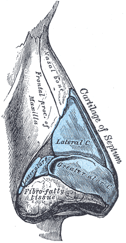
The nasal bridge is the upper, bony part of the human nose, which overlies the nasal bones. Association with epicanthic folds Low-rooted nasal bridges are closely associated with epicanthic folds. A lower nasal bridge is more likely to cause an epicanthic fold, and vice versa. Dysmorphology A lower or higher than average nasal bridge can be a sign of various genetic disorders, such as fetal alcohol syndrome. A flat nasal bridge can be a sign of Down syndrome (Trisomy 21), Fragile X syndrome, 48,XXXY variant Klinefelter syndrome, or Bartarlla-Scott syndrome. An appearance of a widened nasal bridge can be seen with dystopia canthorum, which is a lateral displacement of the inner canthi of the eyes. from UTMB, Dept. of Otolaryngology. DATE: March 17, 2004. RESIDENT PHYSICIAN: Jing Shen. FACUL ... [...More Info...] [...Related Items...] OR: [Wikipedia] [Google] [Baidu] |
Platyspondyly
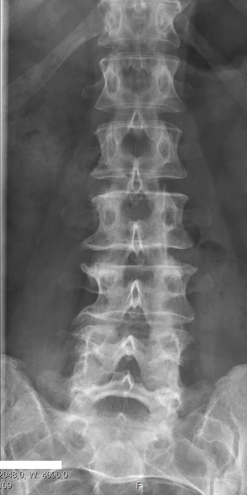
Congenital vertebral anomalies are a collection of malformations of the spine. Most, around 85%, are not clinically significant, but they can cause compression of the spinal cord by deforming the vertebral canal or causing instability. This condition occurs in the womb. Congenital vertebral anomalies include alterations of the shape and number of vertebrae. Lumbarization and sacralization ''Lumbarization'' is an anomaly in the spine. It is defined by the nonfusion of the first and second segments of the sacrum. The lumbar spine subsequently appears to have six vertebrae or segments, not five. This sixth lumbar vertebra is known as a transitional vertebra. Conversely the sacrum appears to have only four segments instead of its designated five segments. Lumbosacral transitional vertebrae consist of the process of the last lumbar vertebra fusing with the first sacral segment. While only around 10 percent of adults have a spinal abnormality due to genetics, a sixth lumbar vertebr ... [...More Info...] [...Related Items...] OR: [Wikipedia] [Google] [Baidu] |
Acetabulum
The acetabulum (), also called the cotyloid cavity, is a concave surface of the pelvis. The head of the femur meets with the pelvis at the acetabulum, forming the hip joint. Structure There are three bones of the ''os coxae'' (hip bone) that come together to form the ''acetabulum''. Contributing a little more than two-fifths of the structure is the ischium, which provides lower and side boundaries to the acetabulum. The ilium forms the upper boundary, providing a little less than two-fifths of the structure of the acetabulum. The rest is formed by the pubis, near the midline. It is bounded by a prominent uneven rim, which is thick and strong above, and serves for the attachment of the acetabular labrum, which reduces its opening, and deepens the surface for formation of the hip joint. At the lower part of the ''acetabulum'' is the acetabular notch, which is continuous with a circular depression, the acetabular fossa, at the bottom of the cavity of the ''acetabulum''. The re ... [...More Info...] [...Related Items...] OR: [Wikipedia] [Google] [Baidu] |
Femur Neck
The femoral neck (femur neck or neck of the femur) is a flattened pyramidal process of bone, connecting the femoral head with the femoral shaft, and forming with the latter a wide angle opening medialward. Structure The neck is flattened from before backward, contracted in the middle, and broader laterally than medially. The vertical diameter of the lateral half is increased by the obliquity of the lower edge, which slopes downward to join the body at the level of the lesser trochanter, so that it measures one-third more than the antero-posterior diameter. The medial half is smaller and of a more circular shape. The anterior surface of the neck is perforated by numerous vascular foramina. Along the upper part of the line of junction of the anterior surface with the head is a shallow groove, best marked in elderly subjects; this groove lodges the orbicular fibers of the capsule of the hip joint. The posterior surface is smooth, and is broader and more concave than the ante ... [...More Info...] [...Related Items...] OR: [Wikipedia] [Google] [Baidu] |
Metacarpals
In human anatomy, the metacarpal bones or metacarpus form the intermediate part of the skeleton, skeletal hand located between the phalanges of the fingers and the carpal bones of the wrist, which forms the connection to the forearm. The metacarpal bones are analogous to the metatarsal bones in the foot. Structure The metacarpals form a transverse arch to which the rigid row of distal carpal bones are fixed. The peripheral metacarpals (those of the thumb and little finger) form the sides of the cup of the palmar gutter and as they are brought together they deepen this concavity. The index metacarpal is the most firmly fixed, while the thumb metacarpal articulates with the trapezium and acts independently from the others. The middle metacarpals are tightly united to the carpus by intrinsic interlocking bone elements at their bases. The ring metacarpal is somewhat more mobile while the fifth metacarpal is semi-independent.Tubiana ''et al'' 1998, p 11 Each metacarpal bone consists o ... [...More Info...] [...Related Items...] OR: [Wikipedia] [Google] [Baidu] |
Brachydactyly
Brachydactyly (Greek βραχύς = "short" plus δάκτυλος = "finger"), is a medical term which literally means "short finger". The shortness is relative to the length of other long bones and other parts of the body. Brachydactyly is an inherited, dominant trait. It most often occurs as an isolated dysmelia, but can also occur with other anomalies as part of many congenital syndromes. Brachydactyly may also be a signal that one is at risk for congenital heart disease due to the association between congenital heart disease and carpenter's syndrome and the link between carpenter's syndrome and brachydactyly Nomograms for normal values of finger length as a ratio to other body measurements have been published. In clinical genetics, the most commonly used index of digit length is the dimensionless ratio of the length of the third (middle) finger to the hand length. Both are expressed in the same units (centimeters, for example) and are measured in an open hand from the finger ... [...More Info...] [...Related Items...] OR: [Wikipedia] [Google] [Baidu] |
Metaphyseal
The metaphysis is the neck portion of a long bone between the epiphysis and the diaphysis. It contains the growth plate, the part of the bone that grows during childhood, and as it grows it ossifies near the diaphysis and the epiphyses. The metaphysis contains a diverse population of cells including mesenchymal stem cells, which give rise to bone and fat cells, as well as hematopoietic stem cells which give rise to a variety of blood cells as well as bone-destroying cells called osteoclasts. Thus the metaphysis contains a highly metabolic set of tissues including trabecular (spongy) bone, blood vessels , as well as Marrow Adipose Tissue (MAT). The metaphysis may be divided anatomically into three components based on tissue content: a cartilaginous component (epiphyseal plate), a bony component (metaphysis) and a fibrous component surrounding the periphery of the plate. The growth plate synchronizes chondrogenesis with osteogenesis or interstitial cartilage growth with both app ... [...More Info...] [...Related Items...] OR: [Wikipedia] [Google] [Baidu] |
Long Bones
The long bones are those that are longer than they are wide. They are one of five types of bones: long, short, flat, irregular and sesamoid. Long bones, especially the femur and tibia, are subjected to most of the load during daily activities and they are crucial for skeletal mobility. They grow primarily by elongation of the diaphysis, with an epiphysis at each end of the growing bone. The ends of epiphyses are covered with hyaline cartilage ("articular cartilage"). The longitudinal growth of long bones is a result of endochondral ossification at the epiphyseal plate. Bone growth in length is stimulated by the production of growth hormone (GH), a secretion of the anterior lobe of the pituitary gland. The long bone category includes the femora, tibiae, and fibulae of the legs; the humeri, radii, and ulnae of the arms; metacarpals and metatarsals of the hands and feet, the phalanges of the fingers and toes, and the clavicles or collar bones. The long bones of the human leg compris ... [...More Info...] [...Related Items...] OR: [Wikipedia] [Google] [Baidu] |
Skeleton
A skeleton is the structural frame that supports the body of an animal. There are several types of skeletons, including the exoskeleton, which is the stable outer shell of an organism, the endoskeleton, which forms the support structure inside the body, and the hydroskeleton, a flexible internal skeleton supported by fluid pressure. Vertebrates are animals with a vertebral column, and their skeletons are typically composed of bone and cartilage. Invertebrates are animals that lack a vertebral column. The skeletons of invertebrates vary, including hard exoskeleton shells, plated endoskeletons, or Sponge spicule, spicules. Cartilage is a rigid connective tissue that is found in the skeletal systems of vertebrates and invertebrates. Etymology The term ''skeleton'' comes . ''Sceleton'' is an archaic form of the word. Classification Skeletons can be defined by several attributes. Solid skeletons consist of hard substances, such as bone, cartilage, or cuticle. These can be further ... [...More Info...] [...Related Items...] OR: [Wikipedia] [Google] [Baidu] |
Gene
In biology, the word gene (from , ; "...Wilhelm Johannsen coined the word gene to describe the Mendelian units of heredity..." meaning ''generation'' or ''birth'' or ''gender'') can have several different meanings. The Mendelian gene is a basic unit of heredity and the molecular gene is a sequence of nucleotides in DNA that is transcribed to produce a functional RNA. There are two types of molecular genes: protein-coding genes and noncoding genes. During gene expression, the DNA is first copied into RNA. The RNA can be directly functional or be the intermediate template for a protein that performs a function. The transmission of genes to an organism's offspring is the basis of the inheritance of phenotypic traits. These genes make up different DNA sequences called genotypes. Genotypes along with environmental and developmental factors determine what the phenotypes will be. Most biological traits are under the influence of polygenes (many different genes) as well as gen ... [...More Info...] [...Related Items...] OR: [Wikipedia] [Google] [Baidu] |


_dorsal_view.png)


