|
Balbarinae
Balbaridae is an extinct family of basal Macropodoidea. The synapomorphies are divided into two areas, the dental and cranial. The dental area of this taxa can be described as having the molar lophodont and brachyodont with a hypo lophid formed by lingually displaced component of posthypo cristid and linked to a buccal crest from the entoconid. Molars have a hypocingulid, first lower molar compressed with the "forelink" absent. First incisor with lingual and dorsal enamel ridgelets. The third lower premolar of some taxa have a posterobuccal cusp (cusp at the back close to the cheek). The skull is defined by four shared characteristics, a large sinuses, postorbital lateral constriction of the skull, a hypertrophy of the mastoid processes and no auditory bulla formed by an inflated tympanic wing of the alisphenoid.Kear, B.P. & Cooke, B.N., 2001:12!20. A review of macropodoid systematics with the inclusion of a new family. Memoirs of the Association of Australasian Palaeontologi ... [...More Info...] [...Related Items...] OR: [Wikipedia] [Google] [Baidu] |
Benjamin Kear
Benjamin ( he, ''Bīnyāmīn''; "Son of (the) right") blue letter bible: https://www.blueletterbible.org/lexicon/h3225/kjv/wlc/0-1/ H3225 - yāmîn - Strong's Hebrew Lexicon (kjv) was the last of the two sons of Jacob and Rachel (Jacob's thirteenth child and twelfth and youngest son) in Jewish, Christian and Islamic tradition. He was also the progenitor of the Israelite Tribe of Benjamin. Unlike Rachel's first son, Joseph, Benjamin was born in Canaan according to biblical narrative. In the Samaritan Pentateuch, Benjamin's name appears as "Binyamēm" ( Samaritan Hebrew: , "son of days"). In the Quran, Benjamin is referred to as a righteous young child, who remained with Jacob when the older brothers plotted against Joseph. Later rabbinic traditions name him as one of four ancient Israelites who died without sin, the other three being Chileab, Jesse and Amram. Name The name is first mentioned in letters from King Sîn-kāšid of Uruk (1801–1771 BC), who called himself “K ... [...More Info...] [...Related Items...] OR: [Wikipedia] [Google] [Baidu] |
Lophodont
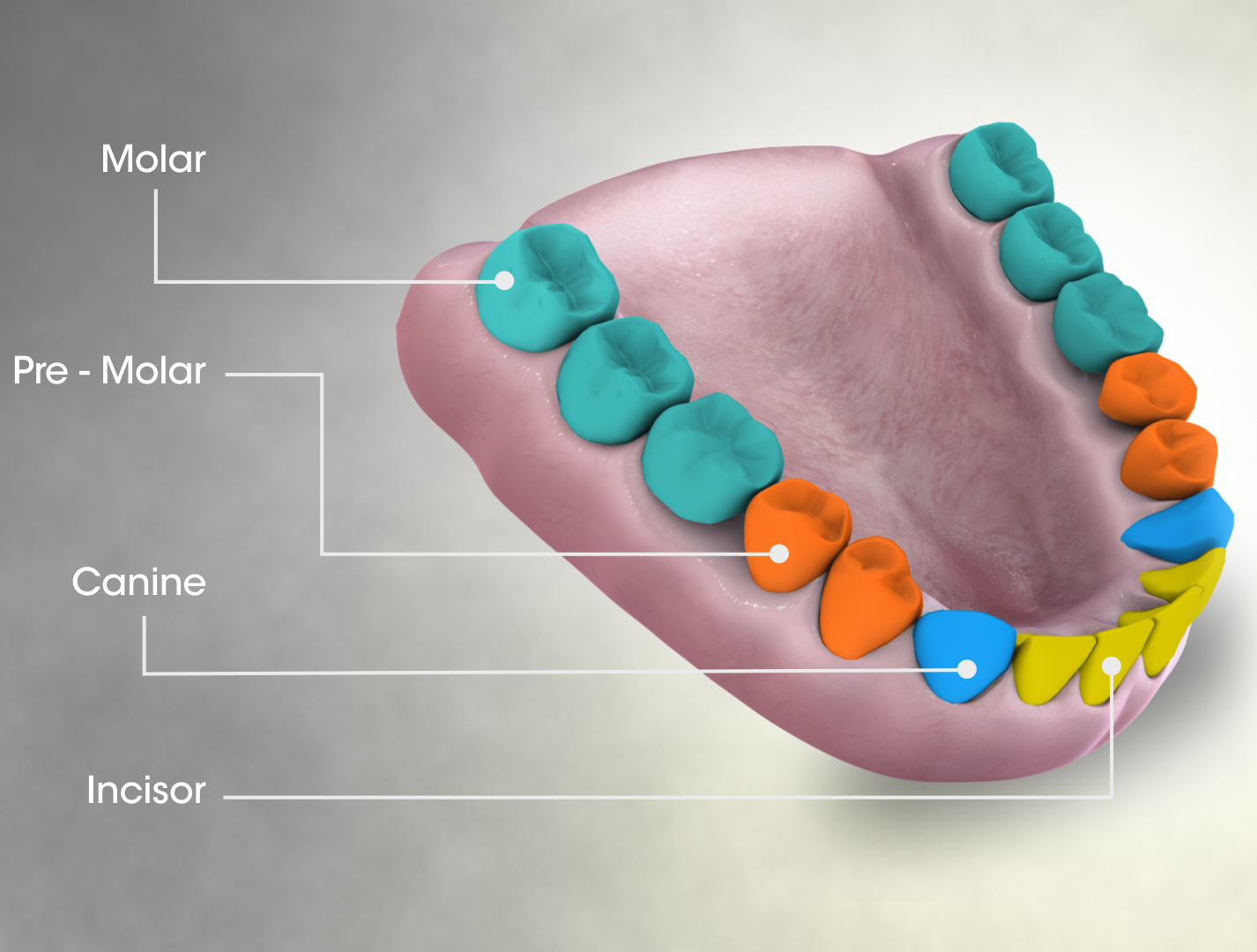
The molars or molar teeth are large, flat teeth at the back of the mouth. They are more developed in mammals. They are used primarily to grind food during chewing. The name ''molar'' derives from Latin, ''molaris dens'', meaning "millstone tooth", from ''mola'', millstone and ''dens'', tooth. Molars show a great deal of diversity in size and shape across mammal groups. The third molar of humans is sometimes vestigial. Human anatomy In humans, the molar teeth have either four or five cusps. Adult humans have 12 molars, in four groups of three at the back of the mouth. The third, rearmost molar in each group is called a wisdom tooth. It is the last tooth to appear, breaking through the front of the gum at about the age of 20, although this varies from individual to individual. Race can also affect the age at which this occurs, with statistical variations between groups. In some cases, it may not even erupt at all. The human mouth contains upper (maxillary) and lower (mandibul ... [...More Info...] [...Related Items...] OR: [Wikipedia] [Google] [Baidu] |
Mastoid Process
The mastoid part of the temporal bone is the posterior (back) part of the temporal bone, one of the bones of the skull. Its rough surface gives attachment to various muscles (via tendons) and it has openings for blood vessels. From its borders, the mastoid part articulates with two other bones. Etymology The word "mastoid" is derived from the Greek word for "breast", a reference to the shape of this bone. Surfaces Outer surface Its outer surface is rough and gives attachment to the occipitalis and posterior auricular muscles. It is perforated by numerous foramina (holes); for example, the mastoid foramen is situated near the posterior border and transmits a vein to the transverse sinus and a small branch of the occipital artery to the dura mater. The position and size of this foramen are very variable; it is not always present; sometimes it is situated in the occipital bone, or in the suture between the temporal and the occipital. Mastoid process The mastoid process is ... [...More Info...] [...Related Items...] OR: [Wikipedia] [Google] [Baidu] |
Hypertrophy
Hypertrophy is the increase in the volume of an organ or tissue due to the enlargement of its component cells. It is distinguished from hyperplasia, in which the cells remain approximately the same size but increase in number.Updated by Linda J. Vorvick. 8/14/1Hyperplasia/ref> Although hypertrophy and hyperplasia are two distinct processes, they frequently occur together, such as in the case of the hormonally-induced proliferation and enlargement of the cells of the uterus during pregnancy. Eccentric hypertrophy is a type of hypertrophy where the walls and chamber of a hollow organ undergo growth in which the overall size and volume are enlarged. It is applied especially to the left ventricle of heart. Sarcomeres are added in series, as for example in dilated cardiomyopathy (in contrast to hypertrophic cardiomyopathy, a type of concentric hypertrophy, where sarcomeres are added in parallel). Gallery File:*+ * Photographic documentation on sexual education - Hypertrophy of bre ... [...More Info...] [...Related Items...] OR: [Wikipedia] [Google] [Baidu] |
Lateral (anatomy)
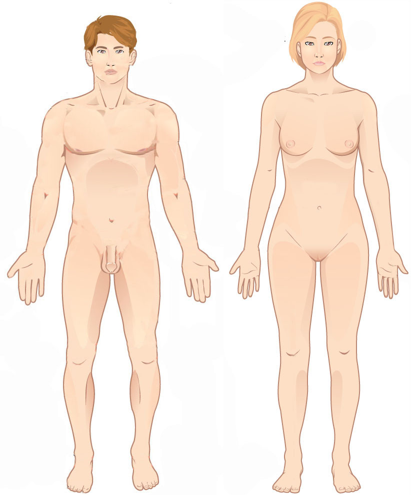
Standard anatomical terms of location are used to unambiguously describe the anatomy of animals, including humans. The terms, typically derived from Latin or Greek roots, describe something in its standard anatomical position. This position provides a definition of what is at the front ("anterior"), behind ("posterior") and so on. As part of defining and describing terms, the body is described through the use of anatomical planes and anatomical axes. The meaning of terms that are used can change depending on whether an organism is bipedal or quadrupedal. Additionally, for some animals such as invertebrates, some terms may not have any meaning at all; for example, an animal that is radially symmetrical will have no anterior surface, but can still have a description that a part is close to the middle ("proximal") or further from the middle ("distal"). International organisations have determined vocabularies that are often used as standard vocabularies for subdisciplines of anatom ... [...More Info...] [...Related Items...] OR: [Wikipedia] [Google] [Baidu] |
Postorbital
The ''postorbital'' is one of the bones in vertebrate skulls which forms a portion of the dermal skull roof and, sometimes, a ring about the orbit. Generally, it is located behind the postfrontal and posteriorly to the orbital fenestra. In some vertebrates, the postorbital is fused with the postfrontal to create a postorbitofrontal. Birds have a separate postorbital as an embryo, but the bone fuses with the frontal Front may refer to: Arts, entertainment, and media Films * ''The Front'' (1943 film), a 1943 Soviet drama film * ''The Front'', 1976 film Music * The Front (band), an American rock band signed to Columbia Records and active in the 1980s and e ... before it hatches. References * Roemer, A. S. 1956. ''Osteology of the Reptiles''. University of Chicago Press. 772 pp. Skull {{Vertebrate anatomy-stub ... [...More Info...] [...Related Items...] OR: [Wikipedia] [Google] [Baidu] |
Premolar
The premolars, also called premolar teeth, or bicuspids, are transitional teeth located between the canine and molar teeth. In humans, there are two premolars per quadrant in the permanent set of teeth, making eight premolars total in the mouth. They have at least two cusps. Premolars can be considered transitional teeth during chewing, or mastication. They have properties of both the canines, that lie anterior and molars that lie posterior, and so food can be transferred from the canines to the premolars and finally to the molars for grinding, instead of directly from the canines to the molars. Human anatomy The premolars in humans are the maxillary first premolar, maxillary second premolar, mandibular first premolar, and the mandibular second premolar. Premolar teeth by definition are permanent teeth distal to the canines, preceded by deciduous molars. Morphology There is always one large buccal cusp, especially so in the mandibular first premolar. The lower second ... [...More Info...] [...Related Items...] OR: [Wikipedia] [Google] [Baidu] |
Dorsal (anatomy)
Standard anatomical terms of location are used to unambiguously describe the anatomy of animals, including humans. The terms, typically derived from Latin or Greek roots, describe something in its standard anatomical position. This position provides a definition of what is at the front ("anterior"), behind ("posterior") and so on. As part of defining and describing terms, the body is described through the use of anatomical planes and anatomical axes. The meaning of terms that are used can change depending on whether an organism is bipedal or quadrupedal. Additionally, for some animals such as invertebrates, some terms may not have any meaning at all; for example, an animal that is radially symmetrical will have no anterior surface, but can still have a description that a part is close to the middle ("proximal") or further from the middle ("distal"). International organisations have determined vocabularies that are often used as standard vocabularies for subdisciplines of anatom ... [...More Info...] [...Related Items...] OR: [Wikipedia] [Google] [Baidu] |
Cingulid
A cingulid is a term used when describing teeth, it refers to a ridge that runs around the base of the crown of a lower tooth (the equivalent on the upper teeth is the cingulum). The presence or absence of a cingulid is often a diagnostic feature for different species of animal, especially among mammals. Some animals don't have a cingulid. Those that do may have them on only some, or all of the teeth, though most often on the molar teeth. It can be on the upper or lower teeth, or both. There are four common descriptions of the position of the cingulid: * Lingual cingulid - a cingulid on the side of the tooth that is next to the tongue *Labial cingulid - a cingulid on the side of the tooth that is next to the lips or cheeks *Distal cingulid - a cingulid on the side of the tooth facing the tooth behind it in the jaw (can also be referred to as a posterior cingulid) *Mesial This is a list of definitions of commonly used terms of location and direction in dentistry. This set of ... [...More Info...] [...Related Items...] OR: [Wikipedia] [Google] [Baidu] |
Entoconid
Many different terms have been proposed for features of the tooth crown in mammals. The structures within the molars receive different names according to their position and morphology. This nomenclature was developed by Henry Fairfield Osborn in 1907 and is, although with many variations, the one that continues today. * The suffix "-cones /-conids" (upper molar/lower molar) is added to the main cusps: Paraconus, Metaconus, Protoconus and Hypoconus on the upper molar, and Paraconid, Metaconid, Protoconid, Hypoconid and Entoconid on the lower molar. This name is used for both bunodont and selenodont molars, that is, as many for "buno" pillar-like cusps as for "selenes" crescent-like cusps. * The suffix "-conule /-conulid" (upper molar/lower molar) is added to the secondary cusps. For example, Metaconule, Hypoconulid. * The suffix "-style/-stylid" (upper molar/lower molar) is added to the peripheral cusps that are found in the cornices or cingulus of the tooth. These cusps are tra ... [...More Info...] [...Related Items...] OR: [Wikipedia] [Google] [Baidu] |
Cheek
The cheeks ( la, buccae) constitute the area of the face below the eyes and between the nose and the left or right ear. "Buccal" means relating to the cheek. In humans, the region is innervated by the buccal nerve. The area between the inside of the cheek and the teeth and gums is called the vestibule or buccal pouch or buccal cavity and forms part of the mouth. In other animals the cheeks may also be referred to as jowls. Structure Humans Cheeks are fleshy in humans, the skin being suspended by the chin and the jaws, and forming the lateral wall of the human mouth, visibly touching the cheekbone below the eye. The inside of the cheek is lined with a mucous membrane (buccal mucosa, part of the oral mucosa). During mastication (chewing), the cheeks and tongue between them serve to keep the food between the teeth. Other animals The cheeks are covered externally by hairy skin, and internally by stratified squamous epithelium. This is mostly smooth, but may have caudally di ... [...More Info...] [...Related Items...] OR: [Wikipedia] [Google] [Baidu] |


