|
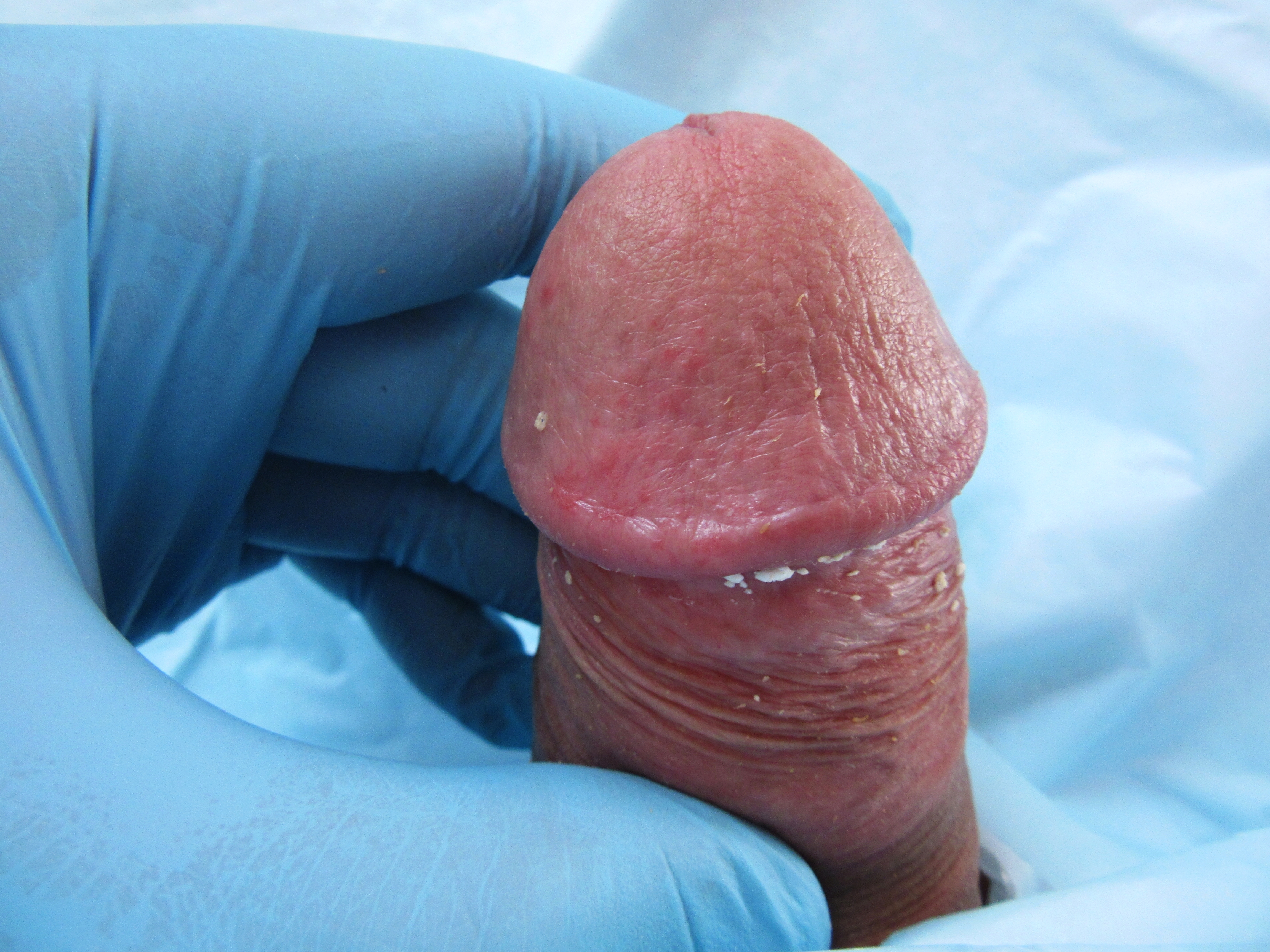
Balanitis Plasmacellularis
Balanitis plasmacellularis is a cutaneous condition characterized by a benign inflammatory skin lesion characterized histologically by a plasma cell infiltrate. A similar condition has been described in women (i.e. "Zoon's vulvitis"), although its existence is controversial due to the possibility of diagnostic error in many of the cases that have been reported in the medical literature. It is named for J.J. Zoon, who characterized it in 1952. See also * Balanitis * Pseudoepitheliomatous keratotic and micaceous balanitis * Skin lesion * List of cutaneous conditions Many skin conditions affect the human integumentary system—the organ system covering the entire surface of the body and composed of skin, hair, nails, and related muscle and glands. The major function of this system is as a barrier against t ... References External links Epidermal nevi, neoplasms, and cysts {{Epidermal-growth-stub ... [...More Info...] [...Related Items...] OR: [Wikipedia] [Google] [Baidu] |
Micrograph
A micrograph or photomicrograph is a photograph or digital image taken through a microscope or similar device to show a magnified image of an object. This is opposed to a macrograph or photomacrograph, an image which is also taken on a microscope but is only slightly magnified, usually less than 10 times. Micrography is the practice or art of using microscopes to make photographs. A micrograph contains extensive details of microstructure. A wealth of information can be obtained from a simple micrograph like behavior of the material under different conditions, the phases found in the system, failure analysis, grain size estimation, elemental analysis and so on. Micrographs are widely used in all fields of microscopy. Types Photomicrograph A light micrograph or photomicrograph is a micrograph prepared using an optical microscope, a process referred to as ''photomicroscopy''. At a basic level, photomicroscopy may be performed simply by connecting a camera to a microscope, th ... [...More Info...] [...Related Items...] OR: [Wikipedia] [Google] [Baidu] |
H&E Stain
Hematoxylin and eosin stain ( or haematoxylin and eosin stain or hematoxylin-eosin stain; often abbreviated as H&E stain or HE stain) is one of the principal tissue stains used in histology. It is the most widely used stain in medical diagnosis and is often the gold standard. For example, when a pathologist looks at a biopsy of a suspected cancer, the histological section is likely to be stained with H&E. H&E is the combination of two histological stains: hematoxylin and eosin. The hematoxylin stains cell nuclei a purplish blue, and eosin stains the extracellular matrix and cytoplasm pink, with other structures taking on different shades, hues, and combinations of these colors. Hence a pathologist can easily differentiate between the nuclear and cytoplasmic parts of a cell, and additionally, the overall patterns of coloration from the stain show the general layout and distribution of cells and provides a general overview of a tissue sample's structure. Thus, pattern recogniti ... [...More Info...] [...Related Items...] OR: [Wikipedia] [Google] [Baidu] |
Benign
Malignancy () is the tendency of a medical condition to become progressively worse. Malignancy is most familiar as a characterization of cancer. A ''malignant'' tumor contrasts with a non-cancerous benign tumor, ''benign'' tumor in that a malignancy is not self-limited in its growth, is capable of invading into adjacent tissues, and may be capable of spreading to distant tissues. A benign tumor has none of those properties. Malignancy in cancers is characterized by anaplasia, invasiveness, and metastasis. Malignant tumors are also characterized by genome instability, so that cancers, as assessed by whole genome sequencing, frequently have between 10,000 and 100,000 mutations in their entire genomes. Cancers usually show tumour heterogeneity, containing multiple subclones. They also frequently have reduced expression of DNA repair enzymes due to Epigenetics#DNA repair epigenetics in cancer, epigenetic methylation of DNA repair genes or altered MicroRNA#DNA repair and cancer, micr ... [...More Info...] [...Related Items...] OR: [Wikipedia] [Google] [Baidu] |
Skin Lesion
A skin condition, also known as cutaneous condition, is any medical condition that affects the integumentary system—the organ system that encloses the body and includes skin, nails, and related muscle and glands. The major function of this system is as a barrier against the external environment. Conditions of the human integumentary system constitute a broad spectrum of diseases, also known as dermatoses, as well as many nonpathologic states (like, in certain circumstances, melanonychia and racquet nails). While only a small number of skin diseases account for most visits to the physician, thousands of skin conditions have been described. Classification of these conditions often presents many nosological challenges, since underlying causes and pathogenetics are often not known. Therefore, most current textbooks present a classification based on location (for example, conditions of the mucous membrane), morphology ( chronic blistering conditions), cause (skin conditions resul ... [...More Info...] [...Related Items...] OR: [Wikipedia] [Google] [Baidu] |
Histology
Histology, also known as microscopic anatomy or microanatomy, is the branch of biology which studies the microscopic anatomy of biological tissues. Histology is the microscopic counterpart to gross anatomy, which looks at larger structures visible without a microscope. Although one may divide microscopic anatomy into ''organology'', the study of organs, ''histology'', the study of tissues, and ''cytology'', the study of cells, modern usage places all of these topics under the field of histology. In medicine, histopathology is the branch of histology that includes the microscopic identification and study of diseased tissue. In the field of paleontology, the term paleohistology refers to the histology of fossil organisms. Biological tissues Animal tissue classification There are four basic types of animal tissues: muscle tissue, nervous tissue, connective tissue, and epithelial tissue. All animal tissues are considered to be subtypes of these four principal tissue types ... [...More Info...] [...Related Items...] OR: [Wikipedia] [Google] [Baidu] |
Plasma Cell
Plasma cells, also called plasma B cells or effector B cells, are white blood cells that originate in the lymphoid organs as B lymphocytes and secrete large quantities of proteins called antibodies in response to being presented specific substances called antigens. These antibodies are transported from the plasma cells by the blood plasma and the lymphatic system to the site of the target antigen (foreign substance), where they initiate its neutralization or destruction. B cells differentiate into plasma cells that produce antibody molecules closely modeled after the receptors of the precursor B cell. Structure Plasma cells are large lymphocytes with abundant cytoplasm and a characteristic appearance on light microscopy. They have basophilic cytoplasm and an eccentric nucleus with heterochromatin in a characteristic cartwheel or clock face arrangement. Their cytoplasm also contains a pale zone that on electron microscopy contains an extensive Golgi apparatus and centrioles ... [...More Info...] [...Related Items...] OR: [Wikipedia] [Google] [Baidu] |
Vulvitis
Vulvitis is inflammation of the vulva, the external female mammalian genitalia that include the labia majora, labia minora, clitoris, and introitus (the entrance to the vagina). It may co-occur as vulvovaginitis with vaginitis, inflammation of the vagina, and may have infectious or non-infectious causes. The warm and moist conditions of the vulva make it easily affected. Vulvitis is prone to occur in any female especially those who have certain sensitivities, infections, allergies, or diseases that make them likely to have vulvitis. Postmenopausal women and prepubescent girls are more prone to be affected by it, as compared to women in their menstruation period. It is so because they have low estrogen levels which makes their vulvar tissue thin and dry. Women having diabetes are also prone to be affected by vulvitis due to the high sugar content in their cells, increasing their vulnerability. Vulvitis is not a disease, it is just an inflammation caused by an infection, allergy or i ... [...More Info...] [...Related Items...] OR: [Wikipedia] [Google] [Baidu] |
Balanitis
Balanitis is inflammation of the glans penis. When the foreskin is also affected, the proper term is balanoposthitis. Balanitis on boys still in diapers must be distinguished from redness caused by ammoniacal dermatitis. The word ''balanitis'' is from the Greek βάλανος'' '', literally meaning 'acorn', used because of the similarity in shape to the glans penis. Signs and symptoms * Small red erosions on the glans (first sign) * Redness of the foreskin * Redness of the penis * Other rashes on the head of the penis * Foul smelling discharge * Painful foreskin and penis Complications Recurrent bouts of balanitis may cause scarring of the preputial orifice; the reduced elasticity may lead to pathologic phimosis. Furthecomplicationsmay include: * Stricture of urethral meatus * Phimosis * Paraphimosis Cause Inflammation has many possible causes, including irritation by environmental substances, physical trauma, and infection such as bacterial, viral, or fungal. Some of these ... [...More Info...] [...Related Items...] OR: [Wikipedia] [Google] [Baidu] |
Pseudoepitheliomatous Keratotic And Micaceous Balanitis
Pseudoepitheliomatous keratotic and micaceous balanitis is a cutaneous condition characterized by skin lesions on the glans penis that are wart-like with scaling. It can present as a cutaneous horn. See also * Balanitis * Balanitis plasmacellularis * Skin lesion A skin condition, also known as cutaneous condition, is any medical condition that affects the integumentary system—the organ system that encloses the body and includes skin, nails, and related muscle and glands. The major function of this s ... References Epidermal nevi, neoplasms, and cysts {{Epidermal-growth-stub ... [...More Info...] [...Related Items...] OR: [Wikipedia] [Google] [Baidu] |
Skin Lesion
A skin condition, also known as cutaneous condition, is any medical condition that affects the integumentary system—the organ system that encloses the body and includes skin, nails, and related muscle and glands. The major function of this system is as a barrier against the external environment. Conditions of the human integumentary system constitute a broad spectrum of diseases, also known as dermatoses, as well as many nonpathologic states (like, in certain circumstances, melanonychia and racquet nails). While only a small number of skin diseases account for most visits to the physician, thousands of skin conditions have been described. Classification of these conditions often presents many nosological challenges, since underlying causes and pathogenetics are often not known. Therefore, most current textbooks present a classification based on location (for example, conditions of the mucous membrane), morphology ( chronic blistering conditions), cause (skin conditions resul ... [...More Info...] [...Related Items...] OR: [Wikipedia] [Google] [Baidu] |
List Of Cutaneous Conditions
Many skin conditions affect the human integumentary system—the organ system covering the entire surface of the body and composed of skin, hair, nails, and related muscle and glands. The major function of this system is as a barrier against the external environment. The skin weighs an average of four kilograms, covers an area of two square metres, and is made of three distinct layers: the epidermis, dermis, and subcutaneous tissue. The two main types of human skin are: glabrous skin, the hairless skin on the palms and soles (also referred to as the "palmoplantar" surfaces), and hair-bearing skin.Burns, Tony; ''et al''. (2006) ''Rook's Textbook of Dermatology CD-ROM''. Wiley-Blackwell. . Within the latter type, the hairs occur in structures called pilosebaceous units, each with hair follicle, sebaceous gland, and associated arrector pili muscle. In the embryo, the epidermis, hair, and glands form from the ectoderm, which is chemically influenced by the underlying mesoderm th ... [...More Info...] [...Related Items...] OR: [Wikipedia] [Google] [Baidu] |


