|
Back Porch Cafe
The human back, also called the dorsum, is the large posterior area of the human body, rising from the top of the buttocks to the back of the neck. It is the surface of the body opposite from the chest and the abdomen. The vertebral column runs the length of the back and creates a central area of recession. The breadth of the back is created by the shoulders at the top and the pelvis at the bottom. Back pain is a common medical condition, generally benign in origin. Structure The central feature of the human back is the vertebral column, specifically the length from the top of the thoracic vertebrae to the bottom of the lumbar vertebrae, which houses the spinal cord in its spinal canal, and which generally has some curvature that gives shape to the back. The ribcage extends from the spine at the top of the back (with the top of the ribcage corresponding to the T1 vertebra), more than halfway down the length of the back, leaving an area with less protection between ... [...More Info...] [...Related Items...] OR: [Wikipedia] [Google] [Baidu] |
Posterior (anatomy)
Standard anatomical terms of location are used to unambiguously describe the anatomy of animals, including humans. The terms, typically derived from Latin or Greek language, Greek roots, describe something in its standard anatomical position. This position provides a definition of what is at the front ("anterior"), behind ("posterior") and so on. As part of defining and describing terms, the body is described through the use of anatomical planes and anatomical axis, anatomical axes. The meaning of terms that are used can change depending on whether an organism is bipedal or quadrupedal. Additionally, for some animals such as invertebrates, some terms may not have any meaning at all; for example, an animal that is radially symmetrical will have no anterior surface, but can still have a description that a part is close to the middle ("proximal") or further from the middle ("distal"). International organisations have determined vocabularies that are often used as standard vocabular ... [...More Info...] [...Related Items...] OR: [Wikipedia] [Google] [Baidu] |
Cutaneous Nerve
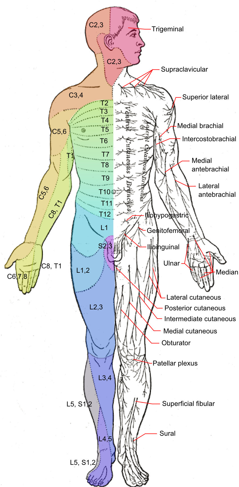
A cutaneous nerve is a nerve that provides nerve supply to the skin. Human anatomy In human anatomy, cutaneous nerves are primarily responsible for providing sensory innervation to the skin. In addition to sympathetic and autonomic afferent (sensory) fibers, most cutaneous nerves also contain sympathetic efferent (visceromotor) fibers, which innervate cutaneous blood vessels, sweat glands, and the arrector pilli muscles of hair follicles. These structures are important to the sympathetic nervous response. There are many cutaneous nerves in the human body, only some of which are named. Some of the larger cutaneous nerves are as follows: Upper body * In the arm (proper) ** Superior lateral cutaneous nerve of arm (Superior LCNOA) ** Inferior lateral cutaneous nerve of arm (Inferior LCNOA) ** Posterior cutaneous nerve of arm (PCNOA) ** Medial cutaneous nerve of arm (MCNOA) * In the forearm ** Lateral cutaneous nerve of forearm (LCNOF) ** Posterior cutaneous nerve of forearm (PCNOF ... [...More Info...] [...Related Items...] OR: [Wikipedia] [Google] [Baidu] |
Longissimus
The longissimus ( la, the longest one) is the muscle lateral to the semispinalis muscles. It is the longest subdivision of the erector spinae muscles that extends forward into the transverse processes of the posterior cervical vertebrae. Structure Longissimus thoracis et lumborum The longissimus thoracis et lumborum is the intermediate and largest of the continuations of the erector spinae. In the lumbar region (longissimus lumborum), where it is as yet blended with the iliocostalis, some of its fibers are attached to the whole length of the posterior surfaces of the transverse processes and the accessory processes of the lumbar vertebrae, and to the anterior layer of the lumbodorsal fascia. In the thoracic region (longissimus thoracis), it is inserted, by rounded tendons, into the tips of the transverse processes of all the thoracic vertebrae, and by fleshy processes into the lower nine or ten ribs between their tubercles and angles. Longissimus cervicis The longissimus cervic ... [...More Info...] [...Related Items...] OR: [Wikipedia] [Google] [Baidu] |
Iliocostalis
Iliocostalis muscle is the muscle immediately lateral to the longissimus that is the nearest to the furrow that separates the epaxial muscles from the hypaxial. It lies very deep to the fleshy portion of the serratus posterior muscle. It laterally flexes the vertebral column to the same side. Structure Iliocostalis muscle has a common origin from the iliac crest, the sacrum, the thoracolumbar fascia, and the spinous processes of the vertebrae from T11 to L5. Iliocostalis cervicis (cervicalis ascendens) arises from the angles of the third, fourth, fifth, and sixth ribs, and is inserted into the posterior tubercles of the transverse processes of the fourth, fifth, and sixth cervical vertebrae. Iliocostalis thoracis (musculus accessorius; iliocostalis thoracis) arises by flattened tendons from the upper borders of the angles of the lower six ribs medial to the tendons of insertion of the iliocostalis lumborum; these become muscular, and are inserted into the upper borders of t ... [...More Info...] [...Related Items...] OR: [Wikipedia] [Google] [Baidu] |
Erector Spinae Muscles
The erector spinae ( ) or spinal erectors is a set of muscles that straighten and rotate the back. The spinal erectors work together with the glutes (gluteus maximus, gluteus medius and gluteus minimus) to maintain stable posture standing or sitting. Structure The erector spinae is not just one muscle, but a group of muscles and tendons which run more or less the length of the spine on the left and the right, from the sacrum, or sacral region, and hips to the base of the skull. They are also known as the sacrospinalis group of muscles. These muscles lie on either side of the spinous processes of the vertebrae and extend throughout the lumbar, thoracic, and cervical regions. The erector spinae is covered in the lumbar and thoracic regions by the thoracolumbar fascia, and in the cervical region by the nuchal ligament. This large muscular and tendinous mass varies in size and structure at different parts of the vertebral column. In the sacral region, it is narrow and pointed, and at ... [...More Info...] [...Related Items...] OR: [Wikipedia] [Google] [Baidu] |
Splenius Cervicis Muscle
The splenius cervicis () (also known as the splenius colli, ) is a muscle in the back of the neck. It arises by a narrow tendinous band from the spinous processes of the third to the sixth thoracic vertebrae; it is inserted, by tendinous fasciculi, into the posterior tubercles of the transverse processes of the upper two or three cervical vertebrae In tetrapods, cervical vertebrae (singular: vertebra) are the vertebrae of the neck, immediately below the skull. Truncal vertebrae (divided into thoracic and lumbar vertebrae in mammals) lie caudal (toward the tail) of cervical vertebrae. In .... Its name is based on the Greek word σπληνίον, ''splenion'' (meaning a bandage) and the Latin word ''cervix'' (meaning a neck). The word ''collum'' also refers to the neck in Latin. The function of the splenius cervicis muscle is extension of the cervical spine, rotation to the ipsilateral side and lateral flexion to the ipsilateral side.R.T. Floyd, Manual of Structural Kinesiolo ... [...More Info...] [...Related Items...] OR: [Wikipedia] [Google] [Baidu] |
Splenius Capitis Muscle
The splenius capitis () () is a broad, straplike muscle in the back of the neck. It pulls on the base of the skull from the vertebrae in the neck and upper thorax. It is involved in movements such as shaking the head. Structure It arises from the lower half of the nuchal ligament, from the spinous process of the seventh cervical vertebra, and from the spinous processes of the upper three or four thoracic vertebrae. The fibers of the muscle are directed upward and laterally and are inserted, under cover of the sternocleidomastoideus, into the mastoid process of the temporal bone, and into the rough surface on the occipital bone just below the lateral third of the superior nuchal line. The splenius capitis is deep to sternocleidomastoideus at the mastoid process, and to the trapezius for its lower portion. It is one of the muscles that forms the floor of the posterior triangle of the neck. The splenius capitis muscle is innervated by the posterior ramus of spinal nerves C3 and ... [...More Info...] [...Related Items...] OR: [Wikipedia] [Google] [Baidu] |
Serratus Posterior Inferior Muscle
The serratus posterior inferior muscle, also known as the posterior serratus muscle, is a muscle of the human body. Structure The muscle is situated at the junction of the thoracic and lumbar regions. It has an irregularly quadrilateral form, broader than the serratus posterior superior muscle, and separated from it by a wide interval. It arises by a thin aponeurosis from the spinous processes of the lower two thoracic and upper two or three lumbar vertebrae. Passing obliquely upward and lateralward, it becomes fleshy, and divides into four flat digitations. These are inserted into the inferior borders of the lower four ribs, a little beyond their angles. The thin aponeurosis of origin is intimately blended with the thoracolumbar fascia, and aponeurosis of the latissimus dorsi muscle. Function The serratus posterior inferior draws the lower ribs backward and downward to assist in rotation and extension of the trunk. This movement of the ribs may also contribute to inhalatio ... [...More Info...] [...Related Items...] OR: [Wikipedia] [Google] [Baidu] |
Serratus Posterior Superior Muscle
The serratus posterior superior muscle is a thin, quadrilateral muscle. It is situated at the upper back part of the thorax, deep to the rhomboid muscles. Structure The serratus posterior superior muscle arises by an aponeurosis from the lower part of the nuchal ligament, from the spinous processes of C7, T1, T2, and sometimes T3, and from the supraspinal ligament. It is inserted, by four fleshy digitations into the upper borders of the second, third, fourth, and fifth ribs past the angle of the rib. Function The serratus posterior superior muscle elevates the second to fifth ribs. This aids deep respiration. Additional images File:Serratus posterior superior muscle animation small.gif, Position of serratus posterior superior muscle (shown in red). File:Serratus posterior superior.jpg, Serratus posterior superior muscles are labeled at center left and center right. See also * Serratus anterior muscle * Serratus posterior inferior muscle The serratus posterior ... [...More Info...] [...Related Items...] OR: [Wikipedia] [Google] [Baidu] |
Ventral Ramus Of Spinal Nerve
The ventral ramus (pl. ''rami'') (Latin for ''branch'') is the anterior division of a spinal nerve. The ventral rami supply the antero-lateral parts of the trunk and the limbs. They are mainly larger than the dorsal rami. Shortly after a spinal nerve exits the intervertebral foramen, it branches into the dorsal ramus, the ventral ramus, and the ramus communicans. Each of these three structures carries both sensory and motor information. Each spinal nerve carries both sensory and motor information, via efferent and afferent nerve fibers - ultimately via the motor cortex in the frontal lobe and to somatosensory cortex in the parietal lobe - but also through the phenomenon of reflex. Spinal nerves are referred to as "mixed nerves". In the thoracic region they remain distinct from each other and each innervates a narrow strip of muscle and skin along the sides, chest, ribs, and abdominal wall. These rami are called the intercostal nerves. In regions other than the thoracic, ventr ... [...More Info...] [...Related Items...] OR: [Wikipedia] [Google] [Baidu] |
Levator Scapulae Muscle
The levator scapulae is a slender skeletal muscle situated at the back and side of the neck. As the Latin name suggests, its main function is to lift the scapula. Anatomy Attachments The muscle descends diagonally from its origin to its insertion. Origin The levator scapulae originates from the posterior tubercles of the transverse processes of cervical vertebrae C1-4. At its origin, it attaches via tendinous slips. Insertion It inserts onto the medial border of the scapula (with its site of insertion extending between the superior angle of the scapula superiorly, and the junction of spine of scapula and medial border of scapula inferiorly). Relations One of the muscles within the floor of the posterior triangle of the neck, the superior part of levator scapulae is covered by sternocleidomastoid and its inferior part by the trapezius. It is bounded in front by the scalenus medius and behind by splenius cervicis. The spinal accessory nerve crosses laterally in the middl ... [...More Info...] [...Related Items...] OR: [Wikipedia] [Google] [Baidu] |
Rhomboid Minor Muscle
In human anatomy, the rhomboid minor is a small skeletal muscle on the back that connects the scapula with the vertebrae of the spinal column. Located inferior to levator scapulae and superior to rhomboid major, it acts together with the latter to keep the scapula pressed against the thoracic wall. It lies deep to trapezius but superficial to the long spinal muscles. Origin and insertion The rhomboid minor arises from the inferior border of the nuchal ligament, from the spinous processes of the seventh cervical and first thoracic vertebrae, and from the intervening supraspinous ligaments. It is inserted into a small area of the medial border of the scapula at the level of the scapular spine. Action Together with the rhomboid major, the rhomboid minor retracts the scapula when trapezius is contracted. Acting as a synergist to the trapezius, the rhomboid major and minor elevate the medial border of the scapula medially and upward, working in tandem with the levator scapu ... [...More Info...] [...Related Items...] OR: [Wikipedia] [Google] [Baidu] |

