|
Bachmann's Bundle
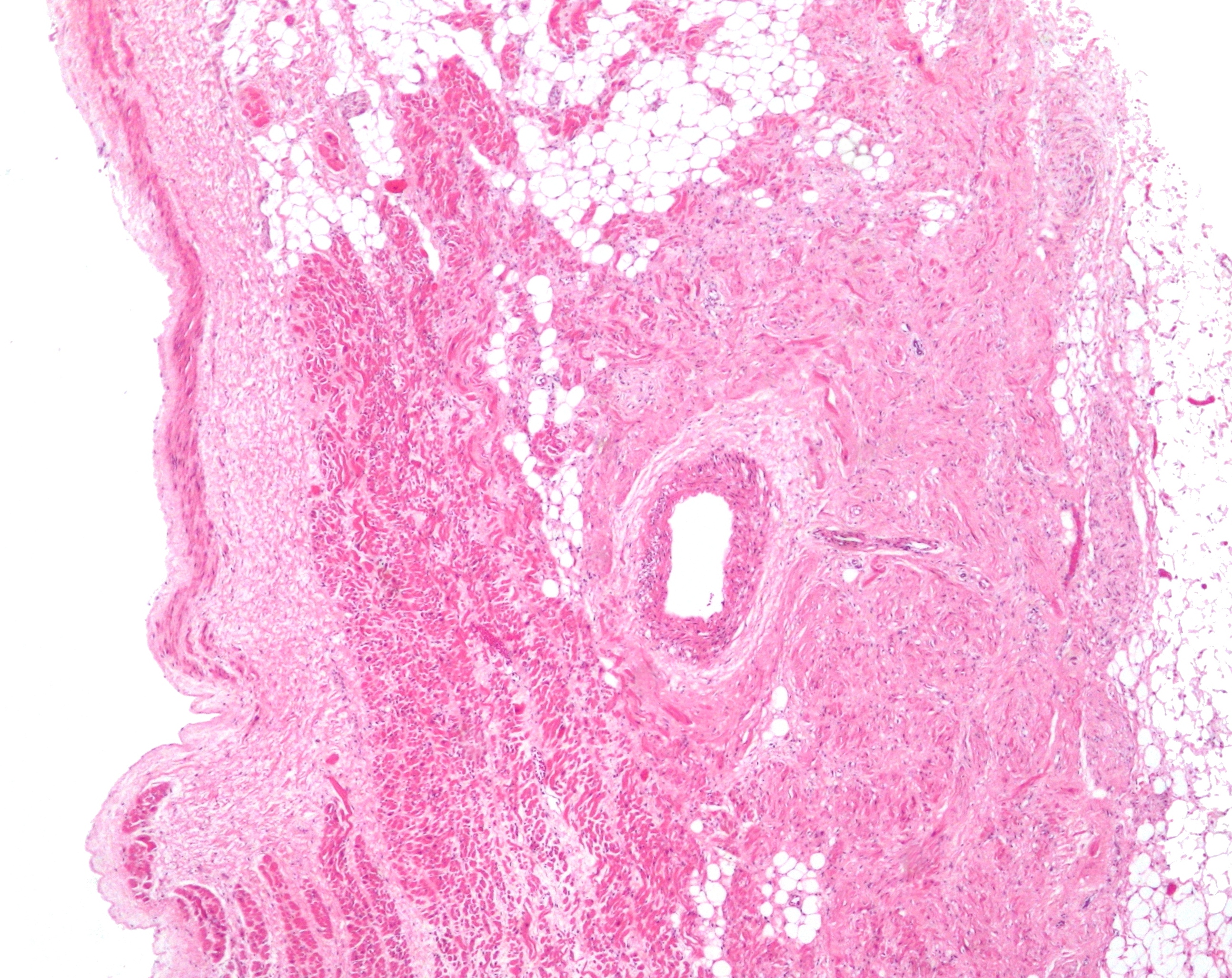
In the heart's conduction system, Bachmann's bundle (also called the Bachmann bundle or the interatrial band) is a branch of the anterior internodal tract that resides on the inner wall of the left atrium. It is a broad band of cardiac muscle that passes from the right atrium, between the superior vena cava and the ascending aorta. Bachmann's bundle is, during normal sinus rhythm, the preferential path for electrical activation of the left atrium. It is therefore considered to be part of the "atrial conduction system" of the heart. History In 1916, Jean George Bachmann conducted canine experiments. He found that clamping the muscular bundle of fibers that connects the atria caused a significant conduction delay. Structure Bachman's bundle receives its blood supply from the sinoatrial nodal artery (right, left or both). Besides Bachmann's bundle, the other three conduction tracts that constitute the ''atrial conduction system'' are known as the anterior, middle, and poste ... [...More Info...] [...Related Items...] OR: [Wikipedia] [Google] [Baidu] |
Heart
The heart is a muscular organ found in most animals. This organ pumps blood through the blood vessels of the circulatory system. The pumped blood carries oxygen and nutrients to the body, while carrying metabolic waste such as carbon dioxide to the lungs. In humans, the heart is approximately the size of a closed fist and is located between the lungs, in the middle compartment of the chest, called the mediastinum. In humans, other mammals, and birds, the heart is divided into four chambers: upper left and right atria and lower left and right ventricles. Commonly, the right atrium and ventricle are referred together as the right heart and their left counterparts as the left heart. Fish, in contrast, have two chambers, an atrium and a ventricle, while most reptiles have three chambers. In a healthy heart, blood flows one way through the heart due to heart valves, which prevent backflow. The heart is enclosed in a protective sac, the pericardium, which also contains ... [...More Info...] [...Related Items...] OR: [Wikipedia] [Google] [Baidu] |
Sinoatrial Nodal Artery
The sinoatrial nodal artery (or sinuatrial nodal artery or sinoatrial artery) is an artery of the heart which supplies the sinoatrial node, the natural pacemaker center of the heart, and arises from the right coronary artery in around 60% of people. In about 40% of cases, the sinoatrial artery is a branch of the left circumflex coronary artery. In less than 1% of humans, the artery has an anomalous origin directly from the coronary sinus, descending aorta, or distal right coronary artery. In more than 50% of human hearts, the artery actually courses close to the superior posterior aspect of the interatrial septum. The origin of the sinoatrial node artery is not related to coronary artery dominance, which means the side (right or left) that provides the circulation to the back of the heart. In contrast, the atrioventricular nodal branch, that is the artery that brings blood to the atrioventricular node, depends on coronary artery dominance. The sinoatrial node surrounds the sinoat ... [...More Info...] [...Related Items...] OR: [Wikipedia] [Google] [Baidu] |
Interatrial Septum
The interatrial septum is the wall of tissue that separates the right and left atria of the heart. Structure The interatrial septum is a that lies between the left atrium and right atrium of the human heart. The interatrial septum lies at angle of 65 degrees from right posterior to left anterior because right atrium is located at the right side of the body while left atrium is located at the left side of the body. Development The interatrial septum forms during the first and second months of fetal development. Formation of the septum occurs in several stages. The first is the development of the septum primum, a crescent-shaped piece of tissue forming the initial divider between the right and left atria. Because of its crescent shape, the septum primum does not fully occlude the space between the left and right atria; the opening that remains is called the ostium primum. During fetal development, this opening allows blood to be shunted from the right atrium to the left. ... [...More Info...] [...Related Items...] OR: [Wikipedia] [Google] [Baidu] |
Sinoatrial Node
The sinoatrial node (also known as the sinuatrial node, SA node or sinus node) is an oval shaped region of special cardiac muscle in the upper back wall of the right atrium made up of cells known as pacemaker cells. The sinus node is approximately fifteen mm long, three mm wide, and one mm thick, located directly below and to the side of the superior vena cava. These cells can produce an electrical impulse an action potential known as a cardiac action potential that travels through the electrical conduction system of the heart, causing it to contract. In a healthy heart, the SA node continuously produces action potentials, setting the rhythm of the heart ( sinus rhythm), and so is known as the heart's natural pacemaker. The rate of action potentials produced (and therefore the heart rate) is influenced by the nerves that supply it. Structure The sinoatrial node is a oval-shaped structure that is approximately fifteen mm long, three mm wide, and one mm thick, located d ... [...More Info...] [...Related Items...] OR: [Wikipedia] [Google] [Baidu] |
Coronary Sinus
In anatomy, the coronary sinus () is a collection of veins joined together to form a large vessel that collects blood from the heart muscle ( myocardium). It delivers deoxygenated blood to the right atrium, as do the superior and inferior venae cavae. It is present in all mammals, including humans. The coronary sinus drains into the right atrium, at the coronary sinus orifice, an opening between the inferior vena cava and the right atrioventricular orifice or tricuspid valve. It returns blood from the heart muscle, and is protected by a semicircular fold of the lining membrane of the auricle, the valve of coronary sinus (or valve of Thebesius). The sinus, before entering the atrium, is considerably dilated - nearly to the size of the end of the little finger. Its wall is partly muscular, and at its junction with the great cardiac vein is somewhat constricted and furnished with a valve, known as the valve of Vieussens consisting of two unequal segments. Structure The coron ... [...More Info...] [...Related Items...] OR: [Wikipedia] [Google] [Baidu] |
Atrioventricular Node
The atrioventricular node or AV node electrically connects the heart's atria and ventricles to coordinate beating in the top of the heart; it is part of the electrical conduction system of the heart. The AV node lies at the lower back section of the interatrial septum near the opening of the coronary sinus, and conducts the normal electrical impulse from the atria to the ventricles. The AV node is quite compact (~1 x 3 x 5 mm).Full Size Picture triangle of-Koch.jpg Retrieved on 2008-12-22 Structure Location The AV node lies at the lower back section of the interatrial septum nea ...[...More Info...] [...Related Items...] OR: [Wikipedia] [Google] [Baidu] |
Posterior (anatomy)
Standard anatomical terms of location are used to unambiguously describe the anatomy of animals, including humans. The terms, typically derived from Latin or Greek roots, describe something in its standard anatomical position. This position provides a definition of what is at the front ("anterior"), behind ("posterior") and so on. As part of defining and describing terms, the body is described through the use of anatomical planes and anatomical axes. The meaning of terms that are used can change depending on whether an organism is bipedal or quadrupedal. Additionally, for some animals such as invertebrates, some terms may not have any meaning at all; for example, an animal that is radially symmetrical will have no anterior surface, but can still have a description that a part is close to the middle ("proximal") or further from the middle ("distal"). International organisations have determined vocabularies that are often used as standard vocabularies for subdisciplines of anatomy ... [...More Info...] [...Related Items...] OR: [Wikipedia] [Google] [Baidu] |
Jean George Bachmann
Jean may refer to: People * Jean (female given name) * Jean (male given name) * Jean (surname) Fictional characters * Jean Grey, a Marvel Comics character * Jean Valjean, fictional character in novel ''Les Misérables'' and its adaptations * Jean Pierre Polnareff, a fictional character from ''JoJo's Bizarre Adventure'' Places * Jean, Nevada, USA; a town * Jean, Oregon, USA Entertainment * Jean (dog), a female collie in silent films * "Jean" (song) (1969), by Rod McKuen, also recorded by Oliver * ''Jean Seberg'' (musical), a 1983 musical by Marvin Hamlisch Other uses * JEAN (programming language) * USS ''Jean'' (ID-1308), American cargo ship c. 1918 * Sternwheeler Jean, a 1938 paddleboat of the Willamette River See also *Jehan * * Gene (other) * Jeanne (other) * Jehanne (other) * Jeans (other) * John (other) John is a common English name and surname: * John (given name) * John (surname) John may also refer to: New ... [...More Info...] [...Related Items...] OR: [Wikipedia] [Google] [Baidu] |
Electrical Conduction System Of The Heart
The cardiac conduction system (CCS) (also called the electrical conduction system of the heart) transmits the signals generated by the sinoatrial node – the heart's pacemaker, to cause the heart muscle to contract, and pump blood through the body's circulatory system. The pacemaking signal travels through the right atrium to the atrioventricular node, along the bundle of His, and through the bundle branches to Purkinje fibers in the walls of the ventricles. The Purkinje fibers transmit the signals more rapidly to stimulate contraction of the ventricles. The conduction system consists of specialized heart muscle cells, situated within the myocardium. There is a skeleton of fibrous tissue that surrounds the conduction system which can be seen on an ECG. Dysfunction of the conduction system can cause irregular heart rhythms including rhythms that are too fast or too slow. Structure Electrical signals arising in the SA node (located in the right atrium) stimulate th ... [...More Info...] [...Related Items...] OR: [Wikipedia] [Google] [Baidu] |
Sinus Rhythm
A sinus rhythm is any cardiac rhythm in which depolarisation of the cardiac muscle begins at the sinus node. It is characterised by the presence of correctly oriented P waves on the electrocardiogram (ECG). Sinus rhythm is necessary, but not sufficient, for normal electrical activity within the heart. The term normal sinus rhythm (NSR) is sometimes used to denote a specific type of sinus rhythm where all other measurements on the ECG also fall within designated normal limits, giving rise to the characteristic appearance of the ECG when the electrical conduction system of the heart is functioning normally; however, other sinus rhythms can be entirely normal in particular patient groups and clinical contexts, so the term is sometimes considered a misnomer and its use is sometimes discouraged. Other types of sinus rhythm that can be normal include sinus tachycardia, sinus bradycardia, and sinus arrhythmia. Sinus rhythms may be present together with various other cardiac arr ... [...More Info...] [...Related Items...] OR: [Wikipedia] [Google] [Baidu] |
Ascending Aorta
The ascending aorta (AAo) is a portion of the aorta commencing at the upper part of the base of the left ventricle, on a level with the lower border of the third costal cartilage behind the left half of the sternum. Structure It passes obliquely upward, forward, and to the right, in the direction of the heart's axis, as high as the upper border of the second right costal cartilage, describing a slight curve in its course, and being situated, about behind the posterior surface of the sternum. The total length is about . Components The aortic root is the portion of the aorta beginning at the aortic annulus and extending to the sinotubular junction. It is sometimes regarded as a part of the ascending aorta, and sometimes regarded as a separate entity from the rest of the ascending aorta. Between each commissure of the aortic valve and opposite the cusps of the aortic valve, three small dilatations called the aortic sinuses. The sinotubular junction is the point in the ascend ... [...More Info...] [...Related Items...] OR: [Wikipedia] [Google] [Baidu] |

