|
BEST2
Bestrophin-2 is a protein that in humans is encoded by the ''BEST2'' gene In biology, the word gene (from , ; "... Wilhelm Johannsen coined the word gene to describe the Mendelian units of heredity..." meaning ''generation'' or ''birth'' or ''gender'') can have several different meanings. The Mendelian gene is a b .... Function This gene is a member of the bestrophin gene family of anion channels. Bestrophin genes share a similar gene structure with highly conserved exon-intron boundaries, but with distinct 3' ends. Bestrophins are transmembrane proteins that contain a homologous region rich in aromatic residues, including an invariant arg-phe-pro motif. Mutation in one of the family members ( bestrophin 1) is associated with vitelliform macular dystrophy. The bestrophin 2 gene is mainly expressed in the non-pigmented ciliary epithelium and colon. References External links * Further reading * * * * * * Ion channels {{gene-19-stub ... [...More Info...] [...Related Items...] OR: [Wikipedia] [Google] [Baidu] |
Bestrophin 1
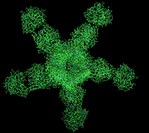
Bestrophin-1 (Best1) is a protein that, in humans, is encoded by the ''BEST1'' gene (RPD ID - 5T5N/4RDQ). The bestrophin family of proteins comprises four evolutionary related genes (BEST1, BEST2, BEST3, and BEST4) that code for integral membrane proteins. This family was first identified in humans by linking a BEST1 mutation with Best vitelliform macular dystrophy (BVMD). Mutations in the BEST1 gene have been identified as the primary cause for at least five different degenerative retinal diseases. The bestrophins are an ancient family of structurally conserved proteins that have been identified in nearly every organism studied from bacteria to humans. In humans, they function as calcium-activated anion channels, each of which has a unique tissue distribution throughout the body. Specifically, the BEST1 gene on chromosome 11q13 encodes the Bestrophin-1 protein in humans whose expression is highest in the retina. Structure Gene The bestrophin genes share a conserved gene ... [...More Info...] [...Related Items...] OR: [Wikipedia] [Google] [Baidu] |
Protein
Proteins are large biomolecules and macromolecules that comprise one or more long chains of amino acid residues. Proteins perform a vast array of functions within organisms, including catalysing metabolic reactions, DNA replication, responding to stimuli, providing structure to cells and organisms, and transporting molecules from one location to another. Proteins differ from one another primarily in their sequence of amino acids, which is dictated by the nucleotide sequence of their genes, and which usually results in protein folding into a specific 3D structure that determines its activity. A linear chain of amino acid residues is called a polypeptide. A protein contains at least one long polypeptide. Short polypeptides, containing less than 20–30 residues, are rarely considered to be proteins and are commonly called peptides. The individual amino acid residues are bonded together by peptide bonds and adjacent amino acid residues. The sequence of amino acid resid ... [...More Info...] [...Related Items...] OR: [Wikipedia] [Google] [Baidu] |
Gene
In biology, the word gene (from , ; "... Wilhelm Johannsen coined the word gene to describe the Mendelian units of heredity..." meaning ''generation'' or ''birth'' or ''gender'') can have several different meanings. The Mendelian gene is a basic unit of heredity and the molecular gene is a sequence of nucleotides in DNA that is transcribed to produce a functional RNA. There are two types of molecular genes: protein-coding genes and noncoding genes. During gene expression, the DNA is first copied into RNA. The RNA can be directly functional or be the intermediate template for a protein that performs a function. The transmission of genes to an organism's offspring is the basis of the inheritance of phenotypic traits. These genes make up different DNA sequences called genotypes. Genotypes along with environmental and developmental factors determine what the phenotypes will be. Most biological traits are under the influence of polygenes (many different genes) as well as g ... [...More Info...] [...Related Items...] OR: [Wikipedia] [Google] [Baidu] |
Vitelliform Macular Dystrophy
Vitelliform macular dystrophy is an irregular autosomal dominant eye disorder which can cause progressive vision loss. This disorder affects the retina, specifically cells in a small area near the center of the retina called the macula. The macula is responsible for sharp central vision, which is needed for detailed tasks such as reading, driving, and recognizing faces. The condition is characterized by yellow (or orange), slightly elevated, round structures similar to the yolk (Latin ''vitellus'') of an egg. Genetics ''Best disease'' is inherited in an autosomal dominant pattern, which means one copy of the altered gene in each cell is sufficient to cause the disorder. In most cases, an affected person has one parent with the condition. The inheritance pattern of adult-onset vitelliform macular dystrophy is definitively autosomal dominant. Many affected people, however, have no history of the disorder in their family and only a small number of affected families have been repo ... [...More Info...] [...Related Items...] OR: [Wikipedia] [Google] [Baidu] |
Colon (anatomy)
The large intestine, also known as the large bowel, is the last part of the gastrointestinal tract and of the digestive system in tetrapods. Water is absorbed here and the remaining waste material is stored in the rectum as feces before being removed by defecation. The colon is the longest portion of the large intestine, and the terms are often used interchangeably but most sources define the large intestine as the combination of the cecum, colon, rectum, and anal canal. Some other sources exclude the anal canal. In humans, the large intestine begins in the right iliac region of the pelvis, just at or below the waist, where it is joined to the end of the small intestine at the cecum, via the ileocecal valve. It then continues as the colon ascending the abdomen, across the width of the abdominal cavity as the transverse colon, and then descending to the rectum and its endpoint at the anal canal. Overall, in humans, the large intestine is about long, which is about one-fi ... [...More Info...] [...Related Items...] OR: [Wikipedia] [Google] [Baidu] |


