|
Avulsion Injury
In medicine, an avulsion is an injury in which a body structure is torn off by either trauma or surgery (from the Latin ''avellere'', meaning "to tear off"). The term most commonly refers to a surface trauma where all layers of the skin have been torn away, exposing the underlying structures (i.e., subcutaneous tissue, muscle, tendons, or bone). This is similar to an abrasion but more severe, as body parts such as an eyelid or an ear can be partially or fully detached from the body. Skin avulsions The most common avulsion injury, skin avulsion often occurs during motor vehicle collisions. The severity of avulsion ranges from skin flaps (minor) to degloving (moderate) and amputation of a finger or limb (severe). Suprafascial avulsions are those in which the depth of the removed skin reaches the subcutaneous tissue layer, while subfascial avulsions extend deeper than the subcutaneous layer.Jeng, S.F., & Wei, F.C. (1997, May). Classification and reconstructive options in foot plant ... [...More Info...] [...Related Items...] OR: [Wikipedia] [Google] [Baidu] |
Avulsion Fractures
An avulsion fracture is a bone fracture which occurs when a fragment of bone tears away from the main mass of bone as a result of physical trauma. This can occur at the ligament by the application of forces external to the body (such as a fall or pull) or at the tendon by a muscular contraction that is stronger than the forces holding the bone together. Generally muscular avulsion is prevented by the neurological limitations placed on muscle contractions. Highly trained athletes can overcome this neurological inhibition of strength and produce a much greater force output capable of breaking or avulsing a bone. Types Dental avulsion Traumatic complete displacement of a tooth from its socket in alveolar bone. It is a serious dental emergency in which prompt management (within 20–40 minutes of injury) affects the prognosis of the tooth. Tuberosity avulsion of the 5th metatarsal left, Proximal fractures of 5th metatarsa The tuberosity avulsion fracture (also known as pseud ... [...More Info...] [...Related Items...] OR: [Wikipedia] [Google] [Baidu] |
Venous
Veins are blood vessels in humans and most other animals that carry blood towards the heart. Most veins carry deoxygenated blood from the tissues back to the heart; exceptions are the pulmonary and umbilical veins, both of which carry oxygenated blood to the heart. In contrast to veins, arteries carry blood away from the heart. Veins are less muscular than arteries and are often closer to the skin. There are valves (called ''pocket valves'') in most veins to prevent backflow. Structure Veins are present throughout the body as tubes that carry blood back to the heart. Veins are classified in a number of ways, including superficial vs. deep, pulmonary vs. systemic, and large vs. small. *Superficial veins are those closer to the surface of the body, and have no corresponding arteries. *Deep veins are deeper in the body and have corresponding arteries. *Perforator veins drain from the superficial to the deep veins. These are usually referred to in the lower limbs and feet. *Communica ... [...More Info...] [...Related Items...] OR: [Wikipedia] [Google] [Baidu] |
Spinal Cord
The spinal cord is a long, thin, tubular structure made up of nervous tissue, which extends from the medulla oblongata in the brainstem to the lumbar region of the vertebral column (backbone). The backbone encloses the central canal of the spinal cord, which contains cerebrospinal fluid. The brain and spinal cord together make up the central nervous system (CNS). In humans, the spinal cord begins at the occipital bone, passing through the foramen magnum and then enters the spinal canal at the beginning of the cervical vertebrae. The spinal cord extends down to between the first and second lumbar vertebrae, where it ends. The enclosing bony vertebral column protects the relatively shorter spinal cord. It is around long in adult men and around long in adult women. The diameter of the spinal cord ranges from in the cervical and lumbar regions to in the thoracic area. The spinal cord functions primarily in the transmission of nerve signals from the motor cortex to the body, ... [...More Info...] [...Related Items...] OR: [Wikipedia] [Google] [Baidu] |
Brachial Plexus
The brachial plexus is a network () of nerves formed by the anterior rami of the lower four cervical nerves and first thoracic nerve ( C5, C6, C7, C8, and T1). This plexus extends from the spinal cord, through the cervicoaxillary canal in the neck, over the first rib, and into the armpit, it supplies afferent and efferent nerve fibers the to chest, shoulder, arm, forearm, and hand. Structure The brachial plexus is divided into five ''roots'', three ''trunks'', six ''divisions'' (three anterior and three posterior), three ''cords'', and five ''branches''. There are five "terminal" branches and numerous other "pre-terminal" or "collateral" branches, such as the subscapular nerve, the thoracodorsal nerve, and the long thoracic nerve, that leave the plexus at various points along its length. A common structure used to identify part of the brachial plexus in cadaver dissections is the M or W shape made by the musculocutaneous nerve, lateral cord, median nerve, medial cord, and ... [...More Info...] [...Related Items...] OR: [Wikipedia] [Google] [Baidu] |
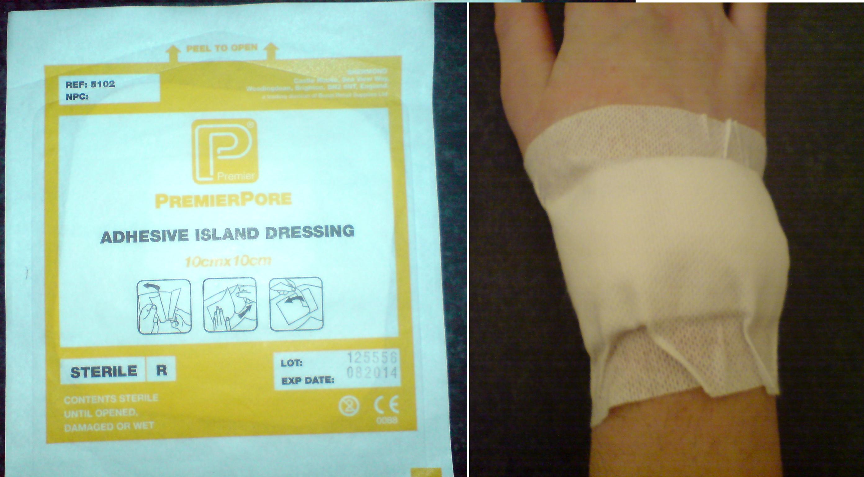
Dressing (medical)
A dressing is a sterile pad or compress applied to a wound to promote healing and protect the wound from further harm. A dressing is designed to be in direct contact with the wound, as distinguished from a bandage, which is most often used to hold a dressing in place. Many modern dressings are self-adhesive. Medical uses A dressing can have a number of purposes, depending on the type, severity and position of the wound, although all purposes are focused on promoting recovery and protecting from further harm. Key purposes of a dressing are: * Stop bleeding – to help to seal the wound to expedite the clotting process; * Protection from infection – to defend the wound against germs and mechanical damage; * Absorb exudate – to soak up blood, plasma, and other fluids exuded from the wound, containing it/them in one place and preventing maceration; * Ease pain – either by a medicated analgesic effect, compression or simply preventing pain from further trauma; * Debride the ... [...More Info...] [...Related Items...] OR: [Wikipedia] [Google] [Baidu] |
Keratin
Keratin () is one of a family of structural fibrous proteins also known as ''scleroproteins''. Alpha-keratin (α-keratin) is a type of keratin found in vertebrates. It is the key structural material making up scales, hair, nails, feathers, horns, claws, hooves, and the outer layer of skin among vertebrates. Keratin also protects epithelial cells from damage or stress. Keratin is extremely insoluble in water and organic solvents. Keratin monomers assemble into bundles to form intermediate filaments, which are tough and form strong unmineralized epidermal appendages found in reptiles, birds, amphibians, and mammals. Excessive keratinization participate in fortification of certain tissues such as in horns of cattle and rhinos, and armadillos' osteoderm. The only other biological matter known to approximate the toughness of keratinized tissue is chitin. Keratin comes in two types, the primitive, softer forms found in all vertebrates and harder, derived forms found only amon ... [...More Info...] [...Related Items...] OR: [Wikipedia] [Google] [Baidu] |
Nail (anatomy)
A nail is a claw-like plate found at the tip of the Finger, fingers and Toe, toes on most primates. Nails correspond to the claws found in other animals. Fingernails and toenails are made of a tough protective protein called alpha-keratin, which is a polymer. Alpha-keratin is found in the hooves, claws, and horns of vertebrates. Structure The nail consists of the nail plate, the nail matrix and the nail bed below it, and the grooves surrounding it. Parts of the nail The matrix, sometimes called the ''matrix unguis'', keratogenous membrane, nail matrix, or onychostroma, is the active Tissue (biology), tissue (or Germ layer, germinal Matrix (biology), matrix) that generates cells, which harden as they move outward from the nail root to the nail plate. It is the part of the nail bed that is beneath the nail and contains nerves, lymph and blood vessels. The matrix produces cells that become the nail plate. The width and thickness of the nail plate is determined by the size, length, ... [...More Info...] [...Related Items...] OR: [Wikipedia] [Google] [Baidu] |
Botox
Botulinum toxin, or botulinum neurotoxin (BoNT), is a neurotoxic protein produced by the bacterium ''Clostridium botulinum'' and related species. It prevents the release of the neurotransmitter acetylcholine from axon endings at the neuromuscular junction, thus causing flaccid paralysis. The toxin causes the disease botulism. The toxin is also used commercially for medical and cosmetic purposes. The seven main types of botulinum toxin are named types A to G (A, B, C1, C2, D, E, F and G). New types are occasionally found. Types A and B are capable of causing disease in humans, and are also used commercially and medically. Types C–G are less common; types E and F can cause disease in humans, while the other types cause disease in other animals. Botulinum toxins are among the most potent toxins known. Intoxication can occur naturally as a result of either wound or intestinal infection or by ingesting formed toxin in food. The estimated human lethal dose of type A toxin is ... [...More Info...] [...Related Items...] OR: [Wikipedia] [Google] [Baidu] |
Microvascular Surgery
Microsurgery is a general term for surgery requiring an operating microscope. The most obvious developments have been procedures developed to allow anastomosis of successively smaller blood vessels and nerves (typically 1 mm in diameter) which have allowed transfer of tissue from one part of the body to another and re-attachment of severed parts. Microsurgical techniques are utilized by several specialties today, such as general surgery, ophthalmology, orthopedic surgery, gynecological surgery, otolaryngology, neurosurgery, oral and maxillofacial surgery, plastic surgery, podiatric surgery and pediatric surgery. History Otolaryngologists were the first physicians to use microsurgical techniques. A Swedish otolaryngologist, Carl-Olof Siggesson Nylén (1892–1978), was the father of microsurgery. In 1921, in the University of Stockholm, he built the first surgical microscope, a modified monocular Brinell-Leitz microscope. At first he used it for operations in animals. In Nove ... [...More Info...] [...Related Items...] OR: [Wikipedia] [Google] [Baidu] |
Eyelid
An eyelid is a thin fold of skin that covers and protects an eye. The levator palpebrae superioris muscle retracts the eyelid, exposing the cornea to the outside, giving vision. This can be either voluntarily or involuntarily. The human eyelid features a row of eyelashes along the eyelid margin, which serve to heighten the protection of the eye from dust and foreign debris, as well as from perspiration. "Palpebral" (and "blepharal") means relating to the eyelids. Its key function is to regularly spread the tears and other secretions on the eye surface to keep it moist, since the cornea must be continuously moist. They keep the eyes from drying out when asleep. Moreover, the blink reflex protects the eye from foreign bodies. The appearance of the human upper eyelid often varies between different populations. The prevalence of an epicanthic fold covering the inner corner of the eye account for the majority of East Asian and Southeast Asian populations, and is also found i ... [...More Info...] [...Related Items...] OR: [Wikipedia] [Google] [Baidu] |
Anaplastologist
Anaplastology (''Gk. ''ana''-again, anew, upon ''plastos''-something made, formed, molded ''logy''-the study of'') is a branch of medicine dealing with the prosthetic rehabilitation of an absent, disfigured or malformed anatomically critical location of the face or Human body, body. The term ''anaplastology'' was coined by Walter G. Spohn and is used worldwide. An anaplastologist (also known as a maxillofacial prosthetist and technologist in the United Kingdom, UK) is an individual who has the knowledge and skill set to provide the service of customizing a facial (craniofacial prosthesis), ocular or somatic prosthesis. In locations around the world where facial, ocular and somatic prostheses are not readily available, a dentist who specializes in maxillofacial prosthetics (prosthodontics), or a dental technician or an ocularist, may also be titled an anaplastologist. In Urban area, urban or more developed locations, an individual referred to as an anaplastologist is one who solely w ... [...More Info...] [...Related Items...] OR: [Wikipedia] [Google] [Baidu] |


.jpg)


.jpg)