|
Auricular Branch Of Occipital Artery
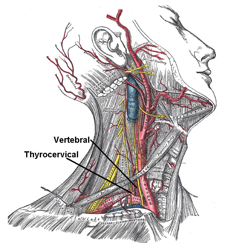
The auricular branch of occipital artery supplies the back of the concha and frequently gives off a branch, which enters the skull through the mastoid foramen and supplies the dura mater, the diploë, and the mastoid cells; this latter branch sometimes arises from the occipital artery The occipital artery arises from the external carotid artery opposite the facial artery. Its path is below the posterior belly of digastric to the occipital region. This artery supplies blood to the back of the scalp and sternocleidomastoid musc ..., and is then known as the mastoid branch. References Arteries of the head and neck {{circulatory-stub ... [...More Info...] [...Related Items...] OR: [Wikipedia] [Google] [Baidu] |
Common Carotid Artery
In anatomy, the left and right common carotid arteries (carotids) ( in Merriam-Webster Online Dictionary '.) are that supply the head and neck with ; they divide in the neck to form the and |
Subclavian Artery
In human anatomy, the subclavian arteries are paired major arteries of the upper thorax, below the clavicle. They receive blood from the aortic arch. The left subclavian artery supplies blood to the left arm and the right subclavian artery supplies blood to the right arm, with some branches supplying the head and thorax. On the left side of the body, the subclavian comes directly off the aortic arch, while on the right side it arises from the relatively short brachiocephalic artery when it bifurcates into the subclavian and the right common carotid artery. The usual branches of the subclavian on both sides of the body are the vertebral artery, the internal thoracic artery, the thyrocervical trunk, the costocervical trunk and the dorsal scapular artery, which may branch off the transverse cervical artery, which is a branch of the thyrocervical trunk. The subclavian becomes the axillary artery at the lateral border of the first rib. Structure From its origin, the subclavian artery t ... [...More Info...] [...Related Items...] OR: [Wikipedia] [Google] [Baidu] |
Occipital Artery
The occipital artery arises from the external carotid artery opposite the facial artery. Its path is below the posterior belly of digastric to the occipital region. This artery supplies blood to the back of the scalp and sternocleidomastoid muscles, and deep muscles in the back and neck. Structure At its origin, it is covered by the posterior belly of the digastricus and the stylohyoideus, and the hypoglossal nerve winds around it from behind forward; higher up, it crosses the internal carotid artery, the internal jugular vein, and the vagus and accessory nerves. It next ascends to the interval between the transverse process of the atlas and the mastoid process of the temporal bone, and passes horizontally backward, grooving the surface of the latter bone, being covered by the sternocleidomastoideus, splenius capitis, longissimus capitis, and digastricus, and resting upon the rectus capitis lateralis, the obliquus superior, and semispinalis capitis. It then changes its course and ... [...More Info...] [...Related Items...] OR: [Wikipedia] [Google] [Baidu] |
Pinna (anatomy)
The auricle or auricula is the visible part of the ear that is outside the head. It is also called the pinna (Latin for "wing" or "fin", plural pinnae), a term that is used more in zoology. Structure The diagram shows the shape and location of most of these components: * ''antihelix'' forms a 'Y' shape where the upper parts are: ** ''Superior crus'' (to the left of the ''fossa triangularis'' in the diagram) ** ''Inferior crus'' (to the right of the ''fossa triangularis'' in the diagram) * ''Antitragus'' is below the ''tragus'' * ''Aperture'' is the entrance to the ear canal * ''Auricular sulcus'' is the depression behind the ear next to the head * ''Concha'' is the hollow next to the ear canal * Conchal angle is the angle that the back of the ''concha'' makes with the side of the head * ''Crus'' of the helix is just above the ''tragus'' * ''Cymba conchae'' is the narrowest end of the ''concha'' * External auditory meatus is the ear canal * ''Fossa triangularis'' is the depres ... [...More Info...] [...Related Items...] OR: [Wikipedia] [Google] [Baidu] |
Mastoid Foramen
The mastoid foramen is a hole in the posterior border of the temporal bone. It transmits an emissary vein between the sigmoid sinus and the suboccipital venous plexus, and a small branch of the occipital artery, the posterior meningeal artery to the dura mater. Structure The mastoid foramen is a hole in the posterior border of the temporal bone of the skull. The opening of the mastoid foramen is an average of 18 mm from the asterion, and around 34 mm from the external auditory meatus. It is typically very narrow. This may be around 2 mm. Variation The position and size of this foramen are very variable. It is not always present. Sometimes, it is duplicated on one side or both sides. Sometimes, it is situated in the occipital bone, or in the suture between the temporal bone and the occipital bone. Function The mastoid foramen transmits: * an emissary vein between the sigmoid sinus and the suboccipital venous plexus or the posterior auricular vein. * a small branch of the o ... [...More Info...] [...Related Items...] OR: [Wikipedia] [Google] [Baidu] |
Dura Mater
In neuroanatomy, dura mater is a thick membrane made of dense irregular connective tissue that surrounds the brain and spinal cord. It is the outermost of the three layers of membrane called the meninges that protect the central nervous system. The other two meningeal layers are the arachnoid mater and the pia mater. It envelops the arachnoid mater, which is responsible for keeping in the cerebrospinal fluid. It is derived primarily from the neural crest cell population, with postnatal contributions of the paraxial mesoderm. Structure The dura mater has several functions and layers. The dura mater is a membrane that envelops the arachnoid mater. It surrounds and supports the dural sinuses (also called dural venous sinuses, cerebral sinuses, or cranial sinuses) and carries blood from the brain toward the heart. Cranial dura mater has two layers called ''lamellae'', a superficial layer (also called the periosteal layer), which serves as the skull's inner periosteum, called the ... [...More Info...] [...Related Items...] OR: [Wikipedia] [Google] [Baidu] |
Diploë
Diploë ( or ) is the spongy cancellous bone separating the inner and outer layers of the cortical bone of the skull. In the cranial bones, the layers of compact cortical tissue are familiarly known as the tables of the skull; the outer one is thick and tough; the inner is thin, dense, and brittle, and hence is termed the ''vitreous table''. The intervening cancellous tissue is called the diploë. In certain regions of the skull The skull is a bone protective cavity for the brain. The skull is composed of four types of bone i.e., cranial bones, facial bones, ear ossicles and hyoid bone. However two parts are more prominent: the cranium and the mandible. In humans, th ... this becomes absorbed so as to leave spaces filled with liquid between the two tables. Etymology From Ancient Greek διπλόη (''diplóē'', “literally, a fold”), noun use of feminine of διπλόος (''diplóos'', “double”) References External links Skull {{musculoskeletal- ... [...More Info...] [...Related Items...] OR: [Wikipedia] [Google] [Baidu] |
Mastoid Cells
The mastoid cells (also called air cells of Lenoir or mastoid cells of Lenoir) are air-filled cavities within the mastoid process of the temporal bone of the cranium. The mastoid cells are a form of skeletal pneumaticity. Infection in these cells is called mastoiditis. The term "cells" refers to enclosed spaces, not cells as living, biological units. Anatomy A section of the mastoid process will show it to be hollowed out into a number of spaces which exhibit great variety in their size and number. At the upper and front part of the process they are large and irregular and contain air, but toward the lower part they diminish in size, while those at the apex of the process are frequently quite small and contain marrow. Occasionally they are entirely absent and the mastoid is solid throughout. Development At birth, the mastoid is not pneumatized, but becomes aerated before age six. Function The air cells are hypothesised to protect the temporal bone and the inner and middle ear ... [...More Info...] [...Related Items...] OR: [Wikipedia] [Google] [Baidu] |
Occipital Artery
The occipital artery arises from the external carotid artery opposite the facial artery. Its path is below the posterior belly of digastric to the occipital region. This artery supplies blood to the back of the scalp and sternocleidomastoid muscles, and deep muscles in the back and neck. Structure At its origin, it is covered by the posterior belly of the digastricus and the stylohyoideus, and the hypoglossal nerve winds around it from behind forward; higher up, it crosses the internal carotid artery, the internal jugular vein, and the vagus and accessory nerves. It next ascends to the interval between the transverse process of the atlas and the mastoid process of the temporal bone, and passes horizontally backward, grooving the surface of the latter bone, being covered by the sternocleidomastoideus, splenius capitis, longissimus capitis, and digastricus, and resting upon the rectus capitis lateralis, the obliquus superior, and semispinalis capitis. It then changes its course and ... [...More Info...] [...Related Items...] OR: [Wikipedia] [Google] [Baidu] |


