|
Aryepiglottic
The aryepiglottic muscle, or aryepiglotticus muscle is an intrinsic muscle of the larynx. The muscle originates from the muscular process of arytenoid cartilage and inserts to the aryepiglottic fold and lateral border of epiglottis. The aryepiglottic muscle is innervated by the inferior laryngeal nerve, a branch of the recurrent laryngeal nerve (a branch of the vagus nerve). The muscle adducts arytenoid cartilages and acts as a sphincter on the laryngeal inlet The laryngeal inlet (laryngeal aditus, laryngeal aperture) is the opening that connects the pharynx and the larynx. Borders Its borders are formed by: * the free curved edge of the epiglottis, anteriorly * the arytenoid cartilages, the cornicula .... Additional images File:Slide14sss.JPG, Aryepiglottic muscle External links * () References Muscles of the head and neck {{muscle-stub ... [...More Info...] [...Related Items...] OR: [Wikipedia] [Google] [Baidu] |
Aryepiglottic Fold
The aryepiglottic folds are triangular folds of mucous membrane of the larynx. They enclose ligamentous and muscular fibres. They extend from the lateral borders of the epiglottis to the arytenoid cartilages, hence the name 'aryepiglottic'. They contain the aryepiglottic muscles and form the upper borders of the quadrangular membrane. They have a role in growling as a form of phonation. They may be narrowed and cause stridor, or be shortened and cause laryngomalacia. Structure The aryepiglottic folds are triangular. They are narrow in front, wide behind, and slope obliquely downward and backward. They originate from the lateral borders of the epiglottis. They insert into the arytenoid cartilages. In front, they are bounded by the epiglottis. Behind, they are bounded by the apices of the arytenoid cartilages, the corniculate cartilages, and the interarytenoid notch. Within the posterior part of each aryepiglottic fold exists a cuneiform cartilage which forms whitish promine ... [...More Info...] [...Related Items...] OR: [Wikipedia] [Google] [Baidu] |
Arytenoid Muscle
The arytenoid muscle is a single muscle of the larynx. It passes from one arytenoid cartilage to the opposite arytenoid cartilage. It has oblique and transverse fibres. It is supplied by the recurrent laryngeal nerve. It approximates the arytenoid cartilages. Continuous electromyography may be used during neck surgeries such as thyroidectomy. Structure The arytenoid muscle fills the posterior concave surface of the arytenoid cartilage. It arises from the posterior surface and lateral border of one arytenoid cartilage. It is inserted into the corresponding parts of the opposite arytenoid cartilage. It consists of oblique and transverse fibres. Nerve supply The arytenoid muscle is supplied by the recurrent laryngeal nerve, a branch of the vagus nerve (CN X). This is a bilateral supply. Function The arytenoid muscle approximates the arytenoid cartilages. This closes the aperture of the glottis, especially at its back part to eliminate the posterior commissure of the vocal c ... [...More Info...] [...Related Items...] OR: [Wikipedia] [Google] [Baidu] |
Epiglottis
The epiglottis is a leaf-shaped flap in the throat that prevents food and water from entering the trachea and the lungs. It stays open during breathing, allowing air into the larynx. During swallowing, it closes to prevent aspiration of food into the lungs, forcing the swallowed liquids or food to go along the oesophagus toward the stomach instead. It is thus the valve that diverts passage to either the trachea or the oesophagus. The epiglottis is made of elastic cartilage covered with a mucous membrane, attached to the entrance of the larynx. It projects upwards and backwards behind the tongue and the hyoid bone. The epiglottis may be inflamed in a condition called epiglottitis, which is most commonly due to the vaccine-preventable bacteria ''Haemophilus influenzae''. Dysfunction may cause the inhalation of food, called aspiration, which may lead to pneumonia or airway obstruction. The epiglottis is also an important landmark for intubation. The epiglottis has been identif ... [...More Info...] [...Related Items...] OR: [Wikipedia] [Google] [Baidu] |
Inferior Laryngeal Nerve
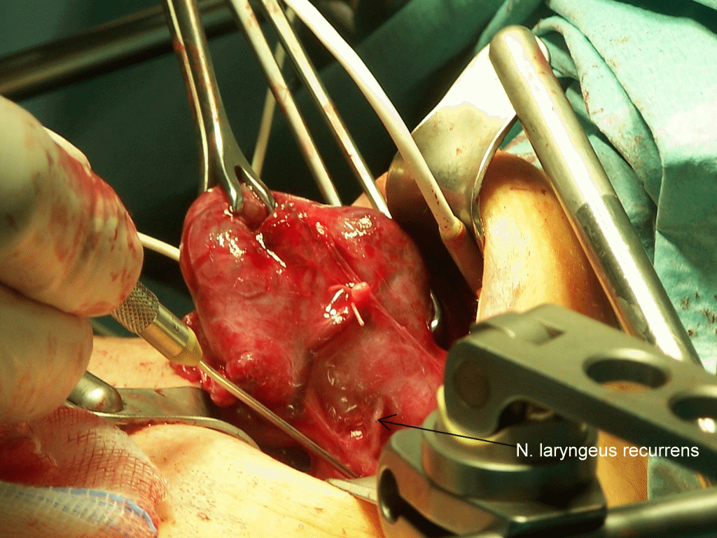
The recurrent laryngeal nerve (RLN) is a branch of the vagus nerve (cranial nerve X) that supplies all the intrinsic muscles of the larynx, with the exception of the cricothyroid muscles. There are two recurrent laryngeal nerves, right and left. The right and left nerves are not symmetrical, with the left nerve looping under the aortic arch, and the right nerve looping under the right subclavian artery then traveling upwards. They both travel alongside the trachea. Additionally, the nerves are among the few nerves that follow a ''recurrent'' course, moving in the opposite direction to the nerve they branch from, a fact from which they gain their name. The recurrent laryngeal nerves supply sensation to the larynx below the vocal cords, give cardiac branches to the deep cardiac plexus, and branch to the trachea, esophagus and the inferior constrictor muscles. The posterior cricoarytenoid muscles, the only muscles that can open the vocal folds, are innervated by this nerve. Th ... [...More Info...] [...Related Items...] OR: [Wikipedia] [Google] [Baidu] |
Recurrent Laryngeal Nerve
The recurrent laryngeal nerve (RLN) is a branch of the vagus nerve (cranial nerve X) that supplies all the intrinsic muscles of the larynx, with the exception of the cricothyroid muscles. There are two recurrent laryngeal nerves, right and left. The right and left nerves are not symmetrical, with the left nerve looping under the aortic arch, and the right nerve looping under the right subclavian artery then traveling upwards. They both travel alongside the trachea. Additionally, the nerves are among the few nerves that follow a ''recurrent'' course, moving in the opposite direction to the nerve they branch from, a fact from which they gain their name. The recurrent laryngeal nerves supply sensation to the larynx below the vocal cords, give cardiac branches to the deep cardiac plexus, and branch to the trachea, esophagus and the inferior constrictor muscles. The posterior cricoarytenoid muscles, the only muscles that can open the vocal folds, are innervated by this nerve. Th ... [...More Info...] [...Related Items...] OR: [Wikipedia] [Google] [Baidu] |
Sagittal Section
The sagittal plane (; also known as the longitudinal plane) is an anatomical plane that divides the body into right and left sections. It is perpendicular to the transverse and coronal planes. The plane may be in the center of the body and divide it into two equal parts ( mid-sagittal), or away from the midline and divide it into unequal parts (para-sagittal). The term ''sagittal'' was coined by Gerard of Cremona. Variations in terminology Examples of sagittal planes include: * The terms ''median plane'' or ''mid-sagittal plane'' are sometimes used to describe the sagittal plane running through the midline. This plane cuts the body into halves (assuming bilateral symmetry), passing through midline structures such as the navel and spine. It is one of the planes which, combined with the Umbilical plane, defines the four quadrants of the human abdomen. * The term ''parasagittal'' is used to describe any plane parallel or adjacent to a given sagittal plane. Specific named parasag ... [...More Info...] [...Related Items...] OR: [Wikipedia] [Google] [Baidu] |
Larynx
The larynx (), commonly called the voice box, is an organ in the top of the neck involved in breathing, producing sound and protecting the trachea against food aspiration. The opening of larynx into pharynx known as the laryngeal inlet is about 4–5 centimeters in diameter. The larynx houses the vocal cords, and manipulates pitch and volume, which is essential for phonation. It is situated just below where the tract of the pharynx splits into the trachea and the esophagus. The word ʻlarynxʼ (plural ʻlaryngesʼ) comes from the Ancient Greek word ''lárunx'' ʻlarynx, gullet, throat.ʼ Structure The triangle-shaped larynx consists largely of cartilages that are attached to one another, and to surrounding structures, by muscles or by fibrous and elastic tissue components. The larynx is lined by a ciliated columnar epithelium except for the vocal folds. The cavity of the larynx extends from its triangle-shaped inlet, to the epiglottis, and to the circular outlet at the ... [...More Info...] [...Related Items...] OR: [Wikipedia] [Google] [Baidu] |
Vertebrate Trachea
The trachea, also known as the windpipe, is a cartilaginous tube that connects the larynx to the bronchi of the lungs, allowing the passage of air, and so is present in almost all air-breathing animals with lungs. The trachea extends from the larynx and branches into the two primary bronchi. At the top of the trachea the cricoid cartilage attaches it to the larynx. The trachea is formed by a number of horseshoe-shaped rings, joined together vertically by overlying ligaments, and by the trachealis muscle at their ends. The epiglottis closes the opening to the larynx during swallowing. The trachea begins to form in the second month of embryo development, becoming longer and more fixed in its position over time. It is epithelium lined with column-shaped cells that have hair-like extensions called cilia, with scattered goblet cells that produce protective mucins. The trachea can be affected by inflammation or infection, usually as a result of a viral illness affecting other parts ... [...More Info...] [...Related Items...] OR: [Wikipedia] [Google] [Baidu] |
Superior Laryngeal Artery
The superior thyroid artery arises from the external carotid artery just below the level of the greater cornu of the hyoid bone and ends in the thyroid gland. Structure From its origin under the anterior border of the sternocleidomastoid the superior thyroid artery runs upward and forward for a short distance in the carotid triangle, where it is covered by the skin, platysma, and fascia; it then arches downward beneath the omohyoid, sternohyoid, and sternothyroid muscles. To its medial side are the inferior pharyngeal constrictor muscle and the external branch of the superior laryngeal nerve. Branches It distributes twigs to the adjacent muscles, and numerous branches to the thyroid gland, connecting with its fellow of the opposite side, and with the inferior thyroid arteries. The branches to the gland are generally two in number. One, the larger, supplies principally the anterior surface; on the isthmus of the gland it connects with the corresponding artery of the opposite si ... [...More Info...] [...Related Items...] OR: [Wikipedia] [Google] [Baidu] |
Superior Thyroid Artery
The superior thyroid artery arises from the external carotid artery just below the level of the greater cornu of the hyoid bone and ends in the thyroid gland. Structure From its origin under the anterior border of the sternocleidomastoid the superior thyroid artery runs upward and forward for a short distance in the carotid triangle, where it is covered by the skin, platysma, and fascia; it then arches downward beneath the omohyoid, sternohyoid, and sternothyroid muscles. To its medial side are the inferior pharyngeal constrictor muscle and the external branch of the superior laryngeal nerve. Branches It distributes twigs to the adjacent muscles, and numerous branches to the thyroid gland, connecting with its fellow of the opposite side, and with the inferior thyroid arteries. The branches to the gland are generally two in number. One, the larger, supplies principally the anterior surface; on the isthmus of the gland it connects with the corresponding artery of the opposite ... [...More Info...] [...Related Items...] OR: [Wikipedia] [Google] [Baidu] |
Vagus Nerve
The vagus nerve, also known as the tenth cranial nerve, cranial nerve X, or simply CN X, is a cranial nerve that interfaces with the parasympathetic control of the heart, lungs, and digestive tract. It comprises two nerves—the left and right vagus nerves—but they are typically referred to collectively as a single subsystem. The vagus is the longest nerve of the autonomic nervous system in the human body and comprises both sensory and motor fibers. The sensory fibers originate from neurons of the nodose ganglion, whereas the motor fibers come from neurons of the dorsal motor nucleus of the vagus and the nucleus ambiguus. The vagus was also historically called the pneumogastric nerve. Structure Upon leaving the medulla oblongata between the olive and the inferior cerebellar peduncle, the vagus nerve extends through the jugular foramen, then passes into the carotid sheath between the internal carotid artery and the internal jugular vein down to the neck, chest, and abdom ... [...More Info...] [...Related Items...] OR: [Wikipedia] [Google] [Baidu] |
Muscles Of Larynx
The larynx (), commonly called the voice box, is an organ in the top of the neck involved in breathing, producing sound and protecting the trachea against food aspiration. The opening of larynx into pharynx known as the laryngeal inlet is about 4–5 centimeters in diameter. The larynx houses the vocal cords, and manipulates pitch and volume, which is essential for phonation. It is situated just below where the tract of the pharynx splits into the trachea and the esophagus. The word ʻlarynxʼ (plural ʻlaryngesʼ) comes from the Ancient Greek word ''lárunx'' ʻlarynx, gullet, throat.ʼ Structure The triangle-shaped larynx consists largely of cartilages that are attached to one another, and to surrounding structures, by muscles or by fibrous and elastic tissue components. The larynx is lined by a ciliated columnar epithelium except for the vocal folds. The cavity of the larynx extends from its triangle-shaped inlet, to the epiglottis, and to the circular outlet at the low ... [...More Info...] [...Related Items...] OR: [Wikipedia] [Google] [Baidu] |
.png)


