|
Ansa Cervicalis
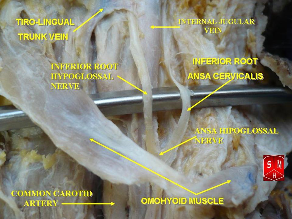
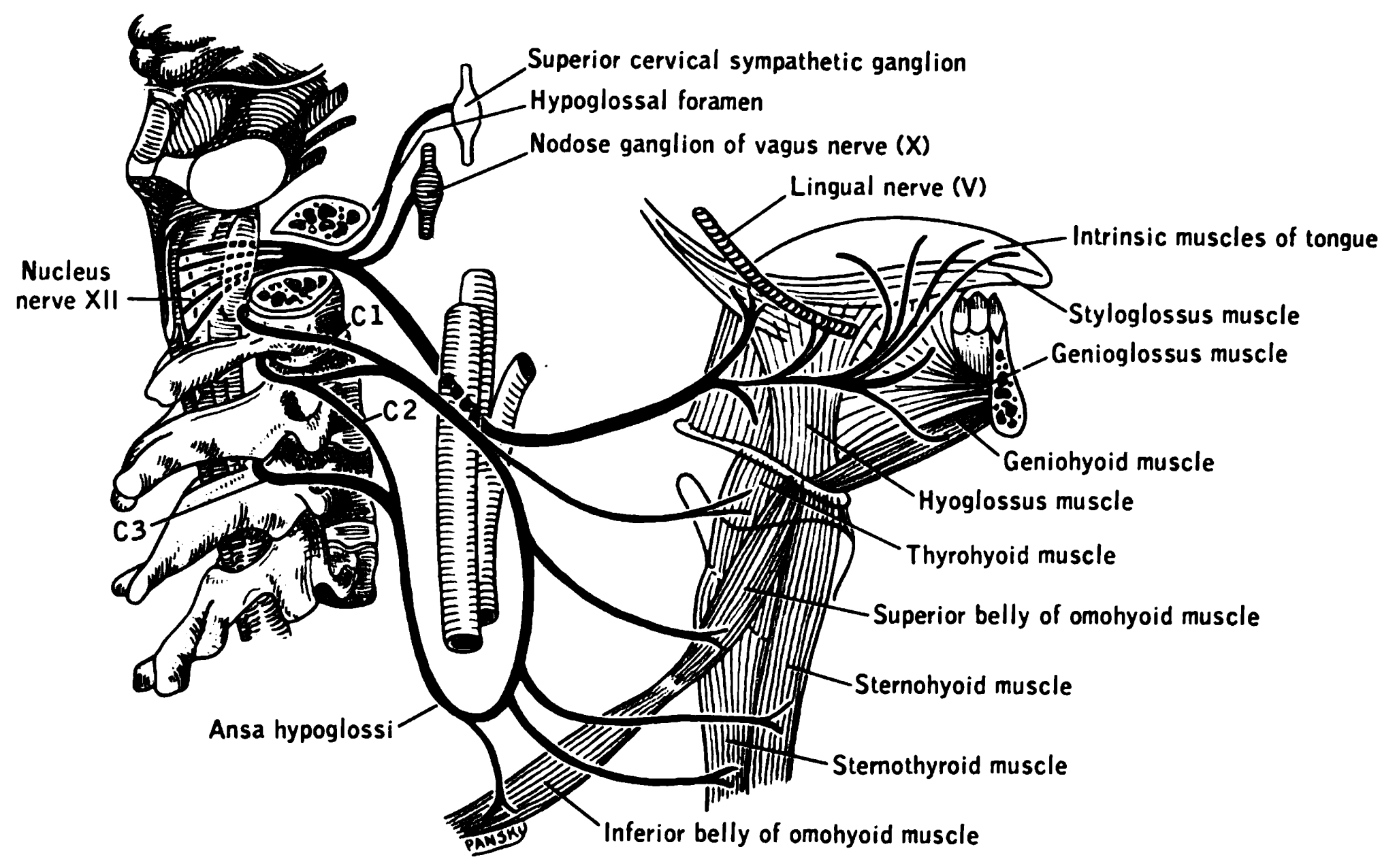
The ansa cervicalis (or ansa hypoglossi in older literature) is a loop of nerves that are part of the cervical plexus. It lies superficial to the internal jugular vein in the carotid triangle. Its name means "handle of the neck" in Latin. Branches from the ansa cervicalis innervate most of the infrahyoid muscles, including the sternothyroid muscle, sternohyoid muscle and the omohyoid muscle. Note that the thyrohyoid muscle, which is also an infrahyoid muscle and the geniohyoid muscle which is a suprahyoid muscle are innervated by cervical spinal nerve 1 via the hypoglossal nerve. Roots Two roots make up the ansa cervicalis, a superior root, and an inferior root. The superior root of the ansa cervicalis is formed from cervical spinal nerve 1 of the cervical plexus. These nerve fibers travel in the hypoglossal nerve before separating in the carotid triangle to form the superior root. The superior root goes around the occipital artery and then descends on the carotid sheath. I ... [...More Info...] [...Related Items...] OR: [Wikipedia] [Google] [Baidu] |
Sternohyoid Muscle
The sternohyoid muscle is a thin, narrow muscle attaching the hyoid bone to the sternum. It is one of the paired strap muscles of the infrahyoid muscles. It is supplied by the ansa cervicalis. It depresses the hyoid bone. Structure The sternohyoid muscle is one of the paired strap muscles of the infrahyoid muscles. It arises from the posterior border of the medial end of the clavicle, the posterior sternoclavicular ligament, and the upper and posterior part of the manubrium of the sternum. Passing upward and medially, it is inserted by short tendinous fibers into the lower border of the body of the hyoid bone. It runs lateral to the trachea. Nerve supply The sternohyoid muscle is supplied by a branch of the ansa cervicalis. Variations The sternohyoid muscle may be doubled, have accessory slips (Cleidohyoideus) or be completely absent in some people. It sometimes presents a transverse tendinous inscription immediately above its origin. Function The sternohyoid muscle p ... [...More Info...] [...Related Items...] OR: [Wikipedia] [Google] [Baidu] |
Cervical Spinal Nerve 1
The cervical spinal nerve 1 (C1) is a spinal nerve of the cervical segment. from spinalcordinjuryzone.com. Published February 23, 2004 Archived Dec 23, 2011. Retrieved June 12, 2018. C1 carries predominantly motor fibres, but also a small meningeal branch that supplies sensation to parts of the dura around the foramen magnum (via dorsal rami). It originates from the spinal column from above the (C1). The dorsal root and gang ... [...More Info...] [...Related Items...] OR: [Wikipedia] [Google] [Baidu] |
Cervical Spinal Nerve 3
The cervical spinal nerve 3 (C3) is a spinal nerve of the cervical segment. Nervous System -- Groups of Nerves It originates from the spinal column from above the cervical vertebra 3
In tetrapods, cervical vertebrae (singular: vertebra) are the vertebrae of the neck, immediately below the skull. Truncal vertebrae (divided into thoracic and lumbar vertebrae in mammals) lie caudal (toward the tail) of cervical vertebrae. In sau ... (C3).
References Spinal nerves {{neuroanatomy-stub ...[...More Info...] [...Related Items...] OR: [Wikipedia] [Google] [Baidu] |
Cervical Spinal Nerve 2
The cervical spinal nerve 2 (C2) is a spinal nerve of the cervical segment. Nervous System -- Groups of Nerves It is a part of the along with the C1 and C3 nerves sometimes forming part of Superior root of the ansa cervicalis. it also connects into the with the C3. It originates from the s ... [...More Info...] [...Related Items...] OR: [Wikipedia] [Google] [Baidu] |
Spinal Nerve
A spinal nerve is a mixed nerve, which carries motor, sensory, and autonomic signals between the spinal cord and the body. In the human body there are 31 pairs of spinal nerves, one on each side of the vertebral column. These are grouped into the corresponding cervical, thoracic, lumbar, sacral and coccygeal regions of the spine. There are eight pairs of cervical nerves, twelve pairs of thoracic nerves, five pairs of lumbar nerves, five pairs of sacral nerves, and one pair of coccygeal nerves. The spinal nerves are part of the peripheral nervous system. Structure Each spinal nerve is a mixed nerve, formed from the combination of nerve fibers from its dorsal and ventral roots. The dorsal root is the afferent sensory root and carries sensory information to the brain. The ventral root is the efferent motor root and carries motor information from the brain. The spinal nerve emerges from the spinal column through an opening (intervertebral foramen) between adjacent vertebrae. ... [...More Info...] [...Related Items...] OR: [Wikipedia] [Google] [Baidu] |
Sternohyoid
The sternohyoid muscle is a thin, narrow muscle attaching the hyoid bone to the sternum. It is one of the paired strap muscles of the infrahyoid muscles. It is supplied by the ansa cervicalis. It depresses the hyoid bone. Structure The sternohyoid muscle is one of the paired strap muscles of the infrahyoid muscles. It arises from the posterior border of the medial end of the clavicle, the posterior sternoclavicular ligament, and the upper and posterior part of the manubrium of the sternum. Passing upward and medially, it is inserted by short tendinous fibers into the lower border of the body of the hyoid bone. It runs lateral to the trachea. Nerve supply The sternohyoid muscle is supplied by a branch of the ansa cervicalis. Variations The sternohyoid muscle may be doubled, have accessory slips (Cleidohyoideus) or be completely absent in some people. It sometimes presents a transverse tendinous inscription immediately above its origin. Function The sternohyoid muscle pe ... [...More Info...] [...Related Items...] OR: [Wikipedia] [Google] [Baidu] |
Sternothyroid
The sternothyroid muscle, or sternothyroideus, is an infrahyoid muscle in the neck. It acts to depress the hyoid bone. It is below the sternohyoid muscle. It is shorter and wider than the sternohyoid. Structure The sternothyroid arises from the posterior surface of the manubrium of the sternum, below the origin of the sternohyoid. It also arises from the edge of the cartilage of the first rib. It is inserted into the oblique line on the lamina of the thyroid cartilage. It is in close contact with its fellow at the lower part of the neck, but diverges somewhat as it ascends. It is occasionally traversed by a transverse or oblique tendinous inscription. Innervation The sternothyroid muscle is innervated by the ansa cervicalis. Variations Doubling; absence; accessory slips to the thyrohyoid, inferior pharyngeal constrictor, or to the carotid sheath. Function The sternothyroid muscle depresses the hyoid bone, along with the other infrahyoid muscle. Clinical significance The u ... [...More Info...] [...Related Items...] OR: [Wikipedia] [Google] [Baidu] |
Carotid Sheath
The carotid sheath is an anatomical term for the fibrous connective tissue that surrounds the vascular compartment of the neck. It is part of the deep cervical fascia of the neck, below the superficial cervical fascia meaning the subcutaneous adipose tissue immediately beneath the skin. The deep cervical fascia of the neck includes four parts: * The investing layer (encloses the SCM and Trapezius) * The carotid sheath (encloses the vascular region of the neck) * The pretracheal fascia (encloses the visceral region of the neck) * The prevertebral fascia (encloses the vertebral region of the neck) Structure The carotid sheath is located at the lateral boundary of the retropharyngeal space at the level of the oropharynx on each side of the neck and deep to the sternocleidomastoid muscle. It extends from the base of the skull to the first rib and sternum, varying between C7 and T4. It merges with the axillary sheath when it reaches the subclavian vein. Contents The four major stru ... [...More Info...] [...Related Items...] OR: [Wikipedia] [Google] [Baidu] |
Occipital Artery
The occipital artery arises from the external carotid artery opposite the facial artery. Its path is below the posterior belly of digastric to the occipital region. This artery supplies blood to the back of the scalp and sternocleidomastoid muscles, and deep muscles in the back and neck. Structure At its origin, it is covered by the posterior belly of the digastricus and the stylohyoideus, and the hypoglossal nerve winds around it from behind forward; higher up, it crosses the internal carotid artery, the internal jugular vein, and the vagus and accessory nerves. It next ascends to the interval between the transverse process of the atlas and the mastoid process of the temporal bone, and passes horizontally backward, grooving the surface of the latter bone, being covered by the sternocleidomastoideus, splenius capitis, longissimus capitis, and digastricus, and resting upon the rectus capitis lateralis, the obliquus superior, and semispinalis capitis. It then changes its course and ... [...More Info...] [...Related Items...] OR: [Wikipedia] [Google] [Baidu] |
Ansa Cervicalis
The ansa cervicalis (or ansa hypoglossi in older literature) is a loop of nerves that are part of the cervical plexus. It lies superficial to the internal jugular vein in the carotid triangle. Its name means "handle of the neck" in Latin. Branches from the ansa cervicalis innervate most of the infrahyoid muscles, including the sternothyroid muscle, sternohyoid muscle and the omohyoid muscle. Note that the thyrohyoid muscle, which is also an infrahyoid muscle and the geniohyoid muscle which is a suprahyoid muscle are innervated by cervical spinal nerve 1 via the hypoglossal nerve. Roots Two roots make up the ansa cervicalis, a superior root, and an inferior root. The superior root of the ansa cervicalis is formed from cervical spinal nerve 1 of the cervical plexus. These nerve fibers travel in the hypoglossal nerve before separating in the carotid triangle to form the superior root. The superior root goes around the occipital artery and then descends on the carotid sheath. I ... [...More Info...] [...Related Items...] OR: [Wikipedia] [Google] [Baidu] |
Hypoglossal Nerve
The hypoglossal nerve, also known as the twelfth cranial nerve, cranial nerve XII, or simply CN XII, is a cranial nerve that innervates all the extrinsic and intrinsic muscles of the tongue except for the palatoglossus, which is innervated by the vagus nerve. CN XII is a nerve with a solely motor function. The nerve arises from the hypoglossal nucleus in the medulla as a number of small rootlets, passes through the hypoglossal canal and down through the neck, and eventually passes up again over the tongue muscles it supplies into the tongue. The nerve is involved in controlling tongue movements required for speech and swallowing, including sticking out the tongue and moving it from side to side. Damage to the nerve or the neural pathways which control it can affect the ability of the tongue to move and its appearance, with the most common sources of damage being injury from trauma or surgery, and motor neuron disease. The first recorded description of the nerve is by Herophil ... [...More Info...] [...Related Items...] OR: [Wikipedia] [Google] [Baidu] |
Suprahyoid Muscles
The suprahyoid muscles are four muscles located above the hyoid bone in the neck. They are the digastric, stylohyoid, geniohyoid, and mylohyoid muscle, mylohyoid muscles. They are all pharyngeal muscles, with the exception of the geniohyoid muscle. The digastric is uniquely named for its two bellies. Its posterior belly rises from the mastoid process of the human skull, cranium and slopes downward and forward. The anterior belly arises from the digastric fossa on the inner surface of the mandibular body, which slopes downward and backward. The two bellies connect at the digastric muscle#intermediate tendon, intermediate tendon. The intermediate tendon passes through a connective tissue loop attached to the hyoid bone. The mylohyoid muscles are thin, flat muscles that form a sling inferior to the tongue supporting the floor of the mouth. The geniohyoids are short, narrow muscles that contact each other in the midline. The stylohyoids are long, thin muscles that are nearly parallel wit ... [...More Info...] [...Related Items...] OR: [Wikipedia] [Google] [Baidu] |


