|
Anoctamin
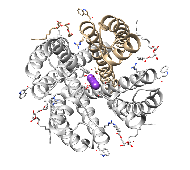
The Calcium-Dependent Chloride Channel (Ca-ClC) proteins (or calcium-activated chloride channels (CaCCs), are heterogeneous groups of ligand-gated ion channels for chloride that have been identified in many epithelial and endothelial cell types as well as in smooth muscle cells. They include proteins from several structurally different families: chloride channel accessory (CLCA), bestrophin (BEST), and calcium-dependent chloride channel anoctamin (ANO or TMEM16) channels ANO1 is highly expressed in human gastrointestinal interstitial cells of Cajal, which are proteins which serve as intestinal pacemakers for peristalsis. In addition to their role as chloride channels some CLCA proteins function as adhesion molecules and may also have roles as tumour suppressors. These eukaryotic proteins are "required for normal electrolyte and fluid secretion, olfactory perception, and neuronal and smooth muscle excitability" in animals. Members of the Ca-CIC family are generally 600 to 1000 a ... [...More Info...] [...Related Items...] OR: [Wikipedia] [Google] [Baidu] |
ANO1
Anoctamin-1 (ANO1) also known as Transmembrane member 16A (TMEM16A) is a protein that, in humans, is encoded by the ''ANO1'' gene. Anoctamin-1 is a voltage-gated calcium-activated anion channel, which acts as a chloride channel and a bicarbonate channel. additionally Anoctamin-1 is apical iodide channel. It is expressed in smooth muscle, epithelial cells, vomeronasal neurons, olfactory sustentacular cells, and is highly expressed in interstitial cells of Cajal (ICC) throughout the gastrointestinal tract. Function ANO1 is a transmembrane protein that functions as a calcium-activated chloride channel. Ca2+, Sr2+, and Ba2+ activate the channel. Structure No atomic resolution structure of this channel has yet been obtained. However, biochemical evidence suggests that the channel assembles as a dimer of two ANO1 polypeptide subunits. From hydropathy plotting, each subunit is thought to encode a molecule with eight transmembrane domains, with a reentrant loop between the fifth ... [...More Info...] [...Related Items...] OR: [Wikipedia] [Google] [Baidu] |
Interstitial Cells Of Cajal
Interstitial cells of Cajal (ICC) are interstitial cells found in the gastrointestinal tract. There are different types of ICC with different functions. ICC and another type of interstitial cell, known as platelet-derived growth factor receptor alpha (PDGFRα) cells, are electrically coupled to smooth muscle cells via gap junctions, that work together as an SIP functional syncytium. Myenteric interstitial cells of Cajal (ICC-MY) serve as pacemaker cells that generate the bioelectrical events known as slow waves. Slow waves conduct to smooth muscle cells and cause phasic contractions. The picture to the right shows an isolated Interstitial cell of Cajal from the Myenteric plexus of the mouse small intestine grown in a primary cell culture. This cell type can be characterized morphologically as having a small cell body often triangular or stellate-shaped with several long processes branching out into secondary and tertiary extensions - these processes often contact smooth muscle ... [...More Info...] [...Related Items...] OR: [Wikipedia] [Google] [Baidu] |
Cryoelectron Microscopy
Cryogenic electron microscopy (cryo-EM) is a cryomicroscopy technique applied on samples cooled to cryogenic temperatures. For biological specimens, the structure is preserved by embedding in an environment of vitreous ice. An aqueous sample solution is applied to a grid-mesh and plunge-frozen in liquid ethane or a mixture of liquid ethane and propane. While development of the technique began in the 1970s, recent advances in detector technology and software algorithms have allowed for the determination of biomolecular structures at near-atomic resolution. This has attracted wide attention to the approach as an alternative to X-ray crystallography or NMR spectroscopy for macromolecular structure determination without the need for crystallization. In 2017, the Nobel Prize in Chemistry was awarded to Jacques Dubochet, Joachim Frank, and Richard Henderson "for developing cryo-electron microscopy for the high-resolution structure determination of biomolecules in solution." ''Nature ... [...More Info...] [...Related Items...] OR: [Wikipedia] [Google] [Baidu] |
Vertebrate
Vertebrates () comprise all animal taxa within the subphylum Vertebrata () ( chordates with backbones), including all mammals, birds, reptiles, amphibians, and fish. Vertebrates represent the overwhelming majority of the phylum Chordata, with currently about 69,963 species described. Vertebrates comprise such groups as the following: * jawless fish, which include hagfish and lampreys * jawed vertebrates, which include: ** cartilaginous fish (sharks, rays, and ratfish) ** bony vertebrates, which include: *** ray-fins (the majority of living bony fish) *** lobe-fins, which include: **** coelacanths and lungfish **** tetrapods (limbed vertebrates) Extant vertebrates range in size from the frog species ''Paedophryne amauensis'', at as little as , to the blue whale, at up to . Vertebrates make up less than five percent of all described animal species; the rest are invertebrates, which lack vertebral columns. The vertebrates traditionally include the hagfish, which do no ... [...More Info...] [...Related Items...] OR: [Wikipedia] [Google] [Baidu] |
National Center For Biotechnology Information
The National Center for Biotechnology Information (NCBI) is part of the United States National Library of Medicine (NLM), a branch of the National Institutes of Health (NIH). It is approved and funded by the government of the United States. The NCBI is located in Bethesda, Maryland, and was founded in 1988 through legislation sponsored by US Congressman Claude Pepper. The NCBI houses a series of databases relevant to biotechnology and biomedicine and is an important resource for bioinformatics tools and services. Major databases include GenBank for DNA sequences and PubMed, a bibliographic database for biomedical literature. Other databases include the NCBI Epigenomics database. All these databases are available online through the Entrez search engine. NCBI was directed by David Lipman, one of the original authors of the BLAST sequence alignment program and a widely respected figure in bioinformatics. GenBank NCBI had responsibility for making available the GenBank DNA seque ... [...More Info...] [...Related Items...] OR: [Wikipedia] [Google] [Baidu] |
Gene (database)
In biology, the word gene (from , ; "...Wilhelm Johannsen coined the word gene to describe the Mendelian units of heredity..." meaning ''generation'' or ''birth'' or ''gender'') can have several different meanings. The Mendelian gene is a basic unit of heredity and the molecular gene is a sequence of nucleotides in DNA that is transcribed to produce a functional RNA. There are two types of molecular genes: protein-coding genes and noncoding genes. During gene expression, the DNA is first copied into RNA. The RNA can be directly functional or be the intermediate template for a protein that performs a function. The transmission of genes to an organism's offspring is the basis of the inheritance of phenotypic traits. These genes make up different DNA sequences called genotypes. Genotypes along with environmental and developmental factors determine what the phenotypes will be. Most biological traits are under the influence of polygenes (many different genes) as well as gene– ... [...More Info...] [...Related Items...] OR: [Wikipedia] [Google] [Baidu] |
Ion Channel
Ion channels are pore-forming membrane proteins that allow ions to pass through the channel pore. Their functions include establishing a resting membrane potential, shaping action potentials and other electrical signals by gating the flow of ions across the cell membrane, controlling the flow of ions across secretory and epithelial cells, and regulating cell volume. Ion channels are present in the membranes of all cells. Ion channels are one of the two classes of ionophoric proteins, the other being ion transporters. The study of ion channels often involves biophysics, electrophysiology, and pharmacology, while using techniques including voltage clamp, patch clamp, immunohistochemistry, X-ray crystallography, fluoroscopy, and RT-PCR. Their classification as molecules is referred to as channelomics. Basic features There are two distinctive features of ion channels that differentiate them from other types of ion transporter proteins: #The rate of ion transport through the ... [...More Info...] [...Related Items...] OR: [Wikipedia] [Google] [Baidu] |
Voltage-gated Ion Channel
Voltage-gated ion channels are a class of transmembrane proteins that form ion channels that are activated by changes in the electrical membrane potential near the channel. The membrane potential alters the conformation of the channel proteins, regulating their opening and closing. Cell membranes are generally impermeable to ions, thus they must diffuse through the membrane through transmembrane protein channels. They have a crucial role in excitable cells such as neuronal and muscle tissues, allowing a rapid and co-ordinated depolarization in response to triggering voltage change. Found along the axon and at the synapse, voltage-gated ion channels directionally propagate electrical signals. Voltage-gated ion-channels are usually ion-specific, and channels specific to sodium (Na+), potassium (K+), calcium (Ca2+), and chloride (Cl−) ions have been identified. The opening and closing of the channels are triggered by changing ion concentration, and hence charge gradient, between ... [...More Info...] [...Related Items...] OR: [Wikipedia] [Google] [Baidu] |
BEST2
Bestrophin-2 is a protein that in humans is encoded by the ''BEST2'' gene. Function This gene is a member of the bestrophin gene family of anion channels. Bestrophin genes share a similar gene structure with highly conserved exon-intron boundaries, but with distinct 3' ends. Bestrophins are transmembrane proteins that contain a homologous region rich in aromatic residues, including an invariant arg-phe-pro motif. Mutation in one of the family members (bestrophin 1) is associated with vitelliform macular dystrophy Vitelliform macular dystrophy is an irregular autosomal dominant eye disorder which can cause progressive vision loss. This disorder affects the retina, specifically cells in a small area near the center of the retina called the macula. The macu .... The bestrophin 2 gene is mainly expressed in the non-pigmented ciliary epithelium and colon. References External links * Further reading * * * * * * Ion channels {{gene-19-stub ... [...More Info...] [...Related Items...] OR: [Wikipedia] [Google] [Baidu] |
BEST1
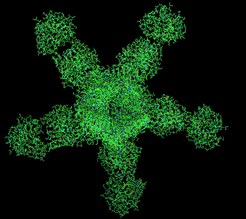
Bestrophin-1 (Best1) is a protein that, in humans, is encoded by the ''BEST1'' gene (RPD ID - 5T5N/4RDQ). The bestrophin family of proteins comprises four evolutionary related genes (BEST1, BEST2, BEST3, and BEST4) that code for integral membrane proteins. This family was first identified in humans by linking a BEST1 mutation with Best vitelliform macular dystrophy (BVMD). Mutations in the BEST1 gene have been identified as the primary cause for at least five different degenerative retinal diseases. The bestrophins are an ancient family of structurally conserved proteins that have been identified in nearly every organism studied from bacteria to humans. In humans, they function as calcium-activated anion channels, each of which has a unique tissue distribution throughout the body. Specifically, the BEST1 gene on chromosome 11q13 encodes the Bestrophin-1 protein in humans whose expression is highest in the retina. Structure Gene The bestrophin genes share a conserved gene ... [...More Info...] [...Related Items...] OR: [Wikipedia] [Google] [Baidu] |


