|
ADAMTS7
A disintegrin and metalloproteinase with thrombospondin motifs 7 (ADAMTS7) is an enzyme that in humans is encoded by the ''ADAMTS7'' gene on chromosome 15. It is ubiquitously expressed in many tissues and cell types. This enzyme catalyzes the degradation of cartilage oligomeric matrix protein (COMP) degradation. ADAMTS7 has been associated with cancer and arthritis in multiple tissue types. The ''ADAMTS7'' gene also contains one of 27 SNPs associated with increased risk of coronary artery disease. Structure Gene The ''ADAMTS7'' gene resides on chromosome 15 at the band 15q24.2 and contains 25 exons. Protein This 1686-amino acid protein belongs to the ADAMTS family and is one of 19 members known in humans. As an ADAMTS protein, ADAMTS7 contains a shared proteinase domain and an ancillary domain. The proteinase domain can be further divided into a signal peptide, a prodomain, a metalloproteinase domain, and a disintegrin-like domain. In particular, the metalloproteinase domain ... [...More Info...] [...Related Items...] OR: [Wikipedia] [Google] [Baidu] |
Thrombospondin
Thrombospondins (TSPs) are a family of secreted glycoproteins with antiangiogenic functions. Due to their dynamic role within the extracellular matrix they are considered matricellular proteins. The first member of the family, thrombospondin 1 (THBS1), was discovered in 1971 by Nancy L. Baenziger. Types The thrombospondins are a family of multifunctional proteins. The family consists of thrombospondins 1-5 and can be divided into 2 subgroups: A, which contains TSP-1 and TSP-2, and B, which contains TSP-3, TSP-4 and TSP-5 (also designated cartilage oligomeric protein or COMP). TSP-1 and TSP-2 are homotrimers, consisting of three identical subunits, whereas TSP-3, TSP-4 and TSP-5 are homopentamers. TSP-1 and TSP-2 are produced by immature astrocytes during brain development, which promotes the development of new synapses. Thrombospondin 1 Thrombospondin 1 (TSP-1) is encoded by THBS1. It was first isolated from platelets that had been stimulated with thrombin, and so was design ... [...More Info...] [...Related Items...] OR: [Wikipedia] [Google] [Baidu] |
ADAMTS
ADAMTS (short for a disintegrin and metalloproteinase with thrombospondin motifs) is a family of multidomain extracellular protease enzymes. 19 members of this family have been identified in humans, the first of which, ADAMTS1, was described in 1997. Known functions of the ADAMTS proteases include processing of procollagens and von Willebrand factor as well as cleavage of aggrecan, versican, brevican and neurocan, making them key remodeling enzymes of the extracellular matrix. They have been demonstrated to have important roles in connective tissue organization, coagulation, inflammation, arthritis, angiogenesis and cell migration. Homologous subfamily of ADAMTSL (ADAMTS-like) proteins, which lack enzymatic activity, has also been described. Most cases of thrombotic thrombocytopenic purpura arise from autoantibody-mediated inhibition of ADAMTS13. Like ADAMs, the name of the ADAMTS family refers to its disintegrin and metalloproteinase activity, and in the case of ADAMTS, the prese ... [...More Info...] [...Related Items...] OR: [Wikipedia] [Google] [Baidu] |
Enzyme
Enzymes () are proteins that act as biological catalysts by accelerating chemical reactions. The molecules upon which enzymes may act are called substrates, and the enzyme converts the substrates into different molecules known as products. Almost all metabolic processes in the cell need enzyme catalysis in order to occur at rates fast enough to sustain life. Metabolic pathways depend upon enzymes to catalyze individual steps. The study of enzymes is called ''enzymology'' and the field of pseudoenzyme analysis recognizes that during evolution, some enzymes have lost the ability to carry out biological catalysis, which is often reflected in their amino acid sequences and unusual 'pseudocatalytic' properties. Enzymes are known to catalyze more than 5,000 biochemical reaction types. Other biocatalysts are catalytic RNA molecules, called ribozymes. Enzymes' specificity comes from their unique three-dimensional structures. Like all catalysts, enzymes increase the reaction ra ... [...More Info...] [...Related Items...] OR: [Wikipedia] [Google] [Baidu] |
Binding Site
In biochemistry and molecular biology, a binding site is a region on a macromolecule such as a protein that binds to another molecule with specificity. The binding partner of the macromolecule is often referred to as a ligand. Ligands may include other proteins (resulting in a protein-protein interaction), enzyme substrates, second messengers, hormones, or allosteric modulators. The binding event is often, but not always, accompanied by a conformational change that alters the protein's function. Binding to protein binding sites is most often reversible (transient and non-covalent), but can also be covalent reversible or irreversible. Function Binding of a ligand to a binding site on protein often triggers a change in conformation in the protein and results in altered cellular function. Hence binding site on protein are critical parts of signal transduction pathways. Types of ligands include neurotransmitters, toxins, neuropeptides, and steroid hormones. Binding sites in ... [...More Info...] [...Related Items...] OR: [Wikipedia] [Google] [Baidu] |
Neointima
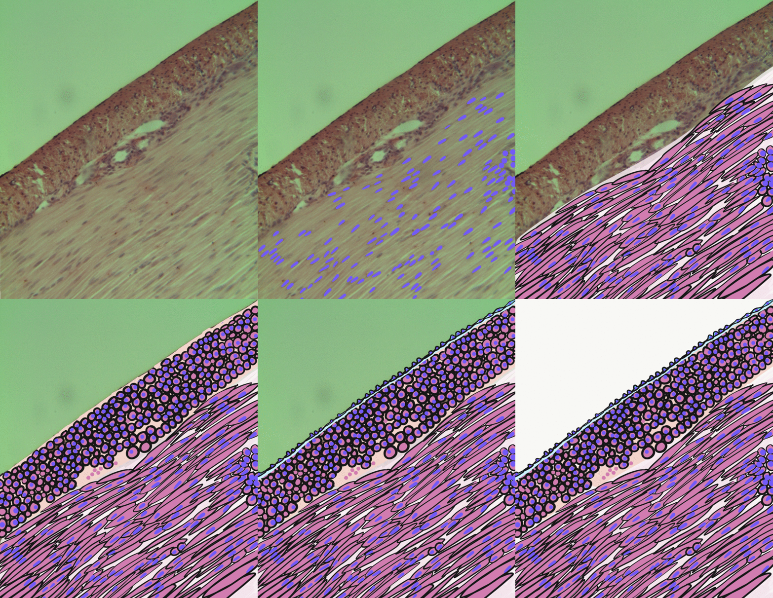
Neointima typically refers to scar tissue that forms within tubular anatomical structures such as blood vessels, as the intima is the innermost lining of these structures. Neointima can form as a result of vascular surgery such as angioplasty Angioplasty, is also known as balloon angioplasty and percutaneous transluminal angioplasty (PTA), is a minimally invasive endovascular procedure used to widen narrowed or obstructed arteries or veins, typically to treat arterial atheroscle ... or stent placement. It is actually due to proliferation of smooth muscle cells in the media giving rise to appearance of fused intima and media References Medical terminology {{circulatory-stub ... [...More Info...] [...Related Items...] OR: [Wikipedia] [Google] [Baidu] |
Restenosis
Restenosis is the recurrence of stenosis, a narrowing of a blood vessel, leading to restricted blood flow. Restenosis usually pertains to an artery or other large blood vessel that has become narrowed, received treatment to clear the blockage and subsequently become renarrowed. This is usually restenosis of an artery, or other blood vessel, or possibly a vessel within an organ. Restenosis is a common adverse event of endovascular procedures. Procedures frequently used to treat the vascular damage from atherosclerosis and related narrowing and renarrowing (restenosis) of blood vessels include vascular surgery, cardiac surgery, and angioplasty. When a stent is used and restenosis occurs, this is called in-stent restenosis or ISR. If it occurs following balloon angioplasty, this is called post-angioplasty restenosis or PARS. The diagnostic threshold for restenosis in both ISR or PARS is ≥50% stenosis. If restenosis occurs after a procedure, follow-up imaging is not the only way to ... [...More Info...] [...Related Items...] OR: [Wikipedia] [Google] [Baidu] |
Atherosclerosis
Atherosclerosis is a pattern of the disease arteriosclerosis in which the wall of the artery develops abnormalities, called lesions. These lesions may lead to narrowing due to the buildup of atheroma, atheromatous plaque. At onset there are usually no symptoms, but if they develop, symptoms generally begin around middle age. When severe, it can result in coronary artery disease, stroke, peripheral artery disease, or kidney problems, depending on which Artery, arteries are affected. The exact cause is not known and is proposed to be multifactorial. Risk factors include dyslipidemia, abnormal cholesterol levels, elevated levels of inflammatory markers, high blood pressure, diabetes, smoking, obesity, family history, genetic, and an unhealthy diet. Atheroma, Plaque is made up of fat, cholesterol, calcium, and other substances found in the blood. The narrowing of Artery, arteries limits the flow of oxygen-rich blood to parts of the body. Diagnosis is based upon a physical exam, ele ... [...More Info...] [...Related Items...] OR: [Wikipedia] [Google] [Baidu] |
Vascular Smooth Muscle
Vascular smooth muscle is the type of smooth muscle that makes up most of the walls of blood vessels. Structure Vascular smooth muscle refers to the particular type of smooth muscle found within, and composing the majority of the wall of blood vessels. Nerve supply Vascular smooth muscle is innervated primarily by the sympathetic nervous system through adrenergic receptors (adrenoceptors). The three types present are: alpha-1, alpha-2 and beta-2 adrenergic receptors, . The main endogenous agonist of these cell receptors is norepinephrine (NE). The adrenergic receptors exert opposite physiologic effects in the vascular smooth muscle under activation: * alpha-1 receptors. Under NE binding alpha-1 receptors cause vasoconstriction ( contraction of the vascular smooth muscle cells decreasing the diameter of the vessels). Thesea receptors are activated in response to shock or low blood pressure as a defensive reaction trying to restore the normal blood pressure. Antagonists ... [...More Info...] [...Related Items...] OR: [Wikipedia] [Google] [Baidu] |
Smooth Muscle Cells
Smooth muscle is an involuntary non-striated muscle, so-called because it has no sarcomeres and therefore no striations (''bands'' or ''stripes''). It is divided into two subgroups, single-unit and multiunit smooth muscle. Within single-unit muscle, the whole bundle or sheet of smooth muscle cells contracts as a syncytium. Smooth muscle is found in the walls of hollow organs, including the stomach, intestines, bladder and uterus; in the walls of passageways, such as blood, and lymph vessels, and in the tracts of the respiratory, urinary, and reproductive systems. In the eyes, the ciliary muscles, a type of smooth muscle, dilate and contract the iris and alter the shape of the lens. In the skin, smooth muscle cells such as those of the arrector pili cause hair to stand erect in response to cold temperature or fear. Structure Gross anatomy Smooth muscle is grouped into two types: single-unit smooth muscle, also known as visceral smooth muscle, and multiunit smooth muscle. M ... [...More Info...] [...Related Items...] OR: [Wikipedia] [Google] [Baidu] |
Epidermal Growth Factor
Epidermal growth factor (EGF) is a protein that stimulates cell growth and differentiation by binding to its receptor, EGFR. Human EGF is 6-k Da and has 53 amino acid residues and three intramolecular disulfide bonds. EGF was originally described as a secreted peptide found in the submaxillary glands of mice and in human urine. EGF has since been found in many human tissues, including platelets, submandibular gland (submaxillary gland), and parotid gland. Initially, human EGF was known as urogastrone. Structure In humans, EGF has 53 amino acids (sequence NSDSECPLSHDGYCLHDGVCMYIEALDKYACNCVVGYIGERCQYRDLKWWELR), with a molecular mass of around 6 kDa. It contains three disulfide bridges (Cys6-Cys20, Cys14-Cys31, Cys33-Cys42). Function EGF, via binding to its cognate receptor, results in cellular proliferation, differentiation, and survival. Salivary EGF, which seems to be regulated by dietary inorganic iodine, also plays an important physiological role in the maintenance of ... [...More Info...] [...Related Items...] OR: [Wikipedia] [Google] [Baidu] |
Two-hybrid Screening
Two-hybrid screening (originally known as yeast two-hybrid system or Y2H) is a molecular biology technique used to discover protein–protein interactions (PPIs) and protein–DNA interactions by testing for physical interactions (such as binding) between two proteins or a single protein and a DNA molecule, respectively. The premise behind the test is the activation of downstream reporter gene(s) by the binding of a transcription factor onto an upstream activating sequence (UAS). For two-hybrid screening, the transcription factor is split into two separate fragments, called the DNA-binding domain (DBD or often also abbreviated as BD) and activating domain (AD). The BD is the domain responsible for binding to the UAS and the AD is the domain responsible for the activation of transcription. The Y2H is thus a protein-fragment complementation assay. History Pioneered by Stanley Fields and Ok-Kyu Song in 1989, the technique was originally designed to detect protein–protein ... [...More Info...] [...Related Items...] OR: [Wikipedia] [Google] [Baidu] |
Extracellular Matrix
In biology, the extracellular matrix (ECM), also called intercellular matrix, is a three-dimensional network consisting of extracellular macromolecules and minerals, such as collagen, enzymes, glycoproteins and hydroxyapatite that provide structural and biochemical support to surrounding cells. Because multicellularity evolved independently in different multicellular lineages, the composition of ECM varies between multicellular structures; however, cell adhesion, cell-to-cell communication and differentiation are common functions of the ECM. The animal extracellular matrix includes the interstitial matrix and the basement membrane. Interstitial matrix is present between various animal cells (i.e., in the intercellular spaces). Gels of polysaccharides and fibrous proteins fill the Interstitial fluid, interstitial space and act as a compression buffer against the stress placed on the ECM. Basement membranes are sheet-like depositions of ECM on which various epithelial cells rest ... [...More Info...] [...Related Items...] OR: [Wikipedia] [Google] [Baidu] |


