|
ACAN (gene)
Aggrecan (ACAN), also known as cartilage-specific proteoglycan core protein (CSPCP) or chondroitin sulfate proteoglycan 1, is a protein that in humans is encoded by the ''ACAN'' gene. This gene is a member of the lectican (chondroitin sulfate proteoglycan) family. The encoded protein is an integral part of the extracellular matrix in cartilagenous tissue and it withstands compression in cartilage. Aggrecan is a proteoglycan, or a protein modified with large carbohydrates; the human form of the protein is 2316 amino acids long and can be expressed in multiple isoforms due to alternative splicing. Aggrecan was named for its ability to form large aggregates in the cartilage tissue (a large aggregating proteoglycan). Structure Aggrecan is a high molecular weight (1x10⁶ < M < 3x10⁶) proteoglycan. It exhibits a bottlebrush structure, in which |
Protein
Proteins are large biomolecules and macromolecules that comprise one or more long chains of amino acid residues. Proteins perform a vast array of functions within organisms, including catalysing metabolic reactions, DNA replication, responding to stimuli, providing structure to cells and organisms, and transporting molecules from one location to another. Proteins differ from one another primarily in their sequence of amino acids, which is dictated by the nucleotide sequence of their genes, and which usually results in protein folding into a specific 3D structure that determines its activity. A linear chain of amino acid residues is called a polypeptide. A protein contains at least one long polypeptide. Short polypeptides, containing less than 20–30 residues, are rarely considered to be proteins and are commonly called peptides. The individual amino acid residues are bonded together by peptide bonds and adjacent amino acid residues. The sequence of amino acid residue ... [...More Info...] [...Related Items...] OR: [Wikipedia] [Google] [Baidu] |
N-terminus
The N-terminus (also known as the amino-terminus, NH2-terminus, N-terminal end or amine-terminus) is the start of a protein or polypeptide, referring to the free amine group (-NH2) located at the end of a polypeptide. Within a peptide, the amine group is bonded to the carboxylic group of another amino acid, making it a chain. That leaves a free carboxylic group at one end of the peptide, called the C-terminus, and a free amine group on the other end called the N-terminus. By convention, peptide sequences are written N-terminus to C-terminus, left to right (in LTR writing systems). This correlates the translation direction to the text direction, because when a protein is translated from messenger RNA, it is created from the N-terminus to the C-terminus, as amino acids are added to the carboxyl end of the protein. Chemistry Each amino acid has an amine group and a carboxylic group. Amino acids link to one another by peptide bonds which form through a dehydration reaction that ... [...More Info...] [...Related Items...] OR: [Wikipedia] [Google] [Baidu] |
Immunoglobulin Domain
The immunoglobulin domain, also known as the immunoglobulin fold, is a type of protein domain that consists of a 2-layer sandwich of 7-9 antiparallel β-strands arranged in two β-sheets with a Greek key topology, consisting of about 125 amino acids. The backbone switches repeatedly between the two β-sheets. Typically, the pattern is (N-terminal β-hairpin in sheet 1)-(β-hairpin in sheet 2)-(β-strand in sheet 1)-(C-terminal β-hairpin in sheet 2). The cross-overs between sheets form an "X", so that the N- and C-terminal hairpins are facing each other. Members of the immunoglobulin superfamily are found in hundreds of proteins of different functions. Examples include antibodies, the giant muscle kinase titin, and receptor tyrosine kinases. Immunoglobulin-like domains may be involved in protein–protein and protein–ligand interactions. Examples Human genes encoding proteins containing the immunoglobulin domain include: * A1BG * ACAM * ADAMTSL1 * ADAMTSL3 * AGER * A ... [...More Info...] [...Related Items...] OR: [Wikipedia] [Google] [Baidu] |
EGF-like Domain
The EGF-like domain is an evolutionary conserved protein domain, which derives its name from the epidermal growth factor where it was first described. It comprises about 30 to 40 amino-acid residues and has been found in a large number of mostly animal proteins. Most occurrences of the EGF-like domain are found in the extracellular domain of membrane-bound proteins or in proteins known to be secreted. An exception to this is the prostaglandin-endoperoxide synthase. The EGF-like domain includes 6 cysteine residues which in the epidermal growth factor have been shown to form 3 disulfide bonds. The structures of 4-disulfide EGF-domains have been solved from the laminin and integrin proteins. The main structure of EGF-like domains is a two-stranded β-sheet followed by a loop to a short C-terminal, two-stranded β-sheet. These two β-sheets are usually denoted as the major (N-terminal) and minor (C-terminal) sheets. EGF-like domains frequently occur in numerous tandem copies in pro ... [...More Info...] [...Related Items...] OR: [Wikipedia] [Google] [Baidu] |
CD44
The CD44 antigen is a cell-surface glycoprotein involved in cell–cell interactions, cell adhesion and migration. In humans, the CD44 antigen is encoded by the ''CD44'' gene on chromosome 11. CD44 has been referred to as HCAM (homing cell adhesion molecule), Pgp-1 (phagocytic glycoprotein-1), Hermes antigen, lymphocyte homing receptor, ECM-III, and HUTCH-1. Tissue distribution and isoforms CD44 is expressed in a large number of mammalian cell types. The standard isoform, designated CD44s, comprising exons 1–5 and 16–20 is expressed in most cell types. CD44 splice variants containing variable exons are designated CD44v. Some epithelial cells also express a larger isoform (CD44E), which includes exons v8–10. Function CD44 participates in a wide variety of cellular functions including lymphocyte activation, recirculation and homing, hematopoiesis, and tumor metastasis. CD44 is a receptor for hyaluronic acid and can also interact with other ligands, such as osteop ... [...More Info...] [...Related Items...] OR: [Wikipedia] [Google] [Baidu] |
Brevican
Brevican core protein is a protein that in humans is encoded by the ''BCAN'' gene. Brevican is a member of the lectican protein family. Brevican is localised to the surface of neurons in the brain. In melanocytic cells, BCAN gene expression may be regulated by MITF Microphthalmia-associated transcription factor also known as class E basic helix-loop-helix protein 32 or bHLHe32 is a protein that in humans is encoded by the ''MITF'' gene. MITF is a basic helix-loop-helix leucine zipper transcription factor .... References Further reading * * * * * * * * * * * * External links * C-type lectins Lecticans Extracellular matrix proteins {{gene-1-stub ... [...More Info...] [...Related Items...] OR: [Wikipedia] [Google] [Baidu] |
Neurocan
Neurocan core protein is a protein that in humans is encoded by the ''NCAN'' gene. Neurocan is a member of the lectican / chondroitin sulfate proteoglycan protein families and consists of neurocan core protein and chondroitin sulfate. It is thought to be involved in the modulation of cell adhesion and migration. Role in bipolar disorder Neurocan is a significant component of the extracellular matrix, and its levels are modulated by a variety of factors, but mice in which the NCAN gene has been knocked out show no easily observable defects in brain development or behavior. However, a genome-wide association study published in 2011 identified Neurocan as a susceptibility factor for bipolar disorder Bipolar disorder, previously known as manic depression, is a mental disorder characterized by periods of depression and periods of abnormally elevated mood that last from days to weeks each. If the elevated mood is severe or associated with .... A more comprehensive study pu ... [...More Info...] [...Related Items...] OR: [Wikipedia] [Google] [Baidu] |
Versican
Versican is a large extracellular matrix proteoglycan that is present in a variety of human tissues. It is encoded by the ''VCAN'' gene. Versican is a large chondroitin sulfate proteoglycan with an apparent molecular mass of more than 1000kDa. In 1989, Zimmermann and Ruoslahti cloned and sequenced the core protein of fibroblast chondroitin sulfate proteoglycan. They designated it versican in recognition of its versatile modular structure. Versican belongs to the lectican protein family, with aggrecan (abundant in cartilage), brevican and neurocan (nervous system proteoglycans) as other members. Versican is also known as chondroitin sulfate proteoglycan core protein 2 or chondroitin sulfate proteoglycan 2 (CSPG2), and PG-M. Structure These proteoglycans share a homologous globular N-terminal, C-terminal, and glycosaminoglycan (GAG) binding regions. The N-terminal (G1) globular domain consists of Ig-like loop and two link modules, and has Hyaluronan (HA) binding properti ... [...More Info...] [...Related Items...] OR: [Wikipedia] [Google] [Baidu] |
Articular Cartilage
Hyaline cartilage is the glass-like (hyaline) and translucent cartilage found on many joint surfaces. It is also most commonly found in the ribs, nose, larynx, and trachea. Hyaline cartilage is pearl-gray in color, with a firm consistency and has a considerable amount of collagen. It contains no nerves or blood vessels, and its structure is relatively simple. Structure Hyaline cartilage is covered externally by a fibrous membrane known as the perichondrium or, when it's along articulating surfaces, the synovial membrane. This membrane contains vessels that provide the cartilage with nutrition through diffusion. Hyaline cartilage matrix is primarily made of type II collagen and chondroitin sulphate, both of which are also found in elastic cartilage. Hyaline cartilage exists on the sternal ends of the ribs, in the larynx, trachea, and bronchi, and on the articulating surfaces of bones. It gives the structures a definite but pliable form. The presence of collagen fibres makes ... [...More Info...] [...Related Items...] OR: [Wikipedia] [Google] [Baidu] |
Type-II Collagen
Type II collagen is the basis for hyaline cartilage, including the articular cartilages at joint surfaces. It is formed by homotrimers of collagen, type II, alpha 1 chains. It makes up 50% of all protein in cartilage and 85–90% of collagen of articular cartilage. Type II collagen is organised into fibrils. This fibrillar network of collagen allows the cartilage to entrap the proteoglycan aggregate, as well as providing tensile strength to the tissue. Oral administration of native type II collagen induces oral tolerance to pathological immune responses and may be useful in arthritis. See also * Type I collagen Type I collagen is the most abundant collagen of the human body. It forms large, eosinophilic fibers known as collagen fibers. It is present in scar tissue, the end product when tissue heals by repair, as well as tendons, ligaments, the endomy ... * Collagen, type III, alpha 1 References External links * Collagens {{gene-12-stub ... [...More Info...] [...Related Items...] OR: [Wikipedia] [Google] [Baidu] |
Chondrocyte
Chondrocytes (, from Greek χόνδρος, ''chondros'' = cartilage + κύτος, ''kytos'' = cell) are the only cells found in healthy cartilage. They produce and maintain the cartilaginous matrix, which consists mainly of collagen and proteoglycans. Although the word ''chondroblast'' is commonly used to describe an immature chondrocyte, the term is imprecise, since the progenitor of chondrocytes (which are mesenchymal stem cells) can differentiate into various cell types, including osteoblasts. Development From least- to terminally-differentiated, the chondrocytic lineage is: # Colony-forming unit-fibroblast # Mesenchymal stem cell / marrow stromal cell # Chondrocyte # Hypertrophic chondrocyte Mesenchymal (mesoderm origin) stem cells are undifferentiated, meaning they can differentiate into a variety of generative cells commonly known as osteochondrogenic (or osteogenic, chondrogenic, osteoprogenitor, etc.) cells. When referring to bone, or in this case cartilage, the origin ... [...More Info...] [...Related Items...] OR: [Wikipedia] [Google] [Baidu] |
Cell Adhesion
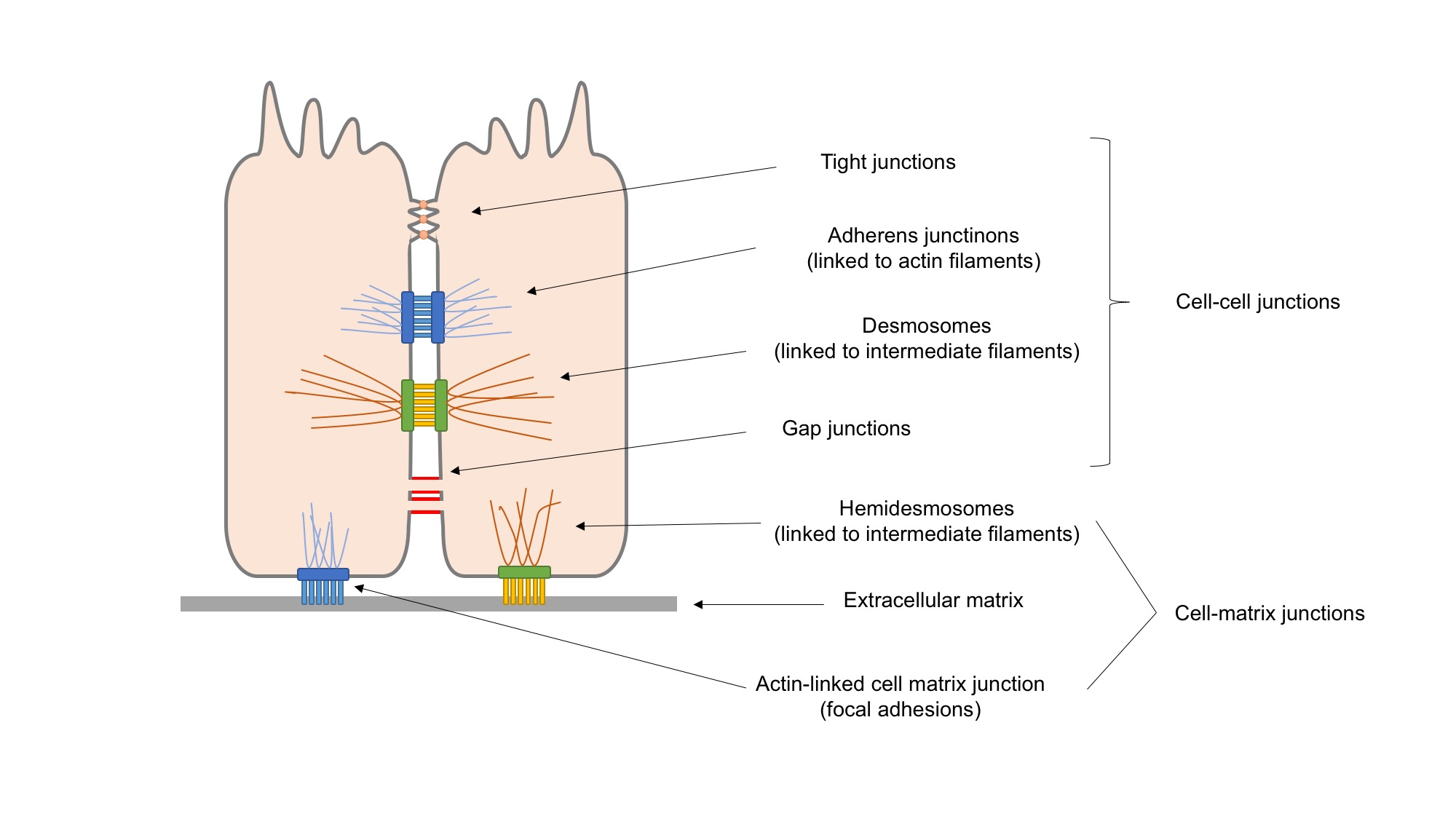
Cell adhesion is the process by which cells interact and attach to neighbouring cells through specialised molecules of the cell surface. This process can occur either through direct contact between cell surfaces such as cell junctions or indirect interaction, where cells attach to surrounding extracellular matrix, a gel-like structure containing molecules released by cells into spaces between them. Cells adhesion occurs from the interactions between cell-adhesion molecules (CAMs), transmembrane proteins located on the cell surface. Cell adhesion links cells in different ways and can be involved in signal transduction for cells to detect and respond to changes in the surroundings. Other cellular processes regulated by cell adhesion include cell migration and tissue development in multicellular organisms. Alterations in cell adhesion can disrupt important cellular processes and lead to a variety of diseases, including cancer and arthritis. Cell adhesion is also essential for in ... [...More Info...] [...Related Items...] OR: [Wikipedia] [Google] [Baidu] |


