|
Axioappendicular Muscles
The Axioappendicular muscles are the muscles that extend between the axial and (superior or inferior) appendicular skeletons. There are two groups, the anterior axioappendicular muscles and the posterior axioappendicular muscles. Anterior axioappendicular muscles The anterior axioappendicular muscles are the: * Pectoralis major * Pectoralis minor * Subclavius * Serratus anterior Posterior axioappendicular muscles The posterior axioappendicular muscles are described as two sub-groups: * Superficial Posterior Axioappendicular muscles (or the extrinsic shoulder muscles) ** Trapezius ** Latissimus dorsi * Deep Posterior Axioappendicular muscles (or the intrinsic shoulder muscles) ** Levator scapulae ** Rhomboid minor ** Rhomboid major The rhomboid major is a skeletal muscle on the back that connects the scapula with the vertebrae of the spinal column. In human anatomy, it acts together with the rhomboid minor to keep the scapula pressed against thoracic wall and to retract th ... [...More Info...] [...Related Items...] OR: [Wikipedia] [Google] [Baidu] |
Axial Skeleton
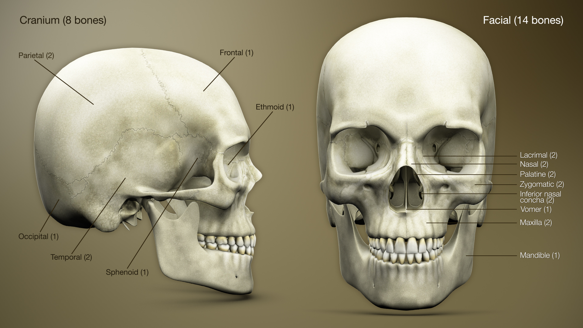
The axial skeleton is the part of the skeleton that consists of the bones of the head and trunk (anatomy), trunk of a vertebrate. In the human skeleton, it consists of 80 bones and is composed of six parts; the human skull, skull (22 bones), also the ossicles of the middle ear, the hyoid bone, the rib cage, sternum and the vertebral column. The axial skeleton together with the appendicular skeleton form the complete skeleton. Another definition of axial skeleton is the bones including the vertebrae, sacrum, coccyx, skull, ribs, and sternum. Structure Flat bones house the brain and other vital organs. This article mainly deals with the axial skeletons of humans; however, it is important to understand the evolutionary lineage of the axial skeleton. The human axial skeleton consists of 81 different bones. It is the medial core of the body and connects the pelvis to the body, where the appendix skeleton attaches. As the skeleton grows older the bones get weaker with the exception ... [...More Info...] [...Related Items...] OR: [Wikipedia] [Google] [Baidu] |
Appendicular Skeleton
The appendicular skeleton is the portion of the skeleton of vertebrates consisting of the bones that support the appendages. There are 126 bones. The appendicular skeleton includes the skeletal elements within the limbs, as well as supporting shoulder girdle and pelvic girdle. ''Encyclopædia Britannica''. Updated 24 August 2014. The word appendicular is the adjective of the noun ''appendage'', which itself means a part that is joined to something larger. The organization of the appendicular system Of the 206 bones in the human skeleton, the appendicular skeleton comprises 126. Functionally it is involved in locomotion (lower limbs) of the |
Pectoralis Major Muscle
The pectoralis major () is a thick, fan-shaped or triangular convergent muscle, situated at the chest of the human body. It makes up the bulk of the chest muscles and lies under the breast. Beneath the pectoralis major is the pectoralis minor, a thin, triangular muscle. The pectoralis major's primary functions are flexion, adduction, and internal rotation of the humerus. The pectoral major may colloquially be referred to as "pecs", "pectoral muscle", or "chest muscle", because it is the largest and most superficial muscle in the chest area. Structure It arises from the anterior surface of the sternal half of the clavicle from breadth of the half of the anterior surface of the sternum, as low down as the attachment of the cartilage of the sixth or seventh rib; from the cartilages of all the true ribs, with the exception, frequently, of the first or seventh, and from the aponeurosis of the abdominal external oblique muscle. From this extensive origin the fibers converge toward the ... [...More Info...] [...Related Items...] OR: [Wikipedia] [Google] [Baidu] |
Pectoralis Minor Muscle
Pectoralis minor muscle () is a thin, triangular muscle, situated at the upper part of the chest, beneath the pectoralis major in the human body. Structure Attachments Pectoralis minor muscle arises from the upper margins and outer surfaces of the third, fourth, and fifth ribs, near their costal cartilages and from the aponeuroses covering the intercostalis. The fibers pass superior and lateral and converge to form a flat tendon. This tendon inserts onto the medial border and upper surface of the coracoid process of the scapula. Relations Pectoralis minor muscle forms part of the anterior wall of the axilla. It is covered anteriorly (superficially) by the clavipectoral fascia. The medial pectoral nerve pierces the pectoralis minor and the clavipectoral fascia. In attaching to the coracoid process, the pectoralis minor forms a 'bridge' - structures passing into the upper limb from the thorax will pass directly underneath.http://www.teachmeanatomy.com/muscles-of-the-pector ... [...More Info...] [...Related Items...] OR: [Wikipedia] [Google] [Baidu] |
Subclavius Muscle
The subclavius is a small triangular muscle, placed between the clavicle and the first rib. Along with the pectoralis major and pectoralis minor muscles, the subclavius muscle makes up the anterior axioappendicular muscles, also known as anterior wall of the axilla. Structure It arises by a short, thick tendon from the first rib and its cartilage at their junction, in front of the costoclavicular ligament. The fleshy fibers proceed obliquely superolaterally, to be inserted into the groove on the under surface of the clavicle. Innervation The nerve to subclavius (or subclavian nerve) innervates the muscle. This arises from the junction of the fifth and sixth cervical nerves, from the superior/upper trunk of the brachial plexus. Variation Insertion into coracoid process instead of clavicle or into both clavicle and coracoid process. Sternoscapular fasciculus to the upper border of scapula. Sternoclavicularis from manubrium to clavicle between pectoralis major and coracoclavicular ... [...More Info...] [...Related Items...] OR: [Wikipedia] [Google] [Baidu] |
Serratus Anterior Muscle
The serratus anterior is a muscle that originates on the surface of the 1st to 8th ribs at the side of the chest and inserts along the entire anterior length of the medial border of the scapula. The serratus anterior acts to pull the scapula forward around the thorax. The muscle is named from Latin: ''serrare'' = to saw, referring to the shape, ''anterior'' = on the front side of the body. Structure Serratus anterior normally originates by nine or ten muscle slips – branches from either the first to ninth ribs or the first to eighth ribs. Because two slips usually arise from the second rib, the number of slips is greater than the number of ribs from which they originate. The muscle is inserted along the medial border of the scapula between the superior and inferior angles along with being inserted along the thoracic vertebrae. The muscle is divided into three named parts depending on their points of insertions: #the serratus anterior superior is inserted near the superior a ... [...More Info...] [...Related Items...] OR: [Wikipedia] [Google] [Baidu] |
Trapezius Muscle
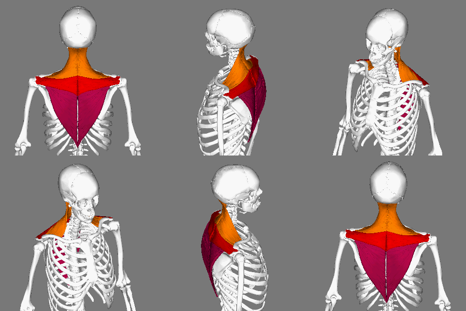
The trapezius is a large paired trapezoid-shaped surface muscle that extends longitudinally from the occipital bone to the lower thoracic vertebrae of the spine and laterally to the spine of the scapula. It moves the scapula and supports the arm. The trapezius has three functional parts: an upper (descending) part which supports the weight of the arm; a middle region (transverse), which retracts the scapula; and a lower (ascending) part which medially rotates and depresses the scapula. Name and history The trapezius muscle resembles a trapezium, also known as a trapezoid, or diamond-shaped quadrilateral. The word "spinotrapezius" refers to the human trapezius, although it is not commonly used in modern texts. In other mammals, it refers to a portion of the analogous muscle. Similarly, the term "tri-axle back plate" was historically used to describe the trapezius muscle. Structure The ''superior'' or ''upper'' (or descending) fibers of the trapezius originate from the sp ... [...More Info...] [...Related Items...] OR: [Wikipedia] [Google] [Baidu] |
Latissimus Dorsi Muscle
The latissimus dorsi () is a large, flat muscle on the back that stretches to the sides, behind the arm, and is partly covered by the trapezius on the back near the midline. The word latissimus dorsi (plural: ''latissimi dorsorum'') comes from Latin and means "broadest uscleof the back", from "latissimus" ( la, broadest)' and "dorsum" ( la, back). The pair of muscles are commonly known as "lats", especially among bodybuilders. The latissimus dorsi is the largest muscle in the upper body. The latissimus dorsi is responsible for extension, adduction, transverse extension also known as horizontal abduction (or horizontal extension), flexion from an extended position, and (medial) internal rotation of the shoulder joint. It also has a synergistic role in extension and lateral flexion of the lumbar spine. Due to bypassing the scapulothoracic joints and attaching directly to the spine, the actions the latissimi dorsi have on moving the arms can also influence the movement of the sca ... [...More Info...] [...Related Items...] OR: [Wikipedia] [Google] [Baidu] |
Levator Scapulae Muscle
The levator scapulae is a slender skeletal muscle situated at the back and side of the neck. As the Latin name suggests, its main function is to lift the scapula. Anatomy Attachments The muscle descends diagonally from its origin to its insertion. Origin The levator scapulae originates from the posterior tubercles of the transverse processes of cervical vertebrae C1-4. At its origin, it attaches via tendinous slips. Insertion It inserts onto the medial border of the scapula (with its site of insertion extending between the superior angle of the scapula superiorly, and the junction of spine of scapula and medial border of scapula inferiorly). Relations One of the muscles within the floor of the posterior triangle of the neck, the superior part of levator scapulae is covered by sternocleidomastoid and its inferior part by the trapezius. It is bounded in front by the scalenus medius and behind by splenius cervicis. The spinal accessory nerve crosses laterally in the middl ... [...More Info...] [...Related Items...] OR: [Wikipedia] [Google] [Baidu] |
Rhomboid Minor Muscle
In human anatomy, the rhomboid minor is a small skeletal muscle on the back that connects the scapula with the vertebrae of the spinal column. Located inferior to levator scapulae and superior to rhomboid major, it acts together with the latter to keep the scapula pressed against the thoracic wall. It lies deep to trapezius but superficial to the long spinal muscles. Origin and insertion The rhomboid minor arises from the inferior border of the nuchal ligament, from the spinous processes of the seventh cervical and first thoracic vertebrae, and from the intervening supraspinous ligaments. It is inserted into a small area of the medial border of the scapula at the level of the scapular spine. Action Together with the rhomboid major, the rhomboid minor retracts the scapula when trapezius is contracted. Acting as a synergist to the trapezius, the rhomboid major and minor elevate the medial border of the scapula medially and upward, working in tandem with the levator scapu ... [...More Info...] [...Related Items...] OR: [Wikipedia] [Google] [Baidu] |
Rhomboid Major Muscle
The rhomboid major is a skeletal muscle on the back that connects the scapula with the vertebrae of the spinal column. In human anatomy, it acts together with the rhomboid minor to keep the scapula pressed against thoracic wall and to retract the scapula toward the vertebral column. Structure The rhomboid major arises from the spinous processes of the thoracic vertebrae T2 to T5 as well as the supraspinous ligament. It inserts on the medial border of the scapula, from about the level of the scapular spine to the scapula's inferior angle. The rhomboid major is considered a superficial back muscle. It is deep to the trapezius, and is located directly inferior to the rhomboid minor. As the word ''rhomboid'' suggests, the rhomboid major is diamond-shaped. The ''major'' in its name indicates that it is the larger of the two rhomboids. Variation The two rhomboids are sometimes fused into a single muscle. Nerve supply The rhomboid major, like the rhomboid minor, is innervated by ... [...More Info...] [...Related Items...] OR: [Wikipedia] [Google] [Baidu] |


