|
Auditory Canal
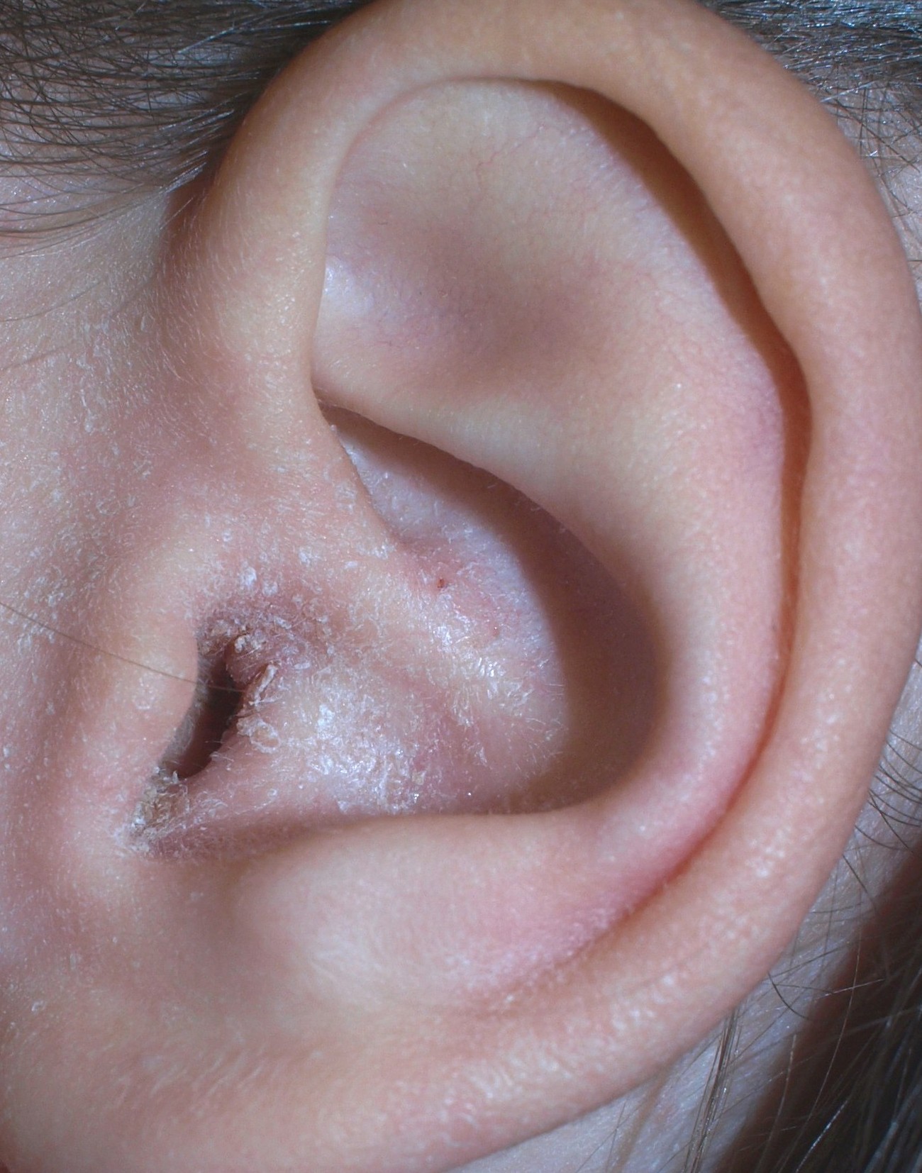
The ear canal (external acoustic meatus, external auditory meatus, EAM) is a pathway running from the outer ear to the middle ear. The adult human ear canal extends from the pinna to the eardrum and is about in length and in diameter. Structure The human ear canal is divided into two parts. The elastic cartilage part forms the outer third of the canal; its anterior and lower wall are cartilaginous, whereas its superior and back wall are fibrous. The cartilage is the continuation of the cartilage framework of pinna. The cartilaginous portion of the ear canal contains small hairs and specialized sweat glands, called apocrine glands, which produce cerumen ( ear wax). The bony part forms the inner two thirds. The bony part is much shorter in children and is only a ring (''annulus tympanicus'') in the newborn. The layer of epithelium encompassing the bony portion of the ear canal is much thinner and therefore, more sensitive in comparison to the cartilaginous portion. Size and sh ... [...More Info...] [...Related Items...] OR: [Wikipedia] [Google] [Baidu] |
First Branchial Arch
The pharyngeal arches, also known as visceral arches'','' are structures seen in the embryonic development of vertebrates that are recognisable precursors for many structures. In fish, the arches are known as the branchial arches, or gill arches. In the human embryo, the arches are first seen during the fourth week of development. They appear as a series of outpouchings of mesoderm on both sides of the developing pharynx. The vasculature of the pharyngeal arches is known as the aortic arches. In fish, the branchial arches support the gills. Structure In vertebrates, the pharyngeal arches are derived from all three germ layers (the primary layers of cells that form during embryogenesis). Neural crest cells enter these arches where they contribute to features of the skull and facial skeleton such as bone and cartilage. However, the existence of pharyngeal structures before neural crest cells evolved is indicated by the existence of neural crest-independent mechanisms of pharyng ... [...More Info...] [...Related Items...] OR: [Wikipedia] [Google] [Baidu] |
Earwax
Earwax, also known by the medical term cerumen, is a brown, orange, red, yellowish or gray waxy substance secreted in the ear canal of humans and other mammals. It protects the skin of the human ear canal, assists in cleaning and lubrication, and provides protection against bacteria, fungi, and water. Earwax consists of dead skin cells, hair, and the secretions of cerumen by the ceruminous and sebaceous glands of the outer ear canal. Major components of earwax are long chain fatty acids, both saturated and unsaturated, alcohols, squalene, and cholesterol. Excess or compacted cerumen is the buildup of ear wax causing a blockage in the ear canal and it can press against the eardrum or block the outside ear canal or hearing aids, potentially causing hearing loss. Physiology Cerumen is produced in the cartilaginous portion which is the outer third portion of the ear canal. It is a mixture of viscous secretions from sebaceous glands and less-viscous ones from modified apocri ... [...More Info...] [...Related Items...] OR: [Wikipedia] [Google] [Baidu] |
Otitis Externa
Otitis externa, also called swimmer's ear, is inflammation of the ear canal. It often presents with ear pain, swelling of the ear canal, and occasionally decreased hearing. Typically there is pain with movement of the outer ear. A high fever is typically not present except in severe cases. Otitis externa may be acute (lasting less than six weeks) or chronic (lasting more than three months). Acute cases are typically due to bacterial infection, and chronic cases are often due to allergies and autoimmune disorders. the most common cause of Otitis externa is bacterial. Risk factors for acute cases include swimming, minor trauma from cleaning, using hearing aids and ear plugs, and other skin problems, such as psoriasis and dermatitis. People with diabetes are at risk of a severe form of ''malignant otitis externa''. Diagnosis is based on the signs and symptoms. Culturing the ear canal may be useful in chronic or severe cases. Acetic acid ear drops may be used as a preventive measur ... [...More Info...] [...Related Items...] OR: [Wikipedia] [Google] [Baidu] |
Tympanostomy Tube
Tympanostomy tube, also known as a grommet or myringotomy tube, is a small tube inserted into the eardrum in order to keep the middle ear aerated for a prolonged period of time, and to prevent the accumulation of fluid in the middle ear. The operation to insert the tube involves a myringotomy and is performed under local or general anesthesia. The tube itself is made in a variety of designs. The most commonly used type is shaped like a grommet. When it is necessary to keep the middle ear ventilated for a very long period, a "T"-shaped tube may be used, as these "T-tubes" can stay in place for 2–4 years. Materials used to construct the tube are most often plastics such as silicone or Teflon. Stainless steel tubes exist, but are no longer in frequent use. Medical uses Inserting grommets is a common surgical procedure for treating children around the world. Grommets are most commonly used to help improve hearing for children who have a condition commonly called "glue ear" (pe ... [...More Info...] [...Related Items...] OR: [Wikipedia] [Google] [Baidu] |
Scar
A scar (or scar tissue) is an area of fibrous tissue that replaces normal skin after an injury. Scars result from the biological process of wound repair in the skin, as well as in other organs, and tissues of the body. Thus, scarring is a natural part of the healing process. With the exception of very minor lesions, every wound (e.g., after accident, disease, or surgery) results in some degree of scarring. An exception to this are animals with complete regeneration, which regrow tissue without scar formation. Scar tissue is composed of the same protein ( collagen) as the tissue that it replaces, but the fiber composition of the protein is different; instead of a random basketweave formation of the collagen fibers found in normal tissue, in fibrosis the collagen cross-links and forms a pronounced alignment in a single direction. This collagen scar tissue alignment is usually of inferior functional quality to the normal collagen randomised alignment. For example, scars in the s ... [...More Info...] [...Related Items...] OR: [Wikipedia] [Google] [Baidu] |
Granuloma
A granuloma is an aggregation of macrophages that forms in response to chronic inflammation. This occurs when the immune system attempts to isolate foreign substances that it is otherwise unable to eliminate. Such substances include infectious organisms including bacteria and fungi, as well as other materials such as foreign objects, keratin, and suture fragments. Definition In pathology, a granuloma is an organized collection of macrophages. In medical practice, doctors occasionally use the term ''granuloma'' in its more literal meaning: "a small nodule". Since a small nodule can represent any tissue from a harmless nevus to a malignant tumor, this use of the term is not very specific. Examples of this use of the term ''granuloma'' are the lesions known as vocal cord granuloma (known as contact granuloma), pyogenic granuloma, and intubation granuloma, all of which are examples of granulation tissue, not granulomas. "Pulmonary hyalinizing granuloma" is a lesion characterized ... [...More Info...] [...Related Items...] OR: [Wikipedia] [Google] [Baidu] |
Myiasis
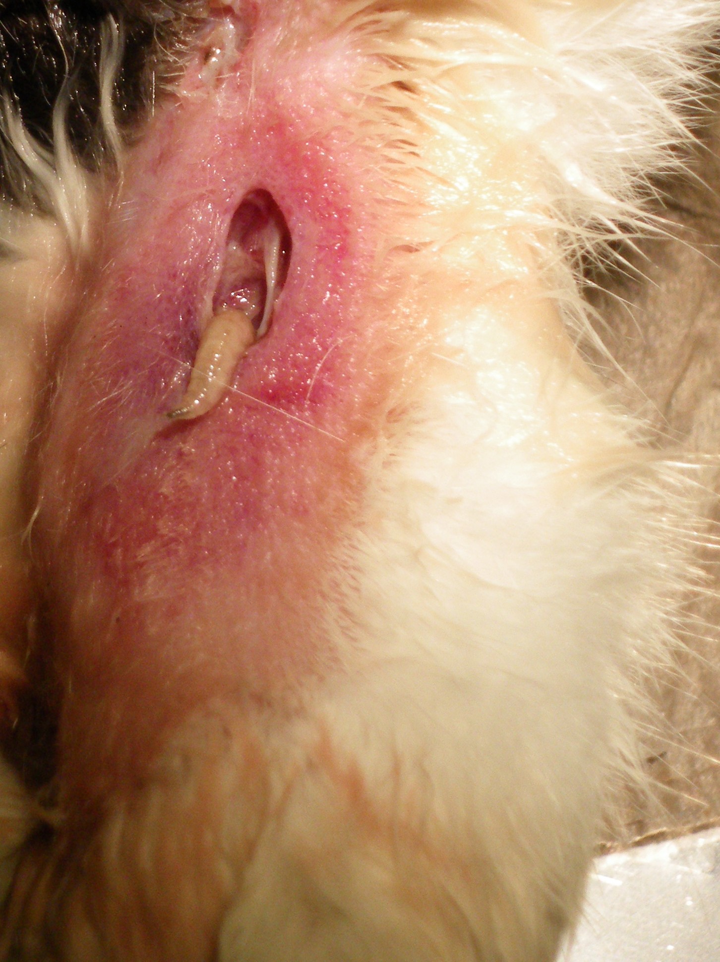
Myiasis is the parasitic infestation of the body of a live animal by fly larvae (maggots) which grow inside the host while feeding on its tissue. Although flies are most commonly attracted to open wounds and urine- or feces-soaked fur, some species (including the most common myiatic flies—the botfly, blowfly, and screwfly) can create an infestation even on unbroken skin and have been known to use moist soil and non-myiatic flies (such as the common housefly) as vector agents for their parasitic larvae. Because some animals (particularly non-native domestic animals) cannot react as effectively as humans to the causes and effects of myiasis, such infestations present a severe and continuing problem for livestock industries worldwide, causing severe economic losses where they are not mitigated by human action. Although typically a far greater issue for animals, myiasis is also a relatively frequent disease for humans in rural tropical regions where myiatic flies thrive, and of ... [...More Info...] [...Related Items...] OR: [Wikipedia] [Google] [Baidu] |
Ear Mite
Ear mites are mites that live in the ears of animals and humans. The most commonly seen species in veterinary medicine is ''Otodectes cynotis'' (Gk. ''oto''=ear, ''dectes''=biter, ''cynotis''=of the dog). This species, despite its name, is also responsible for 90% of ear mite infections in felines. In veterinary practice, ear mite infections in dogs and cats may present as a disease that causes intense itching in one or both ears, which in turn triggers scratching at the affected ear. An unusually dark colored ear wax (cerumen) may also be produced. Cats, as well as dogs with erect ears that have control over ear direction, may be seen with one or both ear pinnas held at an odd or flattened angle. The most common lesion associated with ear mites is an open or crusted ("scabbed") skin wound at the back or base of the ear, caused by abrasion of the skin by hind limb claws, as the ear has been scratched in an attempt to relieve the itching. This lesion often becomes secondarily infe ... [...More Info...] [...Related Items...] OR: [Wikipedia] [Google] [Baidu] |
Otomycosis
Otomycosis is a fungal ear infection, a superficial mycotic infection of the outer ear canal. It is more common in tropical countries. The infection may be either subacute or acute and is characterized by malodorous discharge, inflammation, pruritus, scaling, and severe discomfort. The mycosis results in inflammation, superficial epithelial exfoliation, masses of debris containing hyphae, suppuration, and pain. Diagnosis Otoscopy (exam of the ear) is best done with a binocular microscope that provides adequate lighting, depth perception, and the ability to instrument the ear to comfortably remove the fungus. Findings range from scattered saprophytic fungal colonies of various colors, causing no symptoms, to densely packed fungal debris, often intermixed with cerumen (wax), filling the entire canal and involving the tympanic membrane (eardrum). The fungus can cling to the skin and tympanic membrane, presumably because of invading hyphae, and can require significant time to ... [...More Info...] [...Related Items...] OR: [Wikipedia] [Google] [Baidu] |
Contact Dermatitis
Contact dermatitis is a type of acute or chronic inflammation of the skin caused by exposure to chemical or physical agents. Symptoms of contact dermatitis can include itchy or dry skin, a red rash, bumps, blisters, or swelling. These rashes are not contagious or life-threatening, but can be very uncomfortable. Contact dermatitis results from either exposure to allergens (allergic contact dermatitis), or irritants (irritant contact dermatitis). Allergic contact dermatitis involves a delayed type of hypersensitivity and previous exposure to an allergen to produce a reaction. Irritant contact dermatitis is the most common type and represents 80% of all cases. It is caused by prolonged exposure to irritants, leading to direct injury of the epidermal cells of the skin, which activates an immune response, resulting in an inflammatory cutaneous reaction. Phototoxic dermatitis occurs when the allergen or irritant is activated by sunlight. Diagnosis of allergic contact dermatitis can o ... [...More Info...] [...Related Items...] OR: [Wikipedia] [Google] [Baidu] |
Cholesteatoma
Cholesteatoma is a destructive and expanding growth consisting of keratinizing squamous epithelium in the middle ear and/or mastoid process. Cholesteatomas are not cancerous as the name may suggest, but can cause significant problems because of their erosive and expansile properties. This can result in the destruction of the bones of the middle ear ( ossicles), as well as growth through the base of the skull into the brain. They often become infected and can result in chronically draining ears. Treatment almost always consists of surgical removal. Signs and symptoms Other more common conditions (e.g. otitis externa) may also present with these symptoms, but cholesteatoma is much more serious and should not be overlooked. If a patient presents to a doctor with ear discharge and hearing loss, the doctor should consider cholesteatoma until the disease is definitely excluded. Other less common symptoms (all less than 15%) of cholesteatoma may include pain, balance disruption, tinnitu ... [...More Info...] [...Related Items...] OR: [Wikipedia] [Google] [Baidu] |
Osteoma
An osteoma (plural: "osteomata") is a new piece of bone usually growing on another piece of bone, typically the skull. It is a benign tumor. When the bone tumor grows on other bone it is known as "homoplastic osteoma"; when it grows on other tissue it is called "heteroplastic osteoma". Osteoma represents the most common benign neoplasm of the nose and paranasal sinuses. The cause of osteomata is uncertain, but commonly accepted theories propose embryologic, traumatic, or infectious causes. Osteomata are also found in Gardner's syndrome. Larger craniofacial osteomata may cause facial pain, headache, and infection due to obstructed nasofrontal ducts. Often, craniofacial osteoma presents itself through ocular signs and symptoms (such as proptosis). Variants * "Osteoma cutis, but there is currently no way of detecting if and when this is likely to occur. * " Fibro-osteoma" * " Chondro-osteoma" File:Osteom der Stirnhoehle Roentgen.jpg, Osteoma of the frontal sinus seen on x-ray Fi ... [...More Info...] [...Related Items...] OR: [Wikipedia] [Google] [Baidu] |


.jpg)


