|
Arthrography
An arthrogram is a series of images of a joint after injection of a contrast medium, usually done by fluoroscopy or MRI. The injection is normally done under a local anesthetic such as Novocain or lidocaine. The radiologist or radiographer performs the study using fluoroscopy or x-ray to guide the placement of the needle into the joint and then injects around 10 ml of contrast based on age. There is some burning pain from the anesthetic and a painful bubbling feeling in the joint after the contrast is injected. This only lasts 20 – 30 hours until the Contrast is absorbed. During this time, while it is allowed, it is painful to use the limb for around 10 hours. After that the radiologist can more clearly see what is going on under your skin and can get results out within 24 to 48 hours. Types Conventional arthrography It is used primarily in the evaluation of menisci, cruciate ligaments, articular cartilage, and loose body within a joint. Fluoroscopic allows general view of the ... [...More Info...] [...Related Items...] OR: [Wikipedia] [Google] [Baidu] |
Rotator Cuff Tear
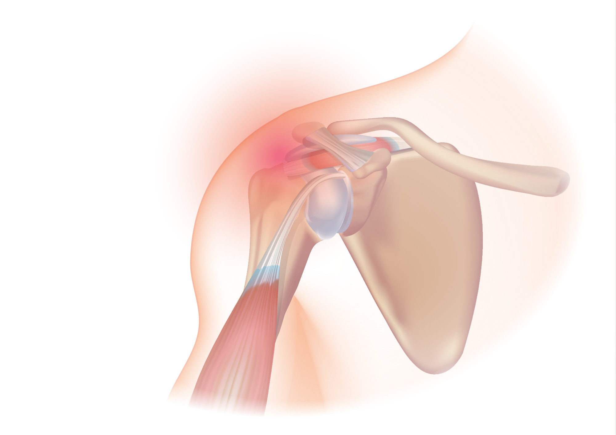
A rotator cuff tear is an injury where one or more of the tendons or muscles of the rotator cuff of the shoulder get torn. Symptoms may include shoulder pain, which is often worse with movement, limited range of motion, or weakness. This may limit people's ability to brush their hair or put on clothing. Clicking may also occur with movement of the arm. Tears may occur as the result of a sudden force or gradually over time. Risk factors include certain repetitive activities, smoking, and a family history of the condition. Diagnosis is based on symptoms, examination, and medical imaging. The rotator cuff is made up of the supraspinatus, infraspinatus, teres minor, and subscapularis. The supraspinatus is the most commonly affected. Treatment may include pain medication such as NSAIDs and specific exercises. It is recommended that people who are unable to raise their arm above 90 degrees after 2 weeks should be further assessed. In severe cases surgery may be tried, however ... [...More Info...] [...Related Items...] OR: [Wikipedia] [Google] [Baidu] |
Joint
A joint or articulation (or articular surface) is the connection made between bones, ossicles, or other hard structures in the body which link an animal's skeletal system into a functional whole.Saladin, Ken. Anatomy & Physiology. 7th ed. McGraw-Hill Connect. Webp.274/ref> They are constructed to allow for different degrees and types of movement. Some joints, such as the knee, elbow, and shoulder, are self-lubricating, almost frictionless, and are able to withstand compression and maintain heavy loads while still executing smooth and precise movements. Other joints such as sutures between the bones of the skull permit very little movement (only during birth) in order to protect the brain and the sense organs. The connection between a tooth and the jawbone is also called a joint, and is described as a fibrous joint known as a gomphosis. Joints are classified both structurally and functionally. Classification The number of joints depends on if sesamoids are included, age of ... [...More Info...] [...Related Items...] OR: [Wikipedia] [Google] [Baidu] |
Radiologist
Radiology ( ) is the medical discipline that uses medical imaging to diagnose diseases and guide their treatment, within the bodies of humans and other animals. It began with radiography (which is why its name has a root referring to radiation), but today it includes all imaging modalities, including those that use no electromagnetic radiation (such as ultrasonography and magnetic resonance imaging), as well as others that do, such as computed tomography (CT), fluoroscopy, and nuclear medicine including positron emission tomography (PET). Interventional radiology is the performance of usually minimally invasive medical procedures with the guidance of imaging technologies such as those mentioned above. The modern practice of radiology involves several different healthcare professions working as a team. The radiologist is a medical doctor who has completed the appropriate post-graduate training and interprets medical images, communicates these findings to other physicians ... [...More Info...] [...Related Items...] OR: [Wikipedia] [Google] [Baidu] |
Radiographer
Radiographers, also known as radiologic technologists, diagnostic radiographers and medical radiation technologists are healthcare professionals who specialise in the imaging of human anatomy for the diagnosis and treatment of pathology. Radiographers are infrequently, and almost always erroneously, known as ''x-ray technicians.'' In countries that use the title ''radiologic technologist'' they are often informally referred to as ''techs'' in the clinical environment; this phrase has emerged in popular culture such as television programmes. The term ''radiographer'' can also refer to a ''therapeutic radiographer'', also known as a radiation therapist. Radiographers are allied health professionals who work in both public healthcare and private healthcare and can be physically located in any setting where appropriate diagnostic equipment is located, most frequently in hospitals. The practice varies from country to country and can even vary between hospitals in the same country. ... [...More Info...] [...Related Items...] OR: [Wikipedia] [Google] [Baidu] |
Medical Imaging
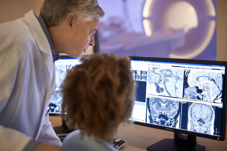
Medical imaging is the technique and process of imaging the interior of a body for clinical analysis and medical intervention, as well as visual representation of the function of some organs or tissues ( physiology). Medical imaging seeks to reveal internal structures hidden by the skin and bones, as well as to diagnose and treat disease. Medical imaging also establishes a database of normal anatomy and physiology to make it possible to identify abnormalities. Although imaging of removed organs and tissues can be performed for medical reasons, such procedures are usually considered part of pathology instead of medical imaging. Measurement and recording techniques that are not primarily designed to produce images, such as electroencephalography (EEG), magnetoencephalography (MEG), electrocardiography (ECG), and others, represent other technologies that produce data susceptible to representation as a parameter graph versus time or maps that contain data about the measurement ... [...More Info...] [...Related Items...] OR: [Wikipedia] [Google] [Baidu] |
Nephrogenic Systemic Fibrosis
Nephrogenic systemic fibrosis is a rare syndrome that involves fibrosis of skin, joints, eyes, and internal organs. NSF is caused by exposure to gadolinium in gadolinium-based MRI contrast agents (GBCAs) in patients with impaired kidney function. Epidemiological studies suggest that the incidence of NSF is unrelated to gender or ethnicity and it is not thought to have a genetic basis. After GBCAs were identified as a cause of the disorder in 2006, and screening and prevention measures put in place, it is now considered rare. Signs and symptoms Clinical features of NSF develop within days to months and, in some cases, years following exposure to some GBCAs. The main symptoms are the thickening and hardening of the skin associated with brawny hyperpigmentation, typically presenting in a symmetric fashion. The skin gradually becomes fibrotic and adheres to the underlying fascia. The symptoms initiate distally in the limbs and progress proximally, sometimes involving the trunk. Joi ... [...More Info...] [...Related Items...] OR: [Wikipedia] [Google] [Baidu] |
Glenoid Labrum
The glenoid labrum (glenoid ligament) is a fibrocartilaginous structure (not a fibrocartilage as previously thought) rim attached around the margin of the glenoid cavity in the shoulder blade. The shoulder joint is considered a ball and socket joint. However, in bony terms the 'socket' (the glenoid fossa of the scapula) is quite shallow and small, covering at most only a third of the 'ball' (the head of the humerus). The socket is deepened by the glenoid labrum, stabilizing the shoulder joint. The labrum is triangular in section; the base is fixed to the circumference of the cavity, while the free edge is thin and sharp. It is continuous above with the tendon of the long head of the biceps brachii, which gives off two fascicles to blend with the fibrous tissue of the labrum. Structure Clinical significance Injury Tearing of the labrum can occur from either acute trauma or repetitive shoulder motion such as in the sports of swimming, baseball and football. Acute trauma ... [...More Info...] [...Related Items...] OR: [Wikipedia] [Google] [Baidu] |
Rotator Cuff
The rotator cuff is a group of muscles and their tendons that act to stabilize the human shoulder and allow for its extensive range of motion. Of the seven scapulohumeral muscles, four make up the rotator cuff. The four muscles are the supraspinatus muscle, the infraspinatus muscle, teres minor muscle, and the subscapularis muscle. Structure Muscles composing rotator cuff The supraspinatus muscle spreads out in a horizontal band to insert on the superior facet of the greater tubercle of the humerus. The greater tubercle projects as the most lateral structure of the humeral head. Medial to this, in turn, is the lesser tubercle of the humeral head. The subscapularis muscle origin is divided from the remainder of the rotator cuff origins as it is deep to the scapula. The four tendons of these muscles converge to form the rotator cuff tendon. These tendinous insertions along with the articular capsule, the coracohumeral ligament, and the glenohumeral ligament complex, b ... [...More Info...] [...Related Items...] OR: [Wikipedia] [Google] [Baidu] |
Acetabular Labrum
The acetabular labrum (glenoidal labrum of the hip joint or cotyloid ligament in older texts) is a ring of cartilage that surrounds the acetabulum of the hip. The anterior portion is most vulnerable when the labrum tears. It provides an articulating surface for the acetabulum, allowing the head of the femur to articulate with the pelvis. Acetabular labrum tear Mechanisms of Injury It is estimated that 75% of acetabular labrum tears have an unknown cause. Tears of the labrum have been credited to a variety of causes such as excessive force, hip dislocation, capsular hip hypermobility, hip dysplasia, and hip degeneration. A tight iliopsoas tendon has also been attributed to labrum tears by causing compression or traction injuries that eventually lead to a labrum tear.Smith, M., Panchal, H., Ruberte, R., & Sekiya, J. (2011). Effect of acetabular labrum tears on hip stability and labral strain in a joint compression model. The American Journal of Sports Medicine, 39, 103S-110S. ... [...More Info...] [...Related Items...] OR: [Wikipedia] [Google] [Baidu] |
Joint
A joint or articulation (or articular surface) is the connection made between bones, ossicles, or other hard structures in the body which link an animal's skeletal system into a functional whole.Saladin, Ken. Anatomy & Physiology. 7th ed. McGraw-Hill Connect. Webp.274/ref> They are constructed to allow for different degrees and types of movement. Some joints, such as the knee, elbow, and shoulder, are self-lubricating, almost frictionless, and are able to withstand compression and maintain heavy loads while still executing smooth and precise movements. Other joints such as sutures between the bones of the skull permit very little movement (only during birth) in order to protect the brain and the sense organs. The connection between a tooth and the jawbone is also called a joint, and is described as a fibrous joint known as a gomphosis. Joints are classified both structurally and functionally. Classification The number of joints depends on if sesamoids are included, age of ... [...More Info...] [...Related Items...] OR: [Wikipedia] [Google] [Baidu] |
Articular Capsule
In anatomy, a joint capsule or articular capsule is an envelope surrounding a synovial joint. Each joint capsule has two parts: an outer fibrous layer or membrane, and an inner synovial layer or membrane. Membranes Each capsule consists of two layers or membranes: * an outer (fibrous membrane, ''fibrous stratum'') composed of avascular white fibrous tissue * an inner ('''', ''synovial stratum'') which is a secreting layer On the inside of the capsule, articular cartilage covers the end surfaces of the bones that articulate within ...[...More Info...] [...Related Items...] OR: [Wikipedia] [Google] [Baidu] |
Magnetic Resonance Imaging
Magnetic resonance imaging (MRI) is a medical imaging technique used in radiology to form pictures of the anatomy and the physiological processes inside the body. MRI scanners use strong magnetic fields, magnetic field gradients, and radio waves to generate images of the organs in the body. MRI does not involve X-rays or the use of ionizing radiation, which distinguishes it from computed tomography (CT) and positron emission tomography (PET) scans. MRI is a medical application of nuclear magnetic resonance (NMR) which can also be used for imaging in other NMR applications, such as NMR spectroscopy. MRI is widely used in hospitals and clinics for medical diagnosis, staging and follow-up of disease. Compared to CT, MRI provides better contrast in images of soft tissues, e.g. in the brain or abdomen. However, it may be perceived as less comfortable by patients, due to the usually longer and louder measurements with the subject in a long, confining tube, although "open ... [...More Info...] [...Related Items...] OR: [Wikipedia] [Google] [Baidu] |


