|
Area Postrema
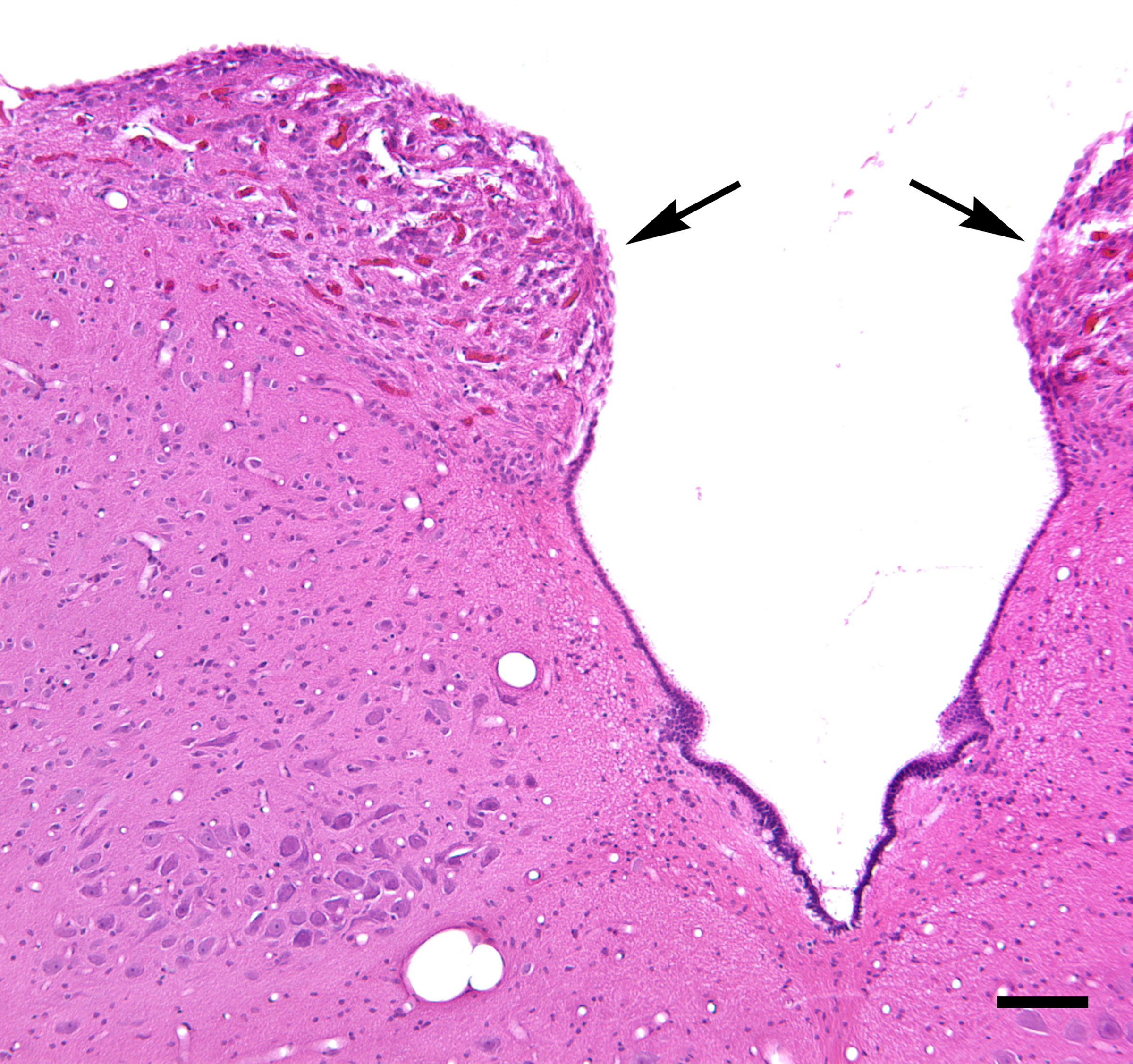
The area postrema, a paired structure in the medulla oblongata of the brainstem, is a circumventricular organ having permeable capillaries and sensory neurons that enable its dual role to detect circulating chemical messengers in the blood and transduce them into neural signals and networks. Its position adjacent to the bilateral nuclei of the solitary tract and role as a sensory transducer allow it to integrate blood-to-brain autonomic functions. Such roles of the area postrema include its detection of circulating hormones involved in vomiting, thirst, hunger, and blood pressure control. Structure The area postrema is a paired protuberance found at the inferoposterior limit of the fourth ventricle. Specialized ependymal cells are found within the area postrema. These cells differ slightly from the majority of ependymal cells (ependymocytes), forming a unicellular epithelial lining of the ventricles and central canal. The area postrema is separated from the vagal trigone by ... [...More Info...] [...Related Items...] OR: [Wikipedia] [Google] [Baidu] |
Medulla Oblongata
The medulla oblongata or simply medulla is a long stem-like structure which makes up the lower part of the brainstem. It is anterior and partially inferior to the cerebellum. It is a cone-shaped neuronal mass responsible for autonomic (involuntary) functions, ranging from vomiting to sneezing. The medulla contains the cardiac, respiratory, vomiting and vasomotor centers, and therefore deals with the autonomic functions of breathing, heart rate and blood pressure as well as the sleep–wake cycle. During embryonic development, the medulla oblongata develops from the myelencephalon. The myelencephalon is a secondary vesicle which forms during the maturation of the rhombencephalon, also referred to as the hindbrain. The bulb is an archaic term for the medulla oblongata. In modern clinical usage, the word bulbar (as in bulbar palsy) is retained for terms that relate to the medulla oblongata, particularly in reference to medical conditions. The word bulbar can refer to the nerves ... [...More Info...] [...Related Items...] OR: [Wikipedia] [Google] [Baidu] |
Ependymal Cells
The ependyma is the thin neuroepithelial ( simple columnar ciliated epithelium) lining of the ventricular system of the brain and the central canal of the spinal cord. The ependyma is one of the four types of neuroglia in the central nervous system (CNS). It is involved in the production of cerebrospinal fluid (CSF), and is shown to serve as a reservoir for neuroregeneration. Structure The ependyma is made up of ependymal cells called ependymocytes, a type of glial cell. These cells line the ventricles in the brain and the central canal of the spinal cord, which become filled with cerebrospinal fluid. These are nervous tissue cells with simple columnar shape, much like that of some mucosal epithelial cells. Early monociliated ependymal cells are differentiated to multiciliated ependymal cells for their function in circulating cerebrospinal fluid. The basal membranes of these cells are characterized by tentacle-like extensions that attach to astrocytes. The apical side is cove ... [...More Info...] [...Related Items...] OR: [Wikipedia] [Google] [Baidu] |
Ventral
Standard anatomical terms of location are used to unambiguously describe the anatomy of animals, including humans. The terms, typically derived from Latin or Greek language, Greek roots, describe something in its standard anatomical position. This position provides a definition of what is at the front ("anterior"), behind ("posterior") and so on. As part of defining and describing terms, the body is described through the use of anatomical planes and anatomical axis, anatomical axes. The meaning of terms that are used can change depending on whether an organism is bipedal or quadrupedal. Additionally, for some animals such as invertebrates, some terms may not have any meaning at all; for example, an animal that is radially symmetrical will have no anterior surface, but can still have a description that a part is close to the middle ("proximal") or further from the middle ("distal"). International organisations have determined vocabularies that are often used as standard vocabular ... [...More Info...] [...Related Items...] OR: [Wikipedia] [Google] [Baidu] |
Morphology (anatomy)
Morphology is a branch of biology dealing with the study of the form and structure of organisms and their specific structural features. This includes aspects of the outward appearance (shape, structure, colour, pattern, size), i.e. external morphology (or eidonomy), as well as the form and structure of the internal parts like bones and organs, i.e. internal morphology (or anatomy). This is in contrast to physiology, which deals primarily with function. Morphology is a branch of life science dealing with the study of gross structure of an organism or taxon and its component parts. History The etymology of the word "morphology" is from the Ancient Greek (), meaning "form", and (), meaning "word, study, research". While the concept of form in biology, opposed to function, dates back to Aristotle (see Aristotle's biology), the field of morphology was developed by Johann Wolfgang von Goethe (1790) and independently by the German anatomist and physiologist Karl Friedrich Burdach ... [...More Info...] [...Related Items...] OR: [Wikipedia] [Google] [Baidu] |
Ventricular System
The ventricular system is a set of four interconnected cavities known as cerebral ventricles in the brain. Within each ventricle is a region of choroid plexus which produces the circulating cerebrospinal fluid (CSF). The ventricular system is continuous with the central canal of the spinal cord from the fourth ventricle, allowing for the flow of CSF to circulate. All of the ventricular system and the central canal of the spinal cord are lined with ependyma, a specialised form of epithelium connected by tight junctions that make up the blood–cerebrospinal fluid barrier. Structure The system comprises four ventricles: * lateral ventricles right and left (one for each hemisphere) * third ventricle * fourth ventricle There are several foramina, openings acting as channels, that connect the ventricles. The interventricular foramina (also called the foramina of Monro) connect the lateral ventricles to the third ventricle through which the cerebrospinal fluid can flow. Ventric ... [...More Info...] [...Related Items...] OR: [Wikipedia] [Google] [Baidu] |
Cerebrospinal Fluid
Cerebrospinal fluid (CSF) is a clear, colorless body fluid found within the tissue that surrounds the brain and spinal cord of all vertebrates. CSF is produced by specialised ependymal cells in the choroid plexus of the ventricles of the brain, and absorbed in the arachnoid granulations. There is about 125 mL of CSF at any one time, and about 500 mL is generated every day. CSF acts as a shock absorber, cushion or buffer, providing basic mechanical and immunological protection to the brain inside the skull. CSF also serves a vital function in the cerebral autoregulation of cerebral blood flow. CSF occupies the subarachnoid space (between the arachnoid mater and the pia mater) and the ventricular system around and inside the brain and spinal cord. It fills the ventricles of the brain, cisterns, and sulci, as well as the central canal of the spinal cord. There is also a connection from the subarachnoid space to the bony labyrinth of the inner ear via the perilymphat ... [...More Info...] [...Related Items...] OR: [Wikipedia] [Google] [Baidu] |
Neurochemical
A neurochemical is a small organic molecule or peptide that participates in neural activity. The science of neurochemistry studies the functions of neurochemicals. Prominent neurochemicals Neurotransmitters and neuromodulators *Glutamate is the most common neurotransmitter. Most neurons secrete with glutamate or GABA. Glutamate is excitatory, meaning that the release of glutamate by one cell usually causes adjacent cells to fire an action potential. (Note: Glutamate is chemically identical to the MSG commonly used to flavor food.) * GABA is an example of an inhibitory neurotransmitter. * Monoamine neurotransmitters: **Dopamine is a monoamine neurotransmitter. It plays a key role in the functioning of the limbic system, which is involved in emotional function and control. It also is involved in cognitive processes associated with movement, arousal, executive function, body temperature regulation, and pleasure and reward, and other processes. **Norepinephrine, also known as noradr ... [...More Info...] [...Related Items...] OR: [Wikipedia] [Google] [Baidu] |
Tanycytes
Tanycytes are special ependymal cells found in the third ventricle of the brain, and on the floor of the fourth ventricle and have processes extending deep into the hypothalamus. It is possible that their function is to transfer chemical signals from the cerebrospinal fluid to the central nervous system. The term ''tanycyte'' comes from the Greek word tanus which means elongated. Structure Tanycytes share some features with radial glial cells and astrocytes. Their form and location have led some authors to regard them as radial glial cells that remain in the hypothalamus throughout life. This has led some to believe that these cells share the same lineage. Even so, tanycytes also display certain characteristics that distinguish them from radial glia cells. Tanycytes in rats begin to develop in the last two days of gestation and continue on until they reach their full differentiation in the first month of life. Radial glia cells on the other hand, are a key component of the embryo ... [...More Info...] [...Related Items...] OR: [Wikipedia] [Google] [Baidu] |
Ependyma
The ependyma is the thin Neuroepithelial cell, neuroepithelial (Simple columnar epithelium, simple columnar ciliated epithelium) lining of the ventricular system of the brain and the central canal of the spinal cord. The ependyma is one of the four types of neuroglia in the central nervous system (CNS). It is involved in the production of cerebrospinal fluid (CSF), and is shown to serve as a reservoir for neuroregeneration. Structure The ependyma is made up of ependymal Cell (biology), cells called ependymocytes, a type of glial cell. These cells line the Ventricular system, ventricles in the brain and the central canal of the spinal cord, which become filled with cerebrospinal fluid. These are nervous tissue cells with simple columnar shape, much like that of some mucosal epithelial cells. Early monociliated ependymal cells are differentiated to multiciliated ependymal cells for their function in circulating cerebrospinal fluid. The epithelial polarity#Basolateral membranes, bas ... [...More Info...] [...Related Items...] OR: [Wikipedia] [Google] [Baidu] |
Obex
OBEX (abbreviation of OBject EXchange, also termed IrOBEX) is a communications protocol that facilitates the exchange of binary objects between devices. It is maintained by the Infrared Data Association but has also been adopted by the Bluetooth Special Interest Group and the SyncML wing of the Open Mobile Alliance (OMA). One of OBEX's earliest popular applications was in the Palm III. This PDA and its many successors use OBEX to exchange business cards, data, even applications. Although OBEX was initially designed for infrared, it has now been adopted by Bluetooth, and is also used over RS-232, USB, WAP and in devices such as Livescribe smartpens. Comparison to HTTP OBEX is similar in design and function to HTTP in providing the client with a reliable transport for connecting to a server and may then request or provide objects. But OBEX differs in many important respects: *HTTP is normally layered above a TCP/IP link. OBEX can also be, but is commonly implemented on an IrLAP/ ... [...More Info...] [...Related Items...] OR: [Wikipedia] [Google] [Baidu] |
Rhomboid Fossa
The rhomboid fossa is a rhombus-shaped depression that is the anterior part of the fourth ventricle. Its anterior wall, formed by the back of the pons and the medulla oblongata, constitutes the floor of the fourth ventricle. It is covered by a thin layer of grey matter continuous with that of the spinal cord; superficial to this is a thin lamina of neuroglia which constitutes the ependyma of the ventricle and supports a layer of ciliated epithelium. Parts The fossa consists of three parts, superior, intermediate, and inferior: ;The superior part :The superior part is triangular in shape and limited laterally by the superior cerebellar peduncle; its apex, directed upward, is continuous with the cerebral aqueduct; its base is represented by an imaginary line at the level of the upper ends of the superior foveae. ;The intermediate part :The intermediate part extends from this level to that of the horizontal portions of the taeniae of the ventricle; it is narrow above where it is li ... [...More Info...] [...Related Items...] OR: [Wikipedia] [Google] [Baidu] |
Dorsal Vagal Nucleus
The dorsal nucleus of vagus nerve (or posterior nucleus of vagus nerve or dorsal vagal nucleus or nucleus dorsalis nervi vagi or nucleus posterior nervi vagi) is a cranial nerve nucleus for the vagus nerve in the medulla that lies ventral to the floor of the fourth ventricle. It mostly serves parasympathetic vagal functions in the gastrointestinal tract, lungs, and other thoracic and abdominal vagal innervations. These functions include, among others, bronchoconstriction and gland secretion. The cell bodies for the preganglionic parasympathetic vagal neurons that innervate the heart reside in the nucleus ambiguus. Additional cell bodies are found in the nucleus ambiguus, which give rise to the branchial efferent motor fibers of the vagus nerve (CN X) terminating in the laryngeal, pharyngeal muscles, and musculus uvulae. Additional images File:Gray694.png, Section of the medulla oblongata at about the middle of the olive. File:Gray696.png, The cranial nerve nuclei schematically ... [...More Info...] [...Related Items...] OR: [Wikipedia] [Google] [Baidu] |


