|
Aquamarina
''Aquamarina'' is a fungal genus in the class Dothideomycetes. It is a monotypic genus, containing the single marine species ''Aquamarina speciosa'', originally found in North Carolina, and distributed in the Atlantic Coast of the United States. The bluish-green species fruits exclusively in the lower parts of dying culms of the saltmarsh plant ''Juncus roemerianus''. Taxonomy, classification, and naming The species was first described by mycologists Jan Kohlmeyer, Brigitte Volkmann-Kohlmeyer, and Ove Eriksson in a 1996 '' Mycological Research'' publication. The generic name is derived from the Latin ''aquamarinus'', meaning "a clear sea-green verging towards blue". The specific epithet ''speciosa'' is Latin for 'beautiful' or 'splendid', and refers to the "beautifully colored ascomata". The relationship of this taxon to other taxa within the Dothideomycetes is unknown (''incertae sedis'') with respect to ordinal and familial placement. Description The leathery aquamarin ... [...More Info...] [...Related Items...] OR: [Wikipedia] [Google] [Baidu] |
Juncus Roemerianus
''Juncus roemerianus'' is a species of flowering plant in the rush family known by the common names black rush, needlerush, and black needlerush. It is native to North America, where its main distribution lies along the coastline of the southeastern United States, including the Gulf Coast. It occurs from New Jersey to Texas, with outlying populations in Connecticut, New York, Mexico, and certain Caribbean islands.Uchytil, Ronald J. (1992)''Juncus roemerianus''.In: Fire Effects Information System, nline U.S. Department of Agriculture, Forest Service, Rocky Mountain Research Station, Fire Sciences Laboratory. Retrieved 1-2-2012. Description This rush is a perennial plant forming tufts of rough, rigid stems and leaves. It is gray-green in color. The plant may appear to be leafless at first glance, but what look like sharp-pointed stems are actually stiff leaves rolled tightly to form pointed cylinders. The true stems are tipped with inflorescences. [...More Info...] [...Related Items...] OR: [Wikipedia] [Google] [Baidu] |
Fungi
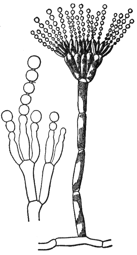
A fungus ( : fungi or funguses) is any member of the group of eukaryotic organisms that includes microorganisms such as yeasts and molds, as well as the more familiar mushrooms. These organisms are classified as a kingdom, separately from the other eukaryotic kingdoms, which by one traditional classification include Plantae, Animalia, Protozoa, and Chromista. A characteristic that places fungi in a different kingdom from plants, bacteria, and some protists is chitin in their cell walls. Fungi, like animals, are heterotrophs; they acquire their food by absorbing dissolved molecules, typically by secreting digestive enzymes into their environment. Fungi do not photosynthesize. Growth is their means of mobility, except for spores (a few of which are flagellated), which may travel through the air or water. Fungi are the principal decomposers in ecological systems. These and other differences place fungi in a single group of related organisms, named the ''Eumycota'' (''t ... [...More Info...] [...Related Items...] OR: [Wikipedia] [Google] [Baidu] |
Aquamarine (color)
Aquamarine is a color that is a light tint of spring green, in between cyan and green on the color wheel. It is named after the mineral aquamarine, a gemstone mainly found in granite rocks. The first recorded use of ''aquamarine'' as a color name in English was in 1598.Maerz, Aloys John, and Morris Rea Paul (1930). ''A Dictionary of Color''. New York: McGraw-Hill. p. 190; color sample of aquamarine: p. 93, plate 35, color sample I3. . File:Aquamarine.jpg, alt=Rough aquamarine, Rough aquamarine File:Aquamarine P1000141.JPG, alt=Aquamarine crystals on muscovite, Aquamarine crystals on muscovite File:Aquamarine brooch (Faberge).jpg, alt=An aquamarine brooch, An aquamarine brooch See also * Aquamarine (other) * List of colors These are the lists of colors; * List of colors: A–F * List of colors: G–M * List of colors: N–Z * List of colors (compact) * List of colors by shade * List of color palettes * List of Crayola crayon colors * List of RAL colors * ... [...More Info...] [...Related Items...] OR: [Wikipedia] [Google] [Baidu] |
Ascospore
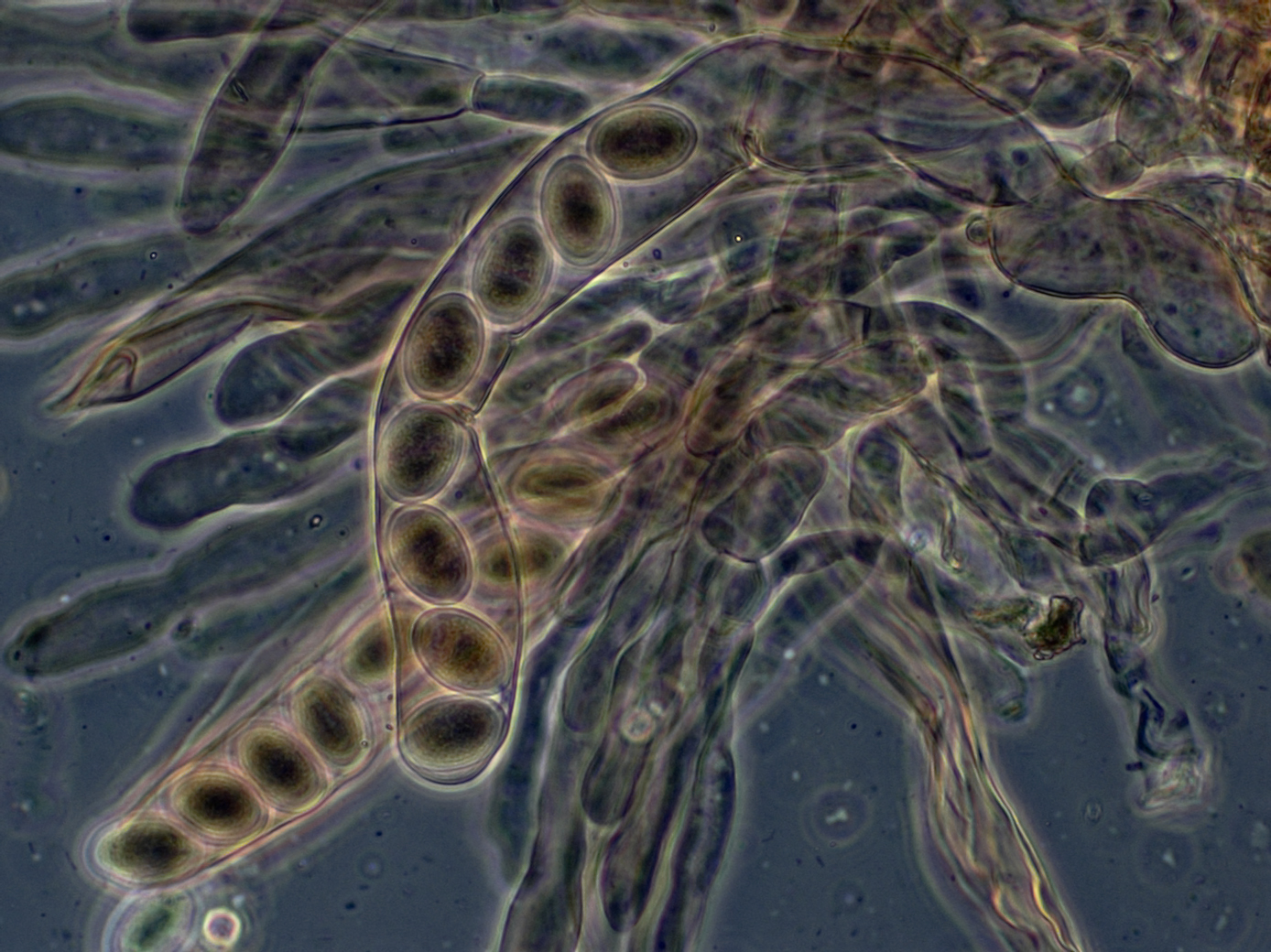
An ascus (; ) is the sexual spore-bearing cell produced in ascomycete fungi. Each ascus usually contains eight ascospores (or octad), produced by meiosis followed, in most species, by a mitotic cell division. However, asci in some genera or species can occur in numbers of one (e.g. ''Monosporascus cannonballus''), two, four, or multiples of four. In a few cases, the ascospores can bud off conidia that may fill the asci (e.g. ''Tympanis'') with hundreds of conidia, or the ascospores may fragment, e.g. some ''Cordyceps'', also filling the asci with smaller cells. Ascospores are nonmotile, usually single celled, but not infrequently may be coenocytic (lacking a septum), and in some cases coenocytic in multiple planes. Mitotic divisions within the developing spores populate each resulting cell in septate ascospores with nuclei. The term ocular chamber, or oculus, refers to the epiplasm (the portion of cytoplasm not used in ascospore formation) that is surrounded by the "bourrelet ... [...More Info...] [...Related Items...] OR: [Wikipedia] [Google] [Baidu] |
Locule
A locule (plural locules) or loculus (plural loculi) (meaning "little place" in Latin) is a small cavity or compartment within an organ or part of an organism (animal, plant, or fungus). In angiosperms (flowering plants), the term ''locule'' usually refers to a chamber within an Ovary (plants), ovary (gynoecium or carpel) of the flower and fruits. Depending on the number of locules in the ovary, fruits can be classified as ''uni-locular'' (unilocular), ''bi-locular'', ''tri-locular'' or ''multi-locular''. The number of locules present in a gynoecium may be equal to or less than the number of carpels. The locules contain the ovules or seeds. The term may also refer to chambers within anthers containing pollen. In Ascomycete fungi, locules are chambers within the hymenium in which the perithecium, perithecia develop. References Plant anatomy Plant morphology Fungal morphology and anatomy {{botany-stub ... [...More Info...] [...Related Items...] OR: [Wikipedia] [Google] [Baidu] |
Amyloid (mycology)
In mycology a tissue or feature is said to be amyloid if it has a positive amyloid reaction when subjected to a crude chemical test using iodine as an ingredient of either Melzer's reagent or Lugol's solution, producing a blue to blue-black staining. The term "amyloid" is derived from the Latin ''amyloideus'' ("starch-like"). It refers to the fact that starch gives a similar reaction, also called an amyloid reaction. The test can be on microscopic features, such as spore walls or hyphal walls, or the apical apparatus or entire ascus wall of an ascus, or be a macroscopic reaction on tissue where a drop of the reagent is applied. Negative reactions, called inamyloid or nonamyloid, are for structures that remain pale yellow-brown or clear. A reaction producing a deep reddish to reddish-brown staining is either termed a dextrinoid reaction (pseudoamyloid is a synonym) or a hemiamyloid reaction. Melzer's reagent reactions Hemiamyloidity Hemiamyloidity in mycology refers to a special ... [...More Info...] [...Related Items...] OR: [Wikipedia] [Google] [Baidu] |
Ascus
An ascus (; ) is the sexual spore-bearing cell produced in ascomycete fungi. Each ascus usually contains eight ascospores (or octad), produced by meiosis followed, in most species, by a mitotic cell division. However, asci in some genera or species can occur in numbers of one (e.g. ''Monosporascus cannonballus''), two, four, or multiples of four. In a few cases, the ascospores can bud off conidia that may fill the asci (e.g. ''Tympanis'') with hundreds of conidia, or the ascospores may fragment, e.g. some ''Cordyceps'', also filling the asci with smaller cells. Ascospores are nonmotile, usually single celled, but not infrequently may be coenocytic (lacking a septum), and in some cases coenocytic in multiple planes. Mitotic divisions within the developing spores populate each resulting cell in septate ascospores with nuclei. The term ocular chamber, or oculus, refers to the epiplasm (the portion of cytoplasm not used in ascospore formation) that is surrounded by the "bourrelet ... [...More Info...] [...Related Items...] OR: [Wikipedia] [Google] [Baidu] |
Parenchyma
Parenchyma () is the bulk of functional substance in an animal organ or structure such as a tumour. In zoology it is the name for the tissue that fills the interior of flatworms. Etymology The term ''parenchyma'' is New Latin from the word παρέγχυμα ''parenchyma'' meaning 'visceral flesh', and from παρεγχεῖν ''parenchyma'' meaning 'to pour in' from παρα- ''para-'' 'beside' + ἐν ''en-'' 'in' + χεῖν ''chyma'' 'to pour'. Originally, Erasistratus and other anatomists used it to refer to certain human tissues. Later, it was also applied to plant tissues by Nehemiah Grew. Structure The parenchyma is the ''functional'' parts of an organ (anatomy), organ, or of a structure such as a tumour in the body. This is in contrast to the Stroma (animal tissue), stroma, which refers to the ''structural'' tissue of organs or of structures, namely, the connective tissues. Brain The brain parenchyma refers to the functional tissue in the brain that is made up of t ... [...More Info...] [...Related Items...] OR: [Wikipedia] [Google] [Baidu] |
Hyaline
A hyaline substance is one with a glassy appearance. The word is derived from el, ὑάλινος, translit=hyálinos, lit=transparent, and el, ὕαλος, translit=hýalos, lit=crystal, glass, label=none. Histopathology Hyaline cartilage is named after its glassy appearance on fresh gross pathology. On light microscopy of H&E stained slides, the extracellular matrix of hyaline cartilage looks homogeneously pink, and the term "hyaline" is used to describe similarly homogeneously pink material besides the cartilage. Hyaline material is usually acellular and proteinaceous. For example, arterial hyaline is seen in aging, high blood pressure, diabetes mellitus and in association with some drugs (e.g. calcineurin inhibitors). It is bright pink with PAS staining. Ichthyology and entomology In ichthyology and entomology, ''hyaline'' denotes a colorless, transparent substance, such as unpigmented fins of fishes or clear insect wings. Resh, Vincent H. and R. T. Cardé, Eds. Encyclo ... [...More Info...] [...Related Items...] OR: [Wikipedia] [Google] [Baidu] |
Peridium
The peridium is the protective layer that encloses a mass of spores in fungi. This outer covering is a distinctive feature of gasteroid fungi. Description Depending on the species, the peridium may vary from being paper-thin to thick and rubbery or even hard. Typically, peridia consist of one to three layers. If there is only a single layer, it is called a peridium. If two layers are present, the outer layer is called the exoperidium and the inner layer the endoperidium. If three layers are present, they are the exoperidium, the mesoperidium and the endoperidium. In the simplest subterranean forms, the peridium remains closed until the spores are mature, and even then shows no special arrangement for dehiscence or opening, but has to decay before the spores are liberated. Puffballs For most fungi, the peridium is ornamented with scales or spines. In species that become raised above ground during their development, generally known as the "puffballs", the peridium is usually di ... [...More Info...] [...Related Items...] OR: [Wikipedia] [Google] [Baidu] |
Hypha
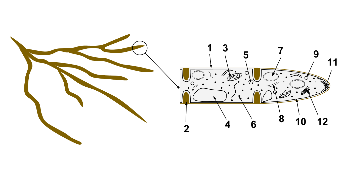
A hypha (; ) is a long, branching, filamentous structure of a fungus, oomycete, or actinobacterium. In most fungi, hyphae are the main mode of vegetative growth, and are collectively called a mycelium. Structure A hypha consists of one or more cells surrounded by a tubular cell wall. In most fungi, hyphae are divided into cells by internal cross-walls called "septa" (singular septum). Septa are usually perforated by pores large enough for ribosomes, mitochondria, and sometimes nuclei to flow between cells. The major structural polymer in fungal cell walls is typically chitin, in contrast to plants and oomycetes that have cellulosic cell walls. Some fungi have aseptate hyphae, meaning their hyphae are not partitioned by septa. Hyphae have an average diameter of 4–6 µm. Growth Hyphae grow at their tips. During tip growth, cell walls are extended by the external assembly and polymerization of cell wall components, and the internal production of new cell membrane. The S ... [...More Info...] [...Related Items...] OR: [Wikipedia] [Google] [Baidu] |
Paraphyses
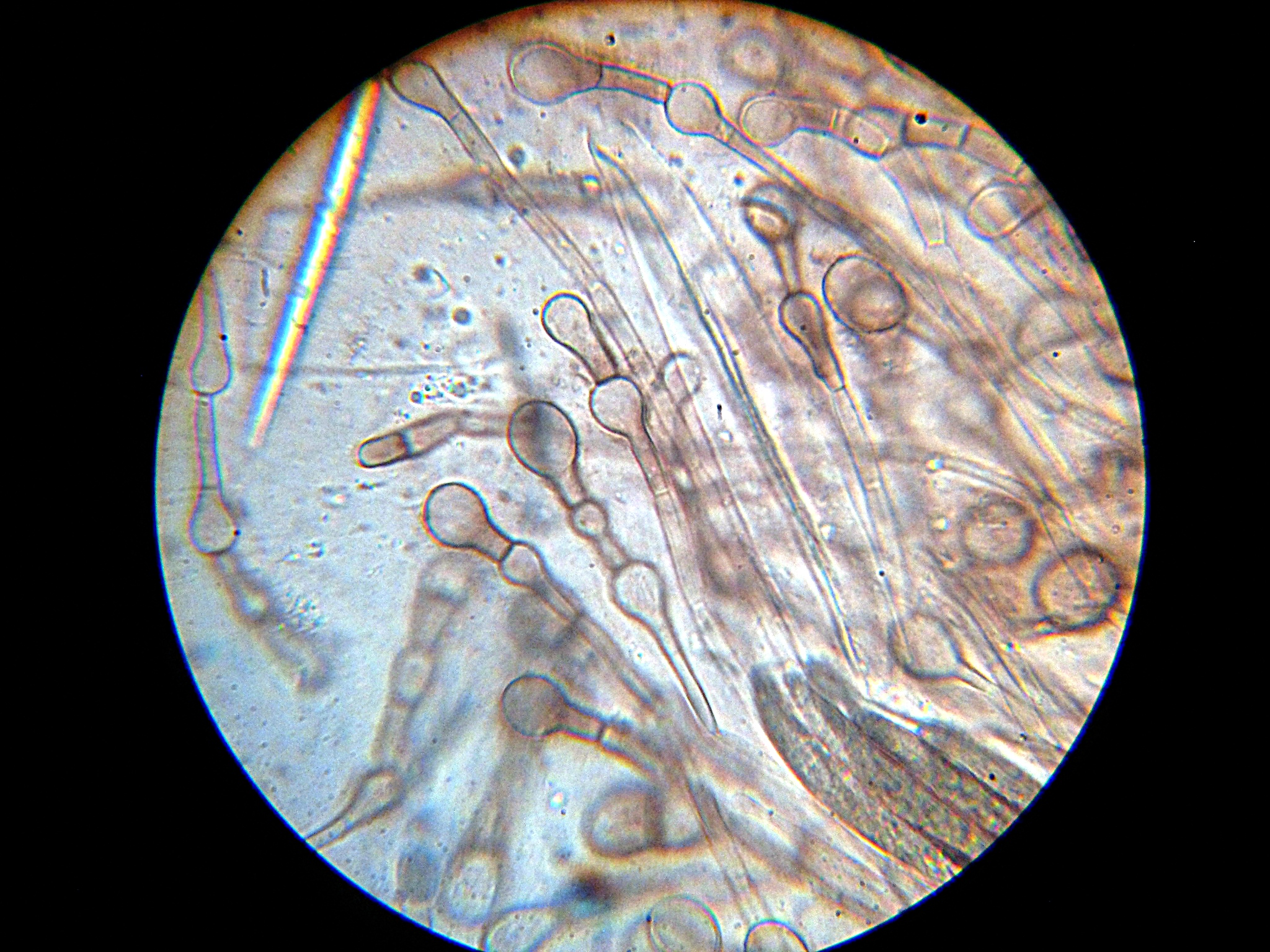
Paraphyses are erect sterile filament-like support structures occurring among the reproductive apparatuses of fungi, ferns, bryophytes and some thallophytes. The singular form of the word is paraphysis. In certain fungi, they are part of the fertile spore-bearing layer. More specifically, paraphyses are sterile filamentous hyphal end cells composing part of the hymenium of Ascomycota and Basidiomycota interspersed among either the asci or basidia respectively, and not sufficiently differentiated to be called cystidia A cystidium (plural cystidia) is a relatively large cell found on the sporocarp of a basidiomycete (for example, on the surface of a mushroom gill), often between clusters of basidia. Since cystidia have highly varied and distinct shapes that ar ..., which are specialized, swollen, often protruding cells. The tips of paraphyses may contain the pigments which colour the hymenium. In ferns and mosses, they are filament-like structures that are found on sporangia ... [...More Info...] [...Related Items...] OR: [Wikipedia] [Google] [Baidu] |


