|
Apoptotic
Apoptosis (from grc, ἀπόπτωσις, apóptōsis, 'falling off') is a form of programmed cell death that occurs in multicellular organisms. Biochemical events lead to characteristic cell changes (morphology) and death. These changes include blebbing, cell shrinkage, nuclear fragmentation, chromatin condensation, DNA fragmentation, and mRNA decay. The average adult human loses between 50 and 70 billion cells each day due to apoptosis. For an average human child between eight and fourteen years old, approximately twenty to thirty billion cells die per day. In contrast to necrosis, which is a form of traumatic cell death that results from acute cellular injury, apoptosis is a highly regulated and controlled process that confers advantages during an organism's life cycle. For example, the separation of fingers and toes in a developing human embryo occurs because cells between the digits undergo apoptosis. Unlike necrosis, apoptosis produces cell fragments called apoptotic bod ... [...More Info...] [...Related Items...] OR: [Wikipedia] [Google] [Baidu] |
Bcl-2 Family
The Bcl-2 familyTC# 1.A.21 consists of a number of evolutionarily-conserved proteins that share Bcl-2 homology (BH) domains. The Bcl-2 family is most notable for their regulation of apoptosis, a form of programmed cell death, at the mitochondrion. The Bcl-2 family proteins consists of members that either promote or inhibit apoptosis, and control apoptosis by governing mitochondrial outer membrane permeabilization (MOMP), which is a key step in the intrinsic pathway of apoptosis. A total of 25 genes in the Bcl-2 family were identified by 2008. Structure Bcl-2 family proteins have a general structure that consists of a hydrophobic α-helix surrounded by amphipathic α-helices. Some members of the family have transmembrane domains at their c-terminus which primarily function to localize them to the mitochondrion. Bcl-x(L) is 233 amino acyl residues (aas) long and exhibits a single very hydrophobic putative transmembrane α-helical segment (residues 210-226) when in the membrane. ... [...More Info...] [...Related Items...] OR: [Wikipedia] [Google] [Baidu] |
Apoptotic DNA Fragmentation
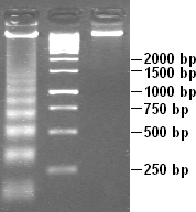
Apoptotic DNA fragmentation is a key feature of apoptosis, a type of programmed cell death. Apoptosis is characterized by the activation of endogenous endonucleases, particularly the caspase-3 activated DNase (CAD), with subsequent cleavage of nuclear DNA into internucleosomal fragments of roughly 180 base pairs (bp) and multiples thereof (360, 540 etc.). The apoptotic DNA fragmentation is being used as a marker of apoptosis and for identification of apoptotic cells either via the DNA laddering assay, the TUNEL assay, or the by detection of cells with fractional DNA content ("sub G1 cells") on DNA content frequency histograms e.g. as in the Nicoletti assay. Mechanism The enzyme responsible for apoptotic DNA fragmentation is the Caspase-Activated DNase (CAD). CAD is normally inhibited by another protein, the Inhibitor of Caspase Activated DNase (ICAD). During apoptosis, the apoptotic effector caspase, caspase-3, cleaves ICAD and thus causes CAD to become activated. CAD ... [...More Info...] [...Related Items...] OR: [Wikipedia] [Google] [Baidu] |
Programmed Cell Death
Programmed cell death (PCD; sometimes referred to as cellular suicide) is the death of a cell as a result of events inside of a cell, such as apoptosis or autophagy. PCD is carried out in a biological process, which usually confers advantage during an organism's lifecycle. For example, the differentiation of fingers and toes in a developing human embryo occurs because cells between the fingers apoptose; the result is that the digits are separate. PCD serves fundamental functions during both plant and animal tissue development. Apoptosis and autophagy are both forms of programmed cell death. Necrosis is the death of a cell caused by external factors such as trauma or infection and occurs in several different forms. Necrosis was long seen as a non-physiological process that occurs as a result of infection or injury, but in the 2000s, a form of programmed necrosis, called necroptosis, was recognized as an alternative form of programmed cell death. It is hypothesized that necroptosis ... [...More Info...] [...Related Items...] OR: [Wikipedia] [Google] [Baidu] |
Fas Receptor
The Fas receptor, also known as Fas, FasR, apoptosis antigen 1 (APO-1 or APT), cluster of differentiation 95 (CD95) or tumor necrosis factor receptor superfamily member 6 (TNFRSF6), is a protein that in humans is encoded by the ''FAS'' gene. Fas was first identified using a monoclonal antibody generated by immunizing mice with the FS-7 cell line. Thus, the name Fas is derived from ''F''S-7-''a''ssociated ''s''urface antigen. The Fas receptor is a death receptor on the surface of cells that leads to programmed cell death (apoptosis) if it binds its ligand, Fas ligand (FasL). It is one of two apoptosis pathways, the other being the mitochondrial pathway. Gene FAS receptor gene is located on the long arm of chromosome 10 (10q24.1) in humans and on chromosome 19 in mice. The gene lies on the plus ( Watson strand) and is 25,255 bases in length organized into nine protein encoding exons. Similar sequences related by evolution (orthologs) are found in most mammals. Protein Prev ... [...More Info...] [...Related Items...] OR: [Wikipedia] [Google] [Baidu] |
Extracellular Vesicle
Extracellular vesicles (EVs) are lipid bilayer-delimited particles that are naturally released from almost all types of cell but, unlike a cell, cannot replicate. EVs range in diameter from near the size of the smallest physically possible unilamellar liposome (around 20-30 nanometers) to as large as 10 microns or more, although the vast majority of EVs are smaller than 200 nm. EVs can be divided according to size and synthesis route into Exosomes, microvesicles and apoptotic bodies. They carry a cargo of proteins, nucleic acids, lipids, metabolites, and even organelles from the parent cell. Most cells that have been studied to date are thought to release EVs, including some archaeal, bacterial, fungal, and plant cells that are surrounded by cell walls. A wide variety of EV subtypes have been proposed, defined variously by size, biogenesis pathway, cargo, cellular source, and function, leading to a historically heterogenous nomenclature including terms like exosomes and ecto ... [...More Info...] [...Related Items...] OR: [Wikipedia] [Google] [Baidu] |
Phagocyte
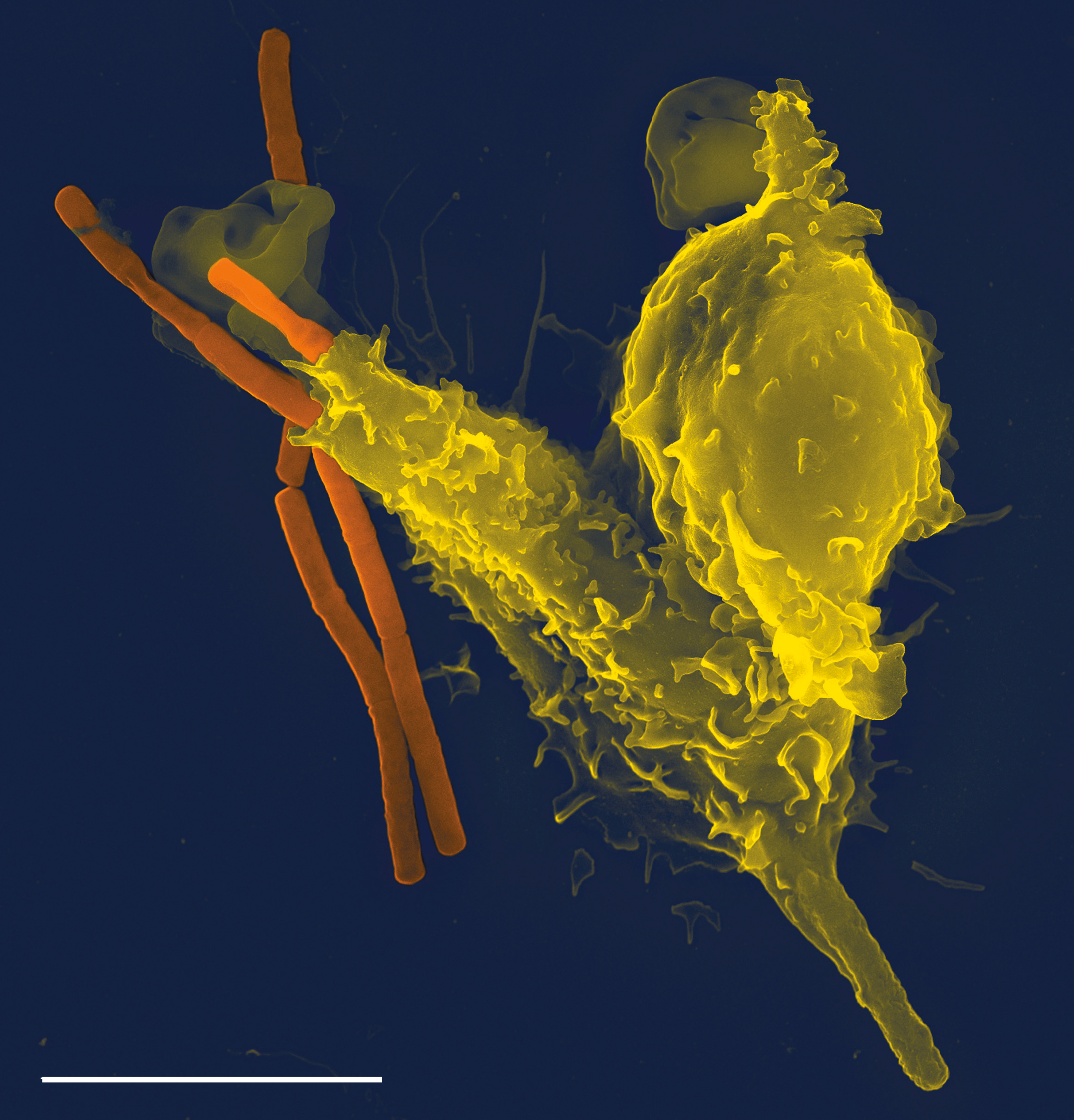
Phagocytes are cells that protect the body by ingesting harmful foreign particles, bacteria, and dead or dying cells. Their name comes from the Greek ', "to eat" or "devour", and "-cyte", the suffix in biology denoting "cell", from the Greek ''kutos'', "hollow vessel". They are essential for fighting infections and for subsequent immunity. Phagocytes are important throughout the animal kingdom and are highly developed within vertebrates. One litre of human blood contains about six billion phagocytes. They were discovered in 1882 by Ilya Ilyich Mechnikov while he was studying starfish larvae.Ilya Mechnikov retrieved on November 28, 2008. Fro ''Physiology or Medicine 1901–1921'' ... [...More Info...] [...Related Items...] OR: [Wikipedia] [Google] [Baidu] |
Bleb (cell Biology)
In cell biology, a bleb is a bulge of the plasma membrane of a cell, characterized by a spherical, bulky morphology. It is characterized by the decoupling of the cytoskeleton from the plasma membrane, degrading the internal structure of the cell, allowing the flexibility required for the cell to separate into individual bulges or pockets of the intercellular matrix. Most commonly, blebs are seen in apoptosis (programmed cell death) but are also seen in other non-apoptotic functions. ''Blebbing'', or ''zeiosis'', is the formation of blebs. Formation Initiation and expansion Bleb growth is driven by intracellular pressure generated in the cytoplasm when the actin cortex undergoes actomyosin contractions. The disruption of the membrane-actin cortex interactions are dependent on the activity of myosin-ATPase Bleb initiation is affected by three main factors: high intracellular pressure, decreased amounts of cortex-membrane linker proteins, and deterioration of the actin cortex. T ... [...More Info...] [...Related Items...] OR: [Wikipedia] [Google] [Baidu] |
Necrosis
Necrosis () is a form of cell injury which results in the premature death of cells in living tissue by autolysis. Necrosis is caused by factors external to the cell or tissue, such as infection, or trauma which result in the unregulated digestion of cell components. In contrast, apoptosis is a naturally occurring programmed and targeted cause of cellular death. While apoptosis often provides beneficial effects to the organism, necrosis is almost always detrimental and can be fatal. Cellular death due to necrosis does not follow the apoptotic signal transduction pathway, but rather various receptors are activated and result in the loss of cell membrane integrity and an uncontrolled release of products of cell death into the extracellular space. This initiates in the surrounding tissue an inflammatory response, which attracts leukocytes and nearby phagocytes which eliminate the dead cells by phagocytosis. However, microbial damaging substances released by leukocytes would crea ... [...More Info...] [...Related Items...] OR: [Wikipedia] [Google] [Baidu] |
Karyorrhexis
Karyorrhexis (from Greek κάρυον ''karyon'', "kernel, seed or nucleus", and ῥῆξις ''rhexis'', "bursting") is the destructive fragmentation of the nucleus of a dying cell whereby its chromatin is distributed irregularly throughout the cytoplasm. It is usually preceded by pyknosis and can occur as a result of either programmed cell death (apoptosis), cellular senescence, or necrosis. In apoptosis, the cleavage of DNA is done by Ca2+ and Mg2+ -dependent endonucleases. Image:nuclear changes.jpg, Morphological characteristics of pyknosis and other forms of nuclear destruction. File:Apoptotic neutrophil with nuclear fragmentation.jpg, Microscopy of an apoptotic neutrophil with nuclear fragmentation (H&E stain) See also *Karyolysis Karyolysis (from Greek κάρυον ''karyon—''kernel, seed, or nucleus), and λύσις ''lysis'' from λύειν ''lyein'', "to separate") is the complete dissolution of the chromatin of a dying cell due to the enzymatic degradation by en ... [...More Info...] [...Related Items...] OR: [Wikipedia] [Google] [Baidu] |
Caspase
Caspases (cysteine-aspartic proteases, cysteine aspartases or cysteine-dependent aspartate-directed proteases) are a family of protease enzymes playing essential roles in programmed cell death. They are named caspases due to their specific cysteine protease activity – a cysteine in its active site nucleophilically attacks and cleaves a target protein only after an aspartic acid residue. As of 2009, there are 12 confirmed caspases in humans and 10 in mice, carrying out a variety of cellular functions. The role of these enzymes in programmed cell death was first identified in 1993, with their functions in apoptosis well characterised. This is a form of programmed cell death, occurring widely during development, and throughout life to maintain cell homeostasis. Activation of caspases ensures that the cellular components are degraded in a controlled manner, carrying out cell death with minimal effect on surrounding tissues. Caspases have other identified roles in programmed cell ... [...More Info...] [...Related Items...] OR: [Wikipedia] [Google] [Baidu] |
Carl Vogt
August Christoph Carl Vogt (; 5 July 18175 May 1895) was a German scientist, philosopher, popularizer of science, and politician who emigrated to Switzerland. Vogt published a number of notable works on zoology, geology and physiology. All his life he was engaged in politics, in the German Frankfurt Parliament of 1848–49 and later in Switzerland. Early life Vogt was born in Giessen, the son of , professor of clinics, and Louise Follenius. His maternal uncle was Charles Follen. From 1833 to 1836, he studied medicine at the University of Giessen, and continued his training in Berne, Switzerland, earning his PhD. in 1839. He then worked with Louis Agassiz in Neuchâtel. Career In 1847 he became professor of zoology at the University of Giessen, and in 1852 professor of geology and afterwards also of zoology at the University of Geneva. His earlier publications were on zoology. He dealt with the Amphibia (1839), Reptiles (1840), with Mollusca and Crustacea (1845) and more gener ... [...More Info...] [...Related Items...] OR: [Wikipedia] [Google] [Baidu] |
Cancer
Cancer is a group of diseases involving abnormal cell growth with the potential to invade or spread to other parts of the body. These contrast with benign tumors, which do not spread. Possible signs and symptoms include a lump, abnormal bleeding, prolonged cough, unexplained weight loss, and a change in bowel movements. While these symptoms may indicate cancer, they can also have other causes. Over 100 types of cancers affect humans. Tobacco use is the cause of about 22% of cancer deaths. Another 10% are due to obesity, poor diet, lack of physical activity or excessive drinking of alcohol. Other factors include certain infections, exposure to ionizing radiation, and environmental pollutants. In the developing world, 15% of cancers are due to infections such as ''Helicobacter pylori'', hepatitis B, hepatitis C, human papillomavirus infection, Epstein–Barr virus and human immunodeficiency virus (HIV). These factors act, at least partly, by changing the genes of ... [...More Info...] [...Related Items...] OR: [Wikipedia] [Google] [Baidu] |


