|
Apical Odontoid Ligament
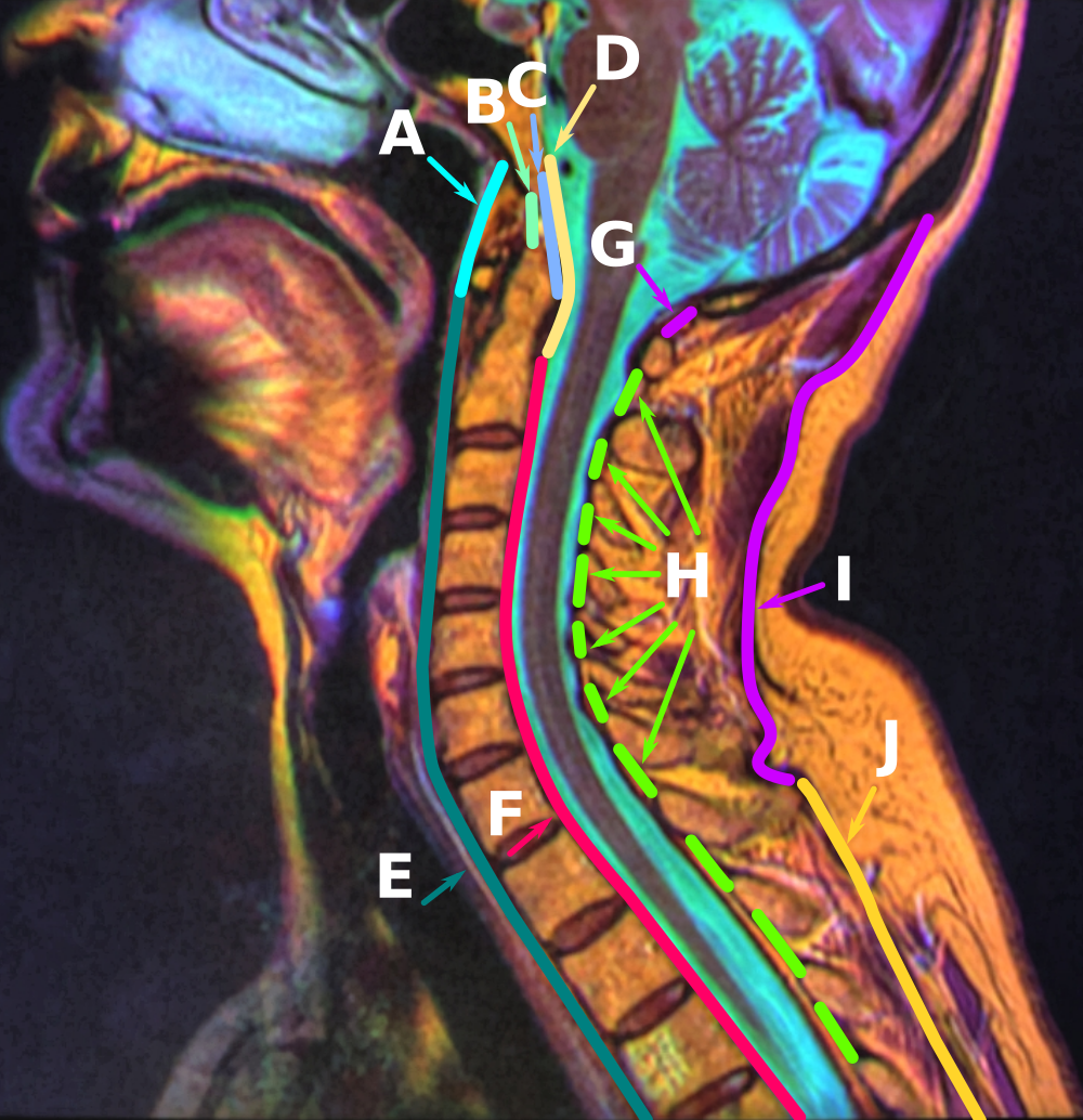
The ligament of apex dentis (or apical odontoid ligament) is a ligament that spans between the axis (anatomy), second cervical vertebra in the neck and the skull. It lies as a fibrous cord in the triangular interval between the alar ligaments, which extends from the tip of the odontoid process on the axis (anatomy), axis to the anterior margin of the foramen magnum, being intimately blended with the deep portion of the anterior atlantooccipital membrane and superior wiktionary:crus, crus of the Transverse ligament of atlas, transverse ligament of the atlas. It is regarded as a rudimentary intervertebral fibrocartilage, and in it traces of the notochord may persist. References External links * Ligaments of the head and neck Bones of the vertebral column {{ligament-stub ... [...More Info...] [...Related Items...] OR: [Wikipedia] [Google] [Baidu] |
Sagittal Section
The sagittal plane (; also known as the longitudinal plane) is an anatomical plane that divides the body into right and left sections. It is perpendicular to the transverse and coronal planes. The plane may be in the center of the body and divide it into two equal parts ( mid-sagittal), or away from the midline and divide it into unequal parts (para-sagittal). The term ''sagittal'' was coined by Gerard of Cremona. Variations in terminology Examples of sagittal planes include: * The terms ''median plane'' or ''mid-sagittal plane'' are sometimes used to describe the sagittal plane running through the midline. This plane cuts the body into halves (assuming bilateral symmetry), passing through midline structures such as the navel and spine. It is one of the planes which, combined with the Umbilical plane, defines the four quadrants of the human abdomen. * The term ''parasagittal'' is used to describe any plane parallel or adjacent to a given sagittal plane. Specific named parasag ... [...More Info...] [...Related Items...] OR: [Wikipedia] [Google] [Baidu] |
Foramen Magnum
The foramen magnum ( la, great hole) is a large, oval-shaped opening in the occipital bone of the skull. It is one of the several oval or circular openings (foramina) in the base of the skull. The spinal cord, an extension of the medulla oblongata, passes through the foramen magnum as it exits the cranial cavity. Apart from the transmission of the medulla oblongata and its membranes, the foramen magnum transmits the vertebral arteries, the anterior and posterior spinal arteries, the tectorial membranes and alar ligaments. It also transmits the accessory nerve into the skull. The foramen magnum is a very important feature in bipedal mammals. One of the attributes of a biped's foramen magnum is a forward shift of the anterior border of the cerebellar tentorium; this is caused by the shortening of the cranial base. Studies on the foramen magnum position have shown a connection to the functional influences of both posture and locomotion. The forward shift of the foramen magnum i ... [...More Info...] [...Related Items...] OR: [Wikipedia] [Google] [Baidu] |
Notochord
In anatomy, the notochord is a flexible rod which is similar in structure to the stiffer cartilage. If a species has a notochord at any stage of its life cycle (along with 4 other features), it is, by definition, a chordate. The notochord consists of inner, vacuolated cells covered by fibrous and elastic sheaths, lies along the anteroposterior axis (''front to back''), is usually closer to the dorsal than the ventral surface of the embryo, and is composed of cells derived from the mesoderm. The most commonly cited functions of the notochord are: as a midline tissue that provides directional signals to surrounding tissue during development, as a skeletal (structural) element, and as a vertebral precursor. In lancelets the notochord persists throughout life as the main structural support of the body. In tunicates the notochord is present only in the larval stage, being completely absent in the adult animal. In these invertebrate chordates, the notochord is not vacuolated. In all ... [...More Info...] [...Related Items...] OR: [Wikipedia] [Google] [Baidu] |
Intervertebral Fibrocartilage
An intervertebral disc (or intervertebral fibrocartilage) lies between adjacent vertebrae in the vertebral column. Each disc forms a fibrocartilaginous joint (a symphysis), to allow slight movement of the vertebrae, to act as a ligament to hold the vertebrae together, and to function as a shock absorber for the spine. Structure Intervertebral discs consist of an outer fibrous ring, the anulus fibrosus disci intervertebralis, which surrounds an inner gel-like center, the nucleus pulposus. The ''anulus fibrosus'' consists of several layers (laminae) of fibrocartilage made up of both type I and type II collagen. Type I is concentrated toward the edge of the ring, where it provides greater strength. The stiff laminae can withstand compressive forces. The fibrous intervertebral disc contains the ''nucleus pulposus'' and this helps to distribute pressure evenly across the disc. This prevents the development of stress concentrations which could cause damage to the underlying vertebrae ... [...More Info...] [...Related Items...] OR: [Wikipedia] [Google] [Baidu] |
Anatomy Of The Neck Sagittal Color MRI
Anatomy () is the branch of biology concerned with the study of the structure of organisms and their parts. Anatomy is a branch of natural science that deals with the structural organization of living things. It is an old science, having its beginnings in prehistoric times. Anatomy is inherently tied to developmental biology, embryology, comparative anatomy, evolutionary biology, and phylogeny, as these are the processes by which anatomy is generated, both over immediate and long-term timescales. Anatomy and physiology, which study the structure and function of organisms and their parts respectively, make a natural pair of related disciplines, and are often studied together. Human anatomy is one of the essential basic sciences that are applied in medicine. The discipline of anatomy is divided into macroscopic and microscopic. Macroscopic anatomy, or gross anatomy, is the examination of an animal's body parts using unaided eyesight. Gross anatomy also includes the branch o ... [...More Info...] [...Related Items...] OR: [Wikipedia] [Google] [Baidu] |
Transverse Ligament Of Atlas
In anatomy, the transverse ligament of the atlas is a ligament which arches across the ring of the atlas (the topmost cervical vertebra, which directly supports the skull), and keeps the odontoid process in contact with the atlas. Anatomy It is concave in front, convex behind, broader and thicker in the middle than at the ends, and firmly attached on either side to a small tubercle on the medial surface of the lateral mass of the atlas.Gray's anatomy, 1918 As it crosses the odontoid process, a small fasciculus (''crus superius'') is prolonged upward, and another (''crus inferius'') downward, from the superficial or posterior fibers of the ligament. The former is attached to the basilar part of the occipital bone, in close relation with the membrana tectoria; the latter is fixed to the posterior surface of the body of the axis; hence, the whole ligament is named the cruciate ligament of the atlas. The transverse ligament divides the ring of the atlas into two unequal parts: of the ... [...More Info...] [...Related Items...] OR: [Wikipedia] [Google] [Baidu] |
Crus
Crus can refer to: *''Crus'', a subgenus of the fly genus ''Metopochetus'' *Crus (lower leg) *Crus, a plural of Cru (wine) *CRUs, an abbreviation of Civil Resettlement Units * Rektorenkonferenz der Schweizer Universitäten (CRUS; English: Rectors' Conference of the Swiss Universities) Crus (plural ''crura'') can also refer to other anatomical structures that are leg-shaped: *crura of antihelix *crus of cerebrum *crus of clitoris *crus of diaphragm *crus of fornix *crus of heart *crus of penis *crura of the stapes * crura of superficial inguinal ring *a leg-like structure of the little skate The little skate (''Leucoraja erinacea'') is a species of skate in the family Rajidae, found from Nova Scotia to North Carolina on sand or gravel habitats. They are one of the dominant members of the demersal fish community in the northwestern ..., used for locomotion See also * Cru (other) {{disambig ... [...More Info...] [...Related Items...] OR: [Wikipedia] [Google] [Baidu] |
Anterior Atlantooccipital Membrane
Standard anatomical terms of location are used to unambiguously describe the anatomy of animals, including humans. The terms, typically derived from Latin or Greek roots, describe something in its standard anatomical position. This position provides a definition of what is at the front ("anterior"), behind ("posterior") and so on. As part of defining and describing terms, the body is described through the use of anatomical planes and anatomical axes. The meaning of terms that are used can change depending on whether an organism is bipedal or quadrupedal. Additionally, for some animals such as invertebrates, some terms may not have any meaning at all; for example, an animal that is radially symmetrical will have no anterior surface, but can still have a description that a part is close to the middle ("proximal") or further from the middle ("distal"). International organisations have determined vocabularies that are often used as standard vocabularies for subdisciplines of anatom ... [...More Info...] [...Related Items...] OR: [Wikipedia] [Google] [Baidu] |
Odontoid Process
In anatomy, the axis (from Latin ''axis'', "axle") or epistropheus is the second cervical vertebra (C2) of the spine, immediately inferior to the atlas, upon which the head rests. The axis' defining feature is its strong odontoid process (bony protrusion) known as the dens, which rises dorsally from the rest of the bone. Structure The body is deeper in front or in the back and is prolonged downward anteriorly to overlap the upper and front part of the third vertebra. It presents a median longitudinal ridge in front, separating two lateral depressions for the attachment of the longus colli muscles. Odontoid Process of Axis (Dens) The dens, also called the odontoid process or the peg, is the most pronounced projecting feature of the axis. The dens exhibits a slight constriction where it joins the main body of the vertebra. The condition where the dens is separated from the body of the axis is called ''os odontoideum'' and may cause nerve and circulation compression syndrome ... [...More Info...] [...Related Items...] OR: [Wikipedia] [Google] [Baidu] |
Occipital Bone
The occipital bone () is a neurocranium, cranial dermal bone and the main bone of the occiput (back and lower part of the skull). It is trapezoidal in shape and curved on itself like a shallow dish. The occipital bone overlies the occipital lobes of the cerebrum. At the base of skull in the occipital bone, there is a large oval opening called the foramen magnum, which allows the passage of the spinal cord. Like the other cranial bones, it is classed as a flat bone. Due to its many attachments and features, the occipital bone is described in terms of separate parts. From its front to the back is the basilar part of occipital bone, basilar part, also called the basioccipital, at the sides of the foramen magnum are the lateral parts of occipital bone, lateral parts, also called the exoccipitals, and the back is named as the squamous part of occipital bone, squamous part. The basilar part is a thick, somewhat quadrilateral piece in front of the foramen magnum and directed towards the ... [...More Info...] [...Related Items...] OR: [Wikipedia] [Google] [Baidu] |
Alar Ligament
In anatomy, the alar ligaments are ligaments which connect the dens (a bony protrusion on the second cervical vertebra) to tubercles on the medial side of the occipital condyle. They are short, tough, fibrous cords that attach on the skull and on the axis, and function to check side-to-side movements of the head when it is turned. Because of their function, the alar ligaments are also known as the "check ligaments of the odontoid". Structure The alar ligaments are two strong, rounded cords of about 0.5 cm in diameter that run from the sides of the foramen magnum of the skull to the dens of the axis, the second cervical vertebra. They span almost horizontally, creating an angle between them of at least 140°. Development The alar ligaments, along with the transverse ligament of the atlas, derive from the axial component of the first cervical sclerotome. Function The function of the alar ligaments is to limit the amount of rotation of the head, and by their action on th ... [...More Info...] [...Related Items...] OR: [Wikipedia] [Google] [Baidu] |
Skull
The skull is a bone protective cavity for the brain. The skull is composed of four types of bone i.e., cranial bones, facial bones, ear ossicles and hyoid bone. However two parts are more prominent: the cranium and the mandible. In humans, these two parts are the neurocranium and the viscerocranium ( facial skeleton) that includes the mandible as its largest bone. The skull forms the anterior-most portion of the skeleton and is a product of cephalisation—housing the brain, and several sensory structures such as the eyes, ears, nose, and mouth. In humans these sensory structures are part of the facial skeleton. Functions of the skull include protection of the brain, fixing the distance between the eyes to allow stereoscopic vision, and fixing the position of the ears to enable sound localisation of the direction and distance of sounds. In some animals, such as horned ungulates (mammals with hooves), the skull also has a defensive function by providing the mount (on the front ... [...More Info...] [...Related Items...] OR: [Wikipedia] [Google] [Baidu] |


