|
Antrodiella Semistipitata
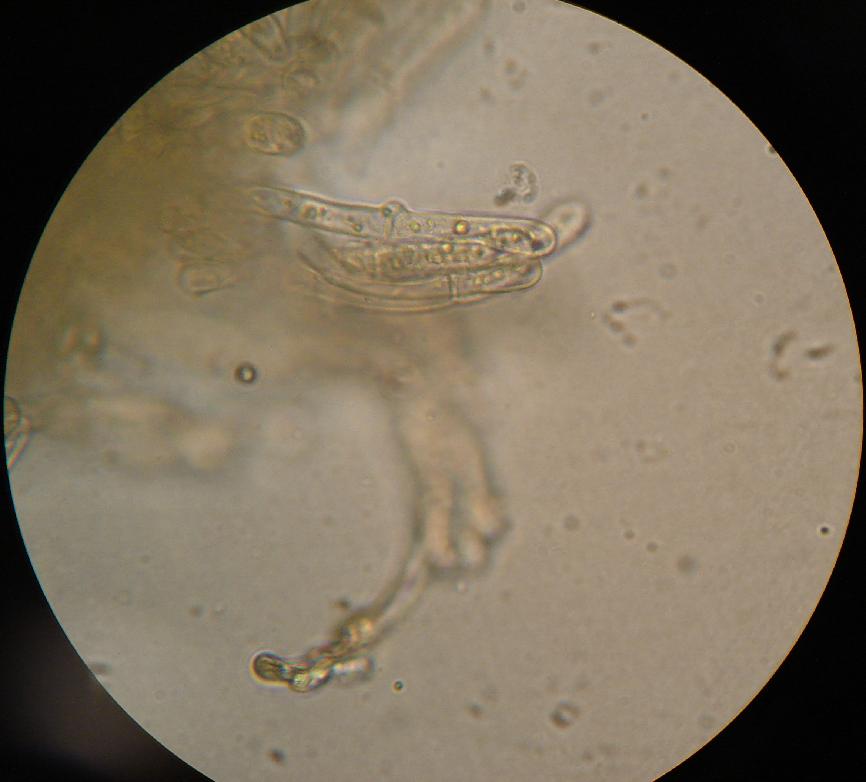
''Antrodiella'' is a genus of fungi in the family Steccherinaceae of the order Polyporales. Taxonomy ''Antrodiella'' was circumscribed by mycologists Leif Ryvarden and I. Johansen in 1980. Of the seven original species it contained, only the type, ''Antrodiella semisupina'', remains in the genus; most of the original species have since been transferred to ''Flaviporus''. ''Antrodiella'' was traditionally placed in the family Phanerochaetaceae until molecular studies were used to determine a more appropriate classification in the Steccherinaceae. The genus is a wastebasket taxon, containing "species that share common macroscopic and microscopic characteristics, but are not necessarily related." Description The fruitbodies of ''Antrodiella'' fungi are either crust-like to effused-reflexed (stretched out on the substrate but with edges curled up to form cap-like structures) in form. They have a waxy and soft fresh texture that becomes dense and hard, and often semitrans ... [...More Info...] [...Related Items...] OR: [Wikipedia] [Google] [Baidu] |
Antrodiella Semisupina
''Antrodiella'' is a genus of fungi in the family Steccherinaceae of the order Polyporales. Taxonomy ''Antrodiella'' was circumscribed by mycologists Leif Ryvarden and I. Johansen in 1980. Of the seven original species it contained, only the type, '' Antrodiella semisupina'', remains in the genus; most of the original species have since been transferred to '' Flaviporus''. ''Antrodiella'' was traditionally placed in the family Phanerochaetaceae until molecular studies were used to determine a more appropriate classification in the Steccherinaceae. The genus is a wastebasket taxon, containing "species that share common macroscopic and microscopic characteristics, but are not necessarily related." Description The fruitbodies of ''Antrodiella'' fungi are either crust-like to effused-reflexed (stretched out on the substrate but with edges curled up to form cap-like structures) in form. They have a waxy and soft fresh texture that becomes dense and hard, and often semitra ... [...More Info...] [...Related Items...] OR: [Wikipedia] [Google] [Baidu] |
Wastebasket Taxon
Wastebasket taxon (also called a wastebin taxon, dustbin taxon or catch-all taxon) is a term used by some taxonomists to refer to a taxon that has the sole purpose of classifying organisms that do not fit anywhere else. They are typically defined by either their designated members' often superficial similarity to each other, or their ''lack'' of one or more distinct character states or by their ''not'' belonging to one or more other taxa. Wastebasket taxa are by definition either paraphyletic or polyphyletic, and are therefore not considered valid taxa under strict cladistic rules of taxonomy. The name of a wastebasket taxon may in some cases be retained as the designation of an evolutionary grade, however. The term was coined in a 1985 essay by Steven Jay Gould. Examples There are many examples of paraphyletic groups, but true "wastebasket" taxa are those that are known not to, and perhaps not intended to, represent natural groups, but are nevertheless used as convenient groups ... [...More Info...] [...Related Items...] OR: [Wikipedia] [Google] [Baidu] |
Ellipsoid
An ellipsoid is a surface that may be obtained from a sphere by deforming it by means of directional scalings, or more generally, of an affine transformation. An ellipsoid is a quadric surface; that is, a surface that may be defined as the zero set of a polynomial of degree two in three variables. Among quadric surfaces, an ellipsoid is characterized by either of the two following properties. Every planar cross section is either an ellipse, or is empty, or is reduced to a single point (this explains the name, meaning "ellipse-like"). It is bounded, which means that it may be enclosed in a sufficiently large sphere. An ellipsoid has three pairwise perpendicular axes of symmetry which intersect at a center of symmetry, called the center of the ellipsoid. The line segments that are delimited on the axes of symmetry by the ellipsoid are called the ''principal axes'', or simply axes of the ellipsoid. If the three axes have different lengths, the figure is a triaxial ellipsoid (r ... [...More Info...] [...Related Items...] OR: [Wikipedia] [Google] [Baidu] |
Basidiospore
A basidiospore is a reproductive spore produced by Basidiomycete fungi, a grouping that includes mushrooms, shelf fungi, rusts, and smuts. Basidiospores typically each contain one haploid nucleus that is the product of meiosis, and they are produced by specialized fungal cells called basidia. Typically, four basidiospores develop on appendages from each basidium, of which two are of one strain and the other two of its opposite strain. In gills under a cap of one common species, there exist millions of basidia. Some gilled mushrooms in the order Agaricales have the ability to release billions of spores. The puffball fungus ''Calvatia gigantea'' has been calculated to produce about five trillion basidiospores. Most basidiospores are forcibly discharged, and are thus considered ballistospores. These spores serve as the main air dispersal units for the fungi. The spores are released during periods of high humidity and generally have a night-time or pre-dawn peak concentration in the ... [...More Info...] [...Related Items...] OR: [Wikipedia] [Google] [Baidu] |
Hymenium
The hymenium is the tissue layer on the hymenophore of a fungal fruiting body where the cells develop into basidia or asci, which produce spores. In some species all of the cells of the hymenium develop into basidia or asci, while in others some cells develop into sterile cells called cystidia (basidiomycetes) or paraphyses (ascomycetes). Cystidia are often important for microscopic identification. The subhymenium consists of the supportive hyphae from which the cells of the hymenium grow, beneath which is the hymenophoral trama, the hyphae that make up the mass of the hymenophore. The position of the hymenium is traditionally the first characteristic used in the classification and identification of mushrooms. Below are some examples of the diverse types which exist among the macroscopic Basidiomycota and Ascomycota. * In agarics, the hymenium is on the vertical faces of the gills. * In boletes and polypores, it is in a spongy mass of downward-pointing tubes. * In puffballs, ... [...More Info...] [...Related Items...] OR: [Wikipedia] [Google] [Baidu] |
Cystidium
A cystidium (plural cystidia) is a relatively large cell found on the sporocarp of a basidiomycete (for example, on the surface of a mushroom gill), often between clusters of basidia. Since cystidia have highly varied and distinct shapes that are often unique to a particular species or genus, they are a useful micromorphological characteristic in the identification of basidiomycetes. In general, the adaptive significance of cystidia is not well understood. Classification of cystidia By position Cystidia may occur on the edge of a lamella (or analogous hymenophoral structure) (cheilocystidia), on the face of a lamella (pleurocystidia), on the surface of the cap (dermatocystidia or pileocystidia), on the margin of the cap (circumcystidia) or on the stipe (caulocystidia). Especially the pleurocystidia and cheilocystidia are important for identification within many genera. Sometimes the cheilocystidia give the gill edge a distinct colour which is visible to the naked eye or wit ... [...More Info...] [...Related Items...] OR: [Wikipedia] [Google] [Baidu] |
Hyaline
A hyaline substance is one with a glassy appearance. The word is derived from el, ὑάλινος, translit=hyálinos, lit=transparent, and el, ὕαλος, translit=hýalos, lit=crystal, glass, label=none. Histopathology Hyaline cartilage is named after its glassy appearance on fresh gross pathology. On light microscopy of H&E stained slides, the extracellular matrix of hyaline cartilage looks homogeneously pink, and the term "hyaline" is used to describe similarly homogeneously pink material besides the cartilage. Hyaline material is usually acellular and proteinaceous. For example, arterial hyaline is seen in aging, high blood pressure, diabetes mellitus and in association with some drugs (e.g. calcineurin inhibitors). It is bright pink with PAS staining. Ichthyology and entomology In ichthyology and entomology, ''hyaline'' denotes a colorless, transparent substance, such as unpigmented fins of fishes or clear insect wings. Resh, Vincent H. and R. T. Cardé, Eds. Encyclo ... [...More Info...] [...Related Items...] OR: [Wikipedia] [Google] [Baidu] |
Clamp Connection
A clamp connection is a hook-like structure formed by growing hyphal cells of certain fungi. It is a characteristic feature of Basidiomycetes fungi. It is created to ensure that each cell, or segment of hypha separated by septa (cross walls), receives a set of differing nuclei, which are obtained through mating of hyphae of differing sexual types. It is used to maintain genetic variation within the hypha much like the mechanisms found in crozier (hook) during sexual reproduction. Formation Clamp connections are formed by the terminal hypha during elongation. Before the clamp connection is formed this terminal segment contains two nuclei. Once the terminal segment is long enough it begins to form the clamp connection. At the same time, each nucleus undergoes mitotic division to produce two daughter nuclei. As the clamp continues to develop it uptakes one of the daughter (green circle) nuclei and separates it from its sister nucleus. While this is occurring the remaining nuclei ... [...More Info...] [...Related Items...] OR: [Wikipedia] [Google] [Baidu] |
Hypha
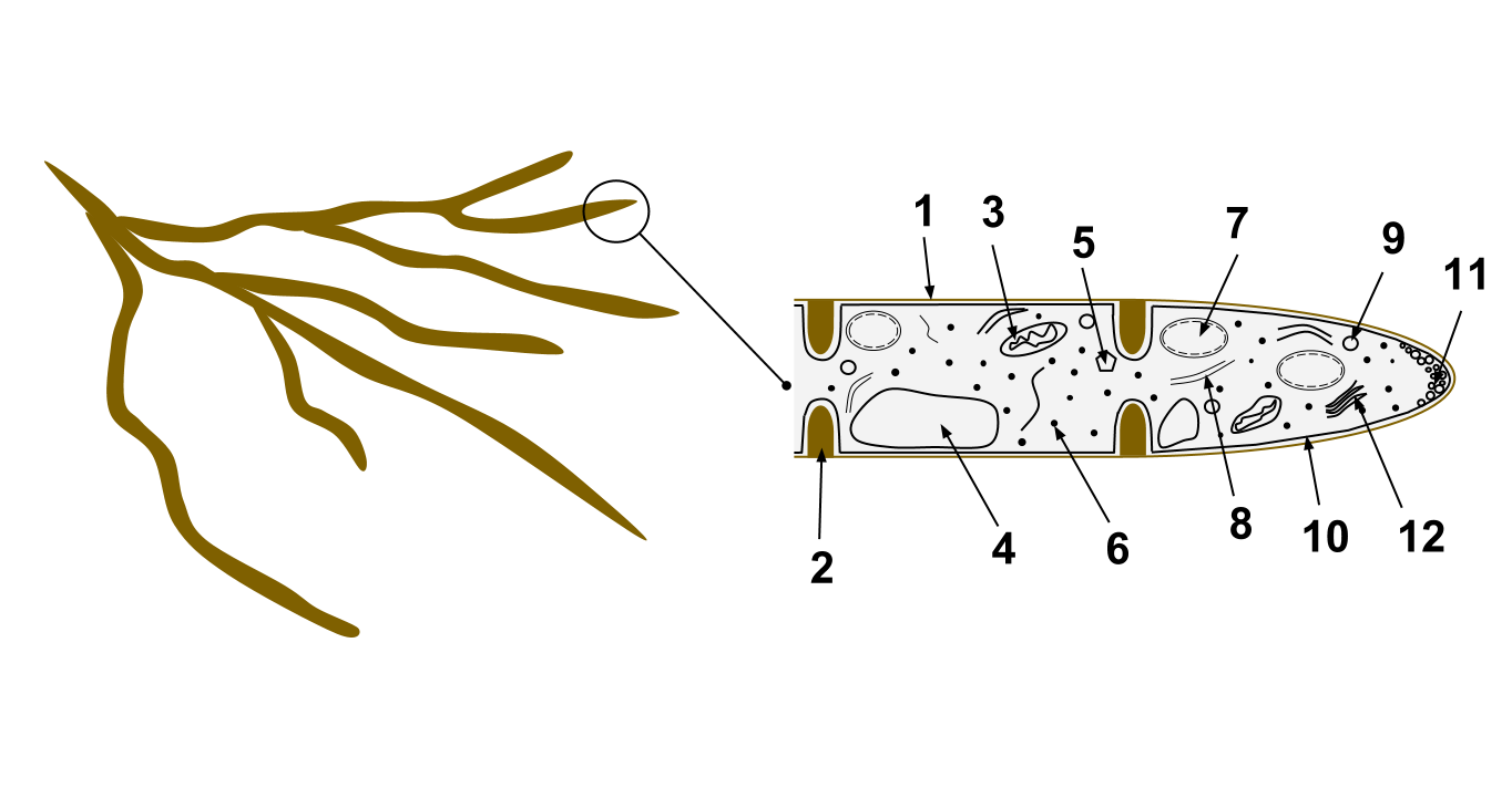
A hypha (; ) is a long, branching, filamentous structure of a fungus, oomycete, or actinobacterium. In most fungi, hyphae are the main mode of vegetative growth, and are collectively called a mycelium. Structure A hypha consists of one or more cells surrounded by a tubular cell wall. In most fungi, hyphae are divided into cells by internal cross-walls called "septa" (singular septum). Septa are usually perforated by pores large enough for ribosomes, mitochondria, and sometimes nuclei to flow between cells. The major structural polymer in fungal cell walls is typically chitin, in contrast to plants and oomycetes that have cellulosic cell walls. Some fungi have aseptate hyphae, meaning their hyphae are not partitioned by septa. Hyphae have an average diameter of 4–6 µm. Growth Hyphae grow at their tips. During tip growth, cell walls are extended by the external assembly and polymerization of cell wall components, and the internal production of new cell membrane. The S ... [...More Info...] [...Related Items...] OR: [Wikipedia] [Google] [Baidu] |
Trama (mycology)
In mycology, the term trama is used in two ways. In the broad sense, it is the inner, fleshy portion of a mushroom's basidiocarp, or fruit body. It is distinct from the outer layer of tissue, known as the pileipellis or cuticle, and from the spore-bearing tissue layer known as the hymenium. In essence, the trama is the tissue that is commonly referred to as the "flesh" of mushrooms and similar fungi.Largent D, Johnson D, Watling R. 1977. ''How to Identify Mushrooms to Genus III: Microscopic Features''. Arcata, CA: Mad River Press. . pp. 60–70. The second use is more specific, and refers to the "hymenophoral trama" that supports the hymenium. It is similarly interior, connective tissue, but it is more specifically the central layer of hyphae running from the underside of the mushroom cap to the lamella or gill, upon which the hymenium rests. Various types have been classified by their structure, including trametoid, cantharelloid, boletoid, and agaricoid, with agaricoid the ... [...More Info...] [...Related Items...] OR: [Wikipedia] [Google] [Baidu] |
Ochre
Ochre ( ; , ), or ocher in American English, is a natural clay earth pigment, a mixture of ferric oxide and varying amounts of clay and sand. It ranges in colour from yellow to deep orange or brown. It is also the name of the colours produced by this pigment, especially a light brownish-yellow. A variant of ochre containing a large amount of hematite, or dehydrated iron oxide, has a reddish tint known as "red ochre" (or, in some dialects, ruddle). The word ochre also describes clays coloured with iron oxide derived during the extraction of tin and copper. Earth pigments Ochre is a family of earth pigments, which includes yellow ochre, red ochre, purple ochre, sienna, and umber. The major ingredient of all the ochres is iron(III) oxide-hydroxide, known as limonite, which gives them a yellow colour. * Yellow ochre, , is a hydrated iron hydroxide (limonite) also called gold ochre. * Red ochre, , takes its reddish colour from the mineral hematite, which is an anhydrous iron ... [...More Info...] [...Related Items...] OR: [Wikipedia] [Google] [Baidu] |
Pileus (mycology)
The pileus is the technical name for the cap, or cap-like part, of a basidiocarp or ascocarp (fungal fruiting body) that supports a spore-bearing surface, the hymenium.Moore-Landecker, E: "Fundamentals of the Fungi", page 560. Prentice Hall, 1972. The hymenium (hymenophore) may consist of lamellae, tubes, or teeth, on the underside of the pileus. A pileus is characteristic of agarics, boletes, some polypores, tooth fungi, and some ascomycetes. Classification Pilei can be formed in various shapes, and the shapes can change over the course of the developmental cycle of a fungus. The most familiar pileus shape is hemispherical or ''convex.'' Convex pilei often continue to expand as they mature until they become flat. Many well-known species have a convex pileus, including the button mushroom, various ''Amanita'' species and boletes. Some, such as the parasol mushroom, have distinct bosses or umbos and are described as ''umbonate''. An umbo is a knobby protrusion at the center of th ... [...More Info...] [...Related Items...] OR: [Wikipedia] [Google] [Baidu] |
.jpg)


