|
Anti-dsDNA
Anti-double stranded DNA (Anti-dsDNA) antibodies are a group of anti-nuclear antibodies (ANA) the target antigen of which is double stranded DNA. Blood tests such as enzyme-linked immunosorbent assay (ELISA) and immunofluorescence are routinely performed to detect anti-dsDNA antibodies in diagnostic laboratories. They are highly diagnostic of systemic lupus erythematosus (SLE) and are implicated in the pathogenesis of lupus nephritis. Discovery The first evidence for antinuclear antibodies arose in 1948 when Hargraves, Richmond and Morton discovered the LE cell. These abnormal cells, which are found in the bone marrow of persons who have SLE are categorised as polymorphonuclear leukocytes with phagocytosed whole nuclei. Subsequently, in 1957, antibodies to dsDNA were the first autoantibodies to be identified in patients with SLE. Antibody production Although the exact mechanism of the generation of dsDNA antibodies is still unknown, it is likely that extracellular DNA is one ca ... [...More Info...] [...Related Items...] OR: [Wikipedia] [Google] [Baidu] |
Anti-nuclear Antibodies
Antinuclear antibodies (ANAs, also known as antinuclear factor or ANF) are autoantibodies that bind to contents of the cell nucleus. In normal individuals, the immune system produces antibodies to foreign proteins (antigens) but not to human proteins (autoantigens). In some cases, antibodies to human antigens are produced. There are many subtypes of ANAs such as anti-Ro antibodies, anti-La antibodies, anti-Sm antibodies, anti-nRNP antibodies, anti-Scl-70 antibodies, anti-dsDNA antibodies, anti-histone antibodies, antibodies to nuclear pore complexes, anti-centromere antibodies and anti-sp100 antibodies. Each of these antibody subtypes binds to different proteins or protein complexes within the nucleus. They are found in many disorders including autoimmunity, cancer and infection, with different prevalences of antibodies depending on the condition. This allows the use of ANAs in the diagnosis of some autoimmune disorders, including systemic lupus erythematosus, Sjögren syndrom ... [...More Info...] [...Related Items...] OR: [Wikipedia] [Google] [Baidu] |
Systemic Lupus Erythematosus
Lupus, technically known as systemic lupus erythematosus (SLE), is an autoimmune disease in which the body's immune system mistakenly attacks healthy tissue in many parts of the body. Symptoms vary among people and may be mild to severe. Common symptoms include painful and swollen joints, fever, chest pain, hair loss, mouth ulcers, swollen lymph nodes, feeling tired, and a red rash which is most commonly on the face. Often there are periods of illness, called flares, and periods of remission during which there are few symptoms. The cause of SLE is not clear. It is thought to involve a mixture of genetics combined with environmental factors. Among identical twins, if one is affected there is a 24% chance the other one will also develop the disease. Female sex hormones, sunlight, smoking, vitamin D deficiency, and certain infections are also believed to increase a person's risk. The mechanism involves an immune response by autoantibodies against a person's own tissues. T ... [...More Info...] [...Related Items...] OR: [Wikipedia] [Google] [Baidu] |
Lupus Nephritis
Lupus nephritis is an inflammation of the kidneys caused by systemic lupus erythematosus (SLE), an autoimmune disease. It is a type of glomerulonephritis in which the glomeruli become inflamed. Since it is a result of SLE, this type of glomerulonephritis is said to be ''secondary'', and has a different pattern and outcome from conditions with a ''primary'' cause originating in the kidney. The diagnosis of lupus nephritis depends on blood tests, urinalysis, X-rays, ultrasound scans of the kidneys, and a kidney biopsy. On urinalysis, a nephritic picture is found and red blood cell casts, red blood cells and proteinuria is found. Classification The World Health Organization has divided lupus nephritis into five stages based on the biopsy. This classification was defined in 1982 and revised in 1995. Class IV disease ( Diffuse proliferative nephritis) is both the most severe, and the most common subtype. Class VI (advanced sclerosing lupus nephritis) is a final class which is included ... [...More Info...] [...Related Items...] OR: [Wikipedia] [Google] [Baidu] |
Pattern Recognition Receptor
Pattern recognition receptors (PRRs) play a crucial role in the proper function of the innate immune system. PRRs are germline-encoded host sensors, which detect molecules typical for the pathogens. They are proteins expressed, mainly, by cells of the innate immune system, such as dendritic cells, macrophages, monocytes, neutrophils and epithelial cells, to identify two classes of molecules: pathogen-associated molecular patterns (PAMPs), which are associated with microbial pathogens, and damage-associated molecular patterns (DAMPs), which are associated with components of host's cells that are released during cell damage or death. They are also called primitive pattern recognition receptors because they evolved before other parts of the immune system, particularly before adaptive immunity. PRRs also mediate the initiation of antigen-specific adaptive immune response and release of inflammatory cytokines. The microbe-specific molecules that are recognized by a given PRR are called p ... [...More Info...] [...Related Items...] OR: [Wikipedia] [Google] [Baidu] |
Dendritic Cells
Dendritic cells (DCs) are antigen-presenting cells (also known as ''accessory cells'') of the mammalian immune system. Their main function is to process antigen material and present it on the cell surface to the T cells of the immune system. They act as messengers between the innate and the adaptive immune systems. Dendritic cells are present in those tissues that are in contact with the external environment, such as the skin (where there is a specialized dendritic cell type called the Langerhans cell) and the inner lining of the nose, lungs, stomach and intestines. They can also be found in an immature state in the blood. Once activated, they migrate to the lymph nodes where they interact with T cells and B cells to initiate and shape the adaptive immune response. At certain development stages they grow branched projections, the ''dendrites'' that give the cell its name (δένδρον or déndron being Greek for 'tree'). While similar in appearance, these are structures disti ... [...More Info...] [...Related Items...] OR: [Wikipedia] [Google] [Baidu] |
Macrophages
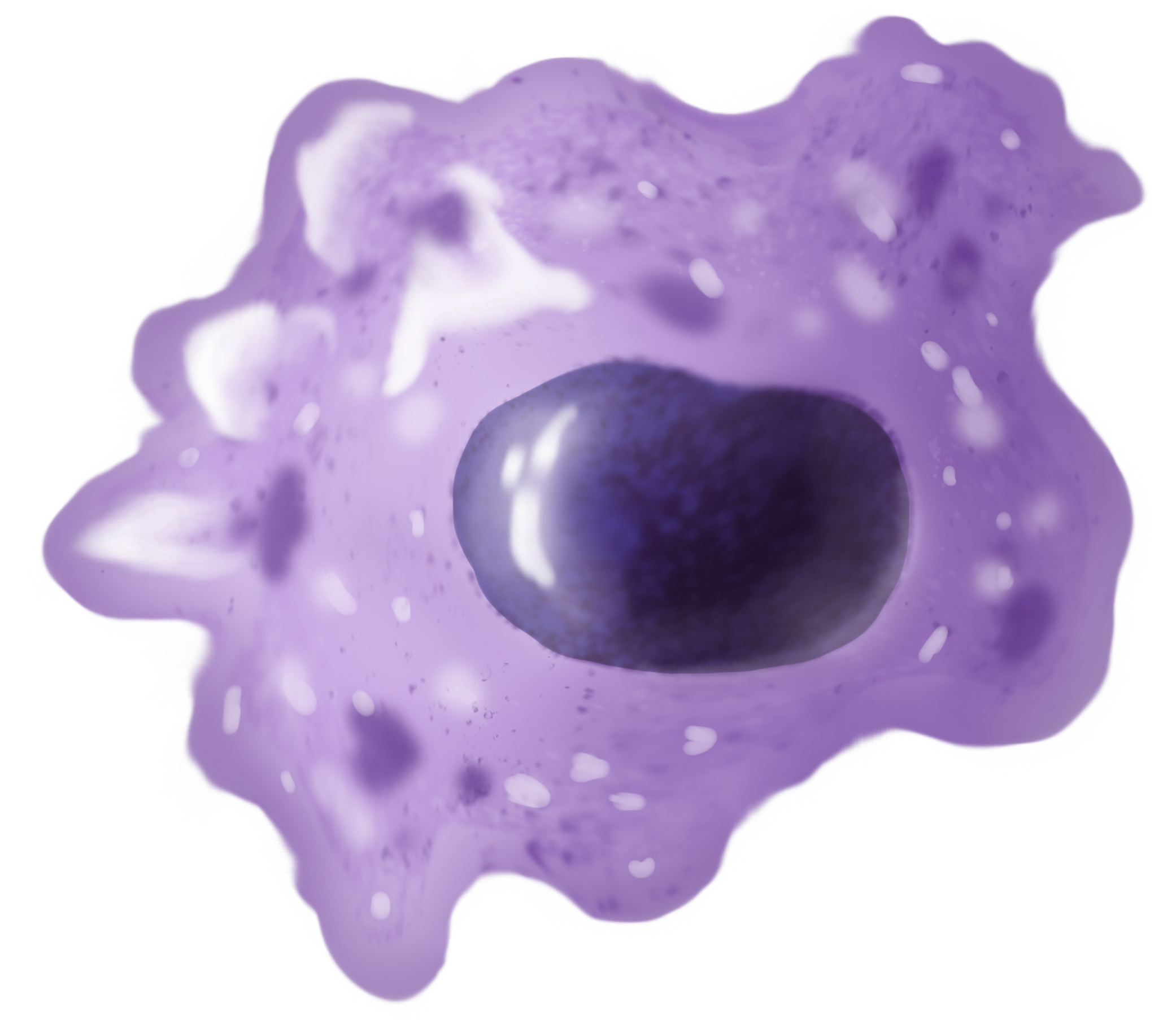
Macrophages (abbreviated as M φ, MΦ or MP) ( el, large eaters, from Greek ''μακρός'' (') = large, ''φαγεῖν'' (') = to eat) are a type of white blood cell of the immune system that engulfs and digests pathogens, such as cancer cells, microbes, cellular debris, and foreign substances, which do not have proteins that are specific to healthy body cells on their surface. The process is called phagocytosis, which acts to defend the host against infection and injury. These large phagocytes are found in essentially all tissues, where they patrol for potential pathogens by amoeboid movement. They take various forms (with various names) throughout the body (e.g., histiocytes, Kupffer cells, alveolar macrophages, microglia, and others), but all are part of the mononuclear phagocyte system. Besides phagocytosis, they play a critical role in nonspecific defense (innate immunity) and also help initiate specific defense mechanisms (adaptive immunity) by recruiting other immun ... [...More Info...] [...Related Items...] OR: [Wikipedia] [Google] [Baidu] |
T Helper Cells
The T helper cells (Th cells), also known as CD4+ cells or CD4-positive cells, are a type of T cell that play an important role in the adaptive immune system. They aid the activity of other immune cells by releasing cytokines. They are considered essential in B cell antibody class switching, breaking cross-tolerance in dendritic cells, in the activation and growth of cytotoxic T cells, and in maximizing bactericidal activity of phagocytes such as macrophages and neutrophils. CD4+ cells are mature Th cells that express the surface protein CD4. Genetic variation in regulatory elements expressed by CD4+ cells determines susceptibility to a broad class of autoimmune diseases. Structure and function Th cells contain and release cytokines to aid other immune cells. Cytokines are small protein mediators that alter the behavior of target cells that express receptors for those cytokines. These cells help polarize the immune response depending on the nature of the immunological insult ... [...More Info...] [...Related Items...] OR: [Wikipedia] [Google] [Baidu] |
Interferon Type 1
The type-I interferons (IFN) are cytokines which play essential roles in inflammation, immunoregulation, tumor cells recognition, and T-cell responses. In the human genome, a cluster of thirteen functional IFN genes is located at the 9p21.3 cytoband over approximately 400 kb including coding genes for IFNα (''IFNA1, IFNA2, IFNA4, IFNA5, IFNA6, IFNA7, IFNA8, IFNA10, IFNA13, IFNA14, IFNA16, IFNA17'' and ''IFNA21''), IFNω (''IFNW1''), IFNɛ (''IFNE''), IFNк (''IFNK'') and IFNβ (''IFNB1''), plus 11 IFN pseudogenes. Interferons bind to interferon receptors. All type I IFNs bind to a specific cell surface receptor complex known as the IFN-α receptor (IFNAR) that consists of IFNAR1 and IFNAR2 chains. Type I IFNs are found in all mammals, and homologous (similar) molecules have been found in birds, reptiles, amphibians and fish species. Sources and functions IFN-α and IFN-β are secreted by many cell types including lymphocytes (NK cells, B-cells and T-cells), macrophages, fi ... [...More Info...] [...Related Items...] OR: [Wikipedia] [Google] [Baidu] |
Plasmacytoid Dendritic Cell
Plasmacytoid dendritic cells (pDCs) are a rare type of immune cell that are known to secrete large quantities of type 1 interferon (IFNs) in response to a viral infection. They circulate in the blood and are found in peripheral lymphoid organs. They develop from bone marrow hematopoietic stem cells and constitute 2% of nucleated cells) and bone marrow and evidence (i.e. cytopenias) of bone marrow failure. Blastic plasmacytoid dendritic cell neoplasm has a high rate of recurrence following initial treatments with various chemotherapy regimens. In consequence, the disease has a poor overall prognosis and newer chemotherapeutic and novel non-chemotherapeutic drug regimens to improve the situation are under study. Role in immunity Upon stimulation and subsequent activation of TLR7 and TLR9, these cells produce large amounts (up to 1,000 times more than other cell type) of type I interferon (mainly IFN-α and IFN-β), which are critical anti-viral compounds mediating a wide range of ef ... [...More Info...] [...Related Items...] OR: [Wikipedia] [Google] [Baidu] |
Toll-like Receptor 9
Toll-like receptor 9 is a protein that in humans is encoded by the ''TLR9'' gene. TLR9 has also been designated as CD289 (cluster of differentiation 289). It is a member of the toll-like receptor (TLR) family. TLR9 is an important receptor expressed in immune system cells including dendritic cells, macrophages, natural killer cells, and other antigen presenting cells. TLR9 preferentially binds DNA present in bacteria and viruses, and triggers signaling cascades that lead to a pro-inflammatory cytokine response. Cancer, infection, and tissue damage can all modulate TLR9 expression and activation. TLR9 is also an important factor in autoimmune diseases, and there is active research into synthetic TLR9 agonists and antagonists that help regulate autoimmune inflammation. Function The TLR family plays a fundamental role in pathogen recognition and activation of innate immunity. TLRs are named for the high degree of conservation in structure and function seen between mammalian TLRs and ... [...More Info...] [...Related Items...] OR: [Wikipedia] [Google] [Baidu] |
Antigen Presenting Cells
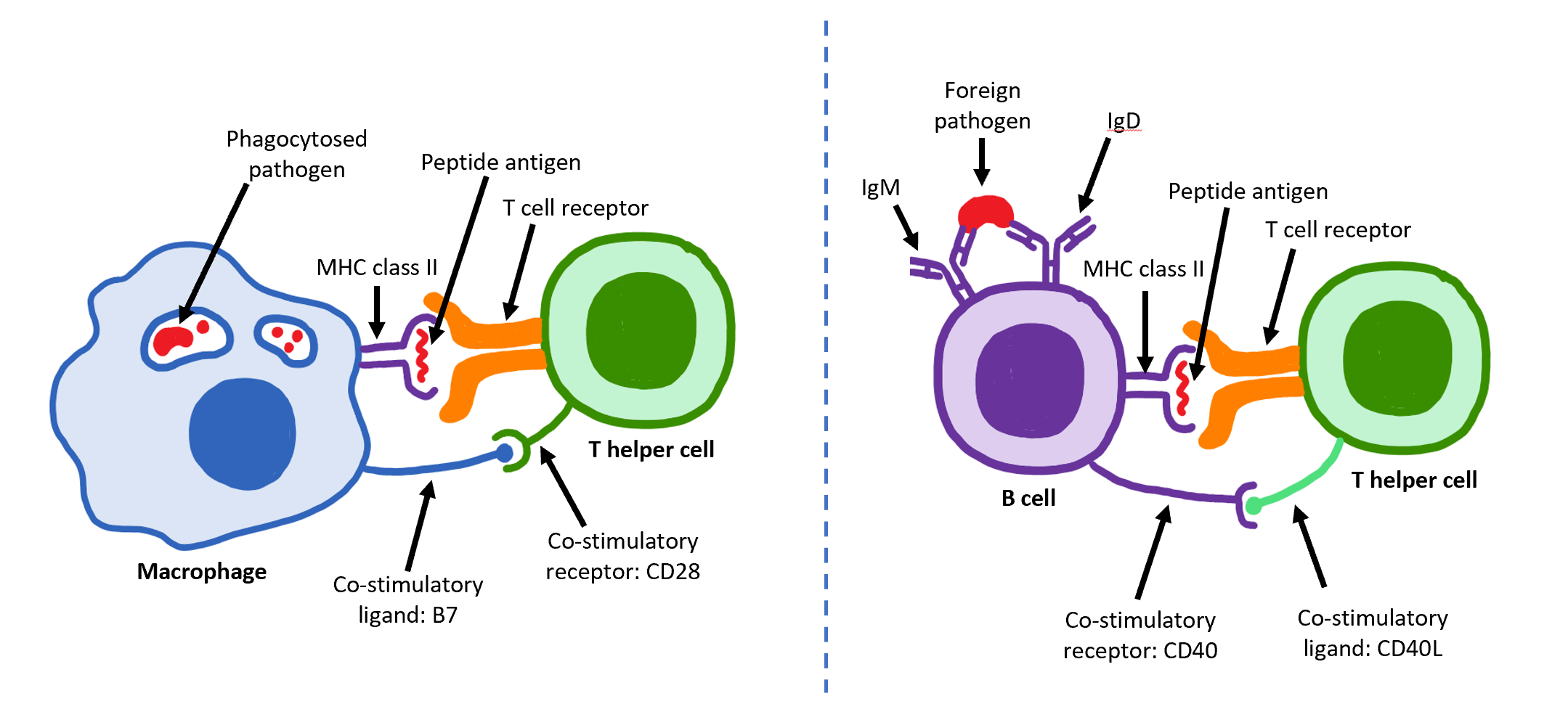
An antigen-presenting cell (APC) or accessory cell is a cell that displays antigen bound by major histocompatibility complex (MHC) proteins on its surface; this process is known as antigen presentation. T cells may recognize these complexes using their T cell receptors (TCRs). APCs process antigens and present them to T-cells. Almost all cell types can present antigens in some way. They are found in a variety of tissue types. Professional antigen-presenting cells, including macrophages, B cells and dendritic cells, present foreign antigens to helper T cells, while virus-infected cells (or cancer cells) can present antigens originating inside the cell to cytotoxic T cells. In addition to the MHC family of proteins, antigen presentation relies on other specialized signaling molecules on the surfaces of both APCs and T cells. Antigen-presenting cells are vital for effective adaptive immune response, as the functioning of both cytotoxic and helper T cells is dependent on APCs. Antigen ... [...More Info...] [...Related Items...] OR: [Wikipedia] [Google] [Baidu] |


