|
Anterior Superior Alveolar Nerve
The anterior superior alveolar nerve (or anterior superior dental nerve), is a branch of the infraorbital nerve, itself a branch of the maxillary nerve (V2). It branches from the infraorbital nerve within the infraorbital canal before the infraorbital nerve exits through the infraorbital foramen. It descends in a canal in the anterior wall of the maxillary sinus, and divides into branches which supply the incisor and canine teeth. It communicates with the middle superior alveolar nerve, and gives off a nasal branch, which passes through a minute canal in the lateral wall of the inferior meatus, and supplies the mucous membrane of the anterior part of the inferior meatus and the floor of the nasal cavity, communicating with the nasal branches from the sphenopalatine ganglion. Dental considerations for this nerve are important. The anterior superior alveolar usually innervates all anterior teeth, loops backwards to join the middle superior alveolar nerve to form the superior denta ... [...More Info...] [...Related Items...] OR: [Wikipedia] [Google] [Baidu] |
Maxillary Nerve
In neuroanatomy, the maxillary nerve (V) is one of the three branches or divisions of the trigeminal nerve, the fifth (CN V) cranial nerve. It comprises the principal functions of sensation from the maxilla, nasal cavity, sinuses, the palate and subsequently that of the mid-face, and is intermediate, both in position and size, between the ophthalmic nerve and the mandibular nerve.Illustrated Anatomy of the Head and Neck, Fehrenbach and Herring, Elsevier, 2012, page 180 Structure It begins at the middle of the trigeminal ganglion as a flattened plexiform band then it passes through the lateral wall of the cavernous sinus. It leaves the skull through the foramen rotundum, where it becomes more cylindrical in form, and firmer in texture. After leaving foramen rotundum it gives two branches to the pterygopalatine ganglion. It then crosses the pterygopalatine fossa, inclines lateralward on the back of the maxilla, and enters the orbit through the inferior orbital fissure. It t ... [...More Info...] [...Related Items...] OR: [Wikipedia] [Google] [Baidu] |
Middle Superior Alveolar Nerve
The middle superior alveolar nerve is a nerve that drops from the infraorbital portion of the maxillary nerve to supply the sinus mucosa, the roots of the maxillary premolar The premolars, also called premolar teeth, or bicuspids, are transitional teeth located between the canine and molar teeth. In humans, there are two premolars per quadrant in the permanent set of teeth, making eight premolars total in the mouth ...s, and the mesiobuccal root of the first maxillary molar. It is not always present; in 72% of cases it is non existent with the anterior superior alveolar nerve innervating the premolars and the posterior superior alveolar nerve innervating the molars, including the mesiobuccal root of the first molar. External links * Maxillary nerve {{Neuroanatomy-stub ... [...More Info...] [...Related Items...] OR: [Wikipedia] [Google] [Baidu] |
Anterior Superior Alveolar Arteries
The anterior superior alveolar arteries originate from the infraorbital artery; they supply the upper incisors and canines; they also supply the mucous membrane of the maxillary sinus. See also * Anterior superior alveolar nerve * Posterior superior alveolar artery The posterior superior alveolar artery (posterior dental artery) is given off from the maxillary, frequently in conjunction with the infraorbital artery just as the trunk of the vessel is passing into the pterygopalatine fossa. Branches Descendi ... Arteries of the head and neck {{Circulatory-stub ... [...More Info...] [...Related Items...] OR: [Wikipedia] [Google] [Baidu] |
Superior Dental Plexus
The superior dental plexus is a nerve plexus which supplies the upper jaw. Formed by posterior superior alveolar nerve, middle superior alveolar nerve, and anterior superior alveolar nerve. See also * Inferior dental plexus The inferior dental plexus is a nerve plexus which supplies the lower jaw. It is branches off of the inferior alveolar nerve and functions as innervation to the mandibular molars, first bicuspid, and part of the second bicuspid. The inferior denta ... Maxillary nerve {{Neuroanatomy-stub ... [...More Info...] [...Related Items...] OR: [Wikipedia] [Google] [Baidu] |
Nasal Cavity
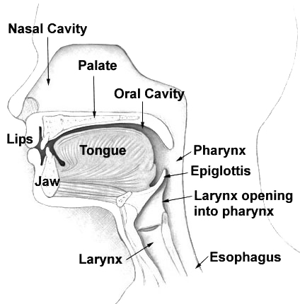
The nasal cavity is a large, air-filled space above and behind the nose in the middle of the face. The nasal septum divides the cavity into two cavities, also known as fossae. Each cavity is the continuation of one of the two nostrils. The nasal cavity is the uppermost part of the respiratory system and provides the nasal passage for inhaled air from the nostrils to the nasopharynx and rest of the respiratory tract. The paranasal sinuses surround and drain into the nasal cavity. Structure The term "nasal cavity" can refer to each of the two cavities of the nose, or to the two sides combined. The lateral wall of each nasal cavity mainly consists of the maxilla. However, there is a deficiency that is compensated for by the perpendicular plate of the palatine bone, the medial pterygoid plate, the labyrinth of ethmoid and the inferior concha. The paranasal sinuses are connected to the nasal cavity through small orifices called ostia. Most of these ostia communicate with ... [...More Info...] [...Related Items...] OR: [Wikipedia] [Google] [Baidu] |
Mucous Membrane
A mucous membrane or mucosa is a membrane that lines various cavities in the body of an organism and covers the surface of internal organs. It consists of one or more layers of epithelial cells overlying a layer of loose connective tissue. It is mostly of endodermal origin and is continuous with the skin at body openings such as the eyes, eyelids, ears, inside the nose, inside the mouth, lips, the genital areas, the urethral opening and the anus. Some mucous membranes secrete mucus, a thick protective fluid. The function of the membrane is to stop pathogens and dirt from entering the body and to prevent bodily tissues from becoming dehydrated. Structure The mucosa is composed of one or more layers of epithelial cells that secrete mucus, and an underlying lamina propria of loose connective tissue. The type of cells and type of mucus secreted vary from organ to organ and each can differ along a given tract. Mucous membranes line the digestive, respiratory and reproduct ... [...More Info...] [...Related Items...] OR: [Wikipedia] [Google] [Baidu] |
Inferior Meatus
In anatomy, the term nasal meatus can refer to any of the three meatuses (passages) through the skulls nasal cavity: the superior meatus (''meatus nasi superior''), middle meatus (''meatus nasi medius''), and inferior meatus (''meatus nasi inferior''). The nasal meatuses are located beneath each of the corresponding nasal conchae. In the case where a fourth, supreme nasal concha is present, there is a fourth supreme nasal meatus. Structure The superior meatus is the smallest of the three. It is a narrow cavity located obliquely below the superior concha. This meatus is short, lies above and extends from the middle part of the middle concha below. From behind, the sphenopalatine foramen opens into the cavity of the superior meatus and the meatus communicates with the posterior ethmoidal cells. Above and at the back of the superior concha is the sphenoethmoidal recess which the sphenoidal sinus opens into. The superior meatus occupies the middle third of the nasal cavity’s la ... [...More Info...] [...Related Items...] OR: [Wikipedia] [Google] [Baidu] |
Nasal Branch
Nasal is an adjective referring to the nose, part of human or animal anatomy. It may also be shorthand for the following uses in combination: * With reference to the human nose: ** Nasal administration, a method of pharmaceutical drug delivery ** Nasal emission, the abnormal passing of oral air through a palatal cleft, or from some other type of pharyngeal inadequacy ** Nasal hair, the hair in the nose * With reference to phonetics: ** Nasalization, the production of a sound with a lowered velum, allowing some of the air to escape through the nose; the resulting being either: *** a nasal consonant, or *** a nasal vowel * With reference to the nose of humans or other animals: ** Nasal bone, two small oblong bones placed side by side at the middle and upper part of the face, and form, by their junction, "the bridge" of the nose ** Nasal cavity, a large air filled space above and behind the nose in the middle of the face ** Nasal concha, a long, narrow and curled bone shelf which pro ... [...More Info...] [...Related Items...] OR: [Wikipedia] [Google] [Baidu] |
Canine Teeth
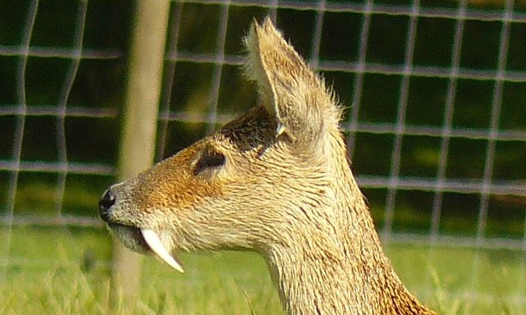
In mammalian oral anatomy, the canine teeth, also called cuspids, dog teeth, or (in the context of the upper jaw) fangs, eye teeth, vampire teeth, or vampire fangs, are the relatively long, pointed teeth. They can appear more flattened however, causing them to resemble incisors and leading them to be called ''incisiform''. They developed and are used primarily for firmly holding food in order to tear it apart, and occasionally as weapons. They are often the largest teeth in a mammal's mouth. Individuals of most species that develop them normally have four, two in the upper jaw and two in the lower, separated within each jaw by incisors; humans and dogs are examples. In most species, canines are the anterior-most teeth in the maxilla The maxilla (plural: ''maxillae'' ) in vertebrates is the upper fixed (not fixed in Neopterygii) bone of the jaw formed from the fusion of two maxillary bones. In humans, the upper jaw includes the hard palate in the front of the mouth. The ... [...More Info...] [...Related Items...] OR: [Wikipedia] [Google] [Baidu] |
Mandibular Nerve
In neuroanatomy, the mandibular nerve (V) is the largest of the three divisions of the trigeminal nerve, the fifth cranial nerve (CN V). Unlike the other divisions of the trigeminal nerve ( ophthalmic nerve, maxillary nerve) which contain only afferent fibers, the mandibular nerve contains both afferent and efferent fibers. These nerve fibers innervate structures of the lower jaw and face, such as the tongue, lower lip, and chin. The mandibular nerve also innervates the muscles of mastication. Structure The large sensory root emerges from the lateral part of the trigeminal ganglion and exits the cranial cavity through the foramen ovale. Portio minor, the small motor root of the trigeminal nerve, passes under the trigeminal ganglion and through the foramen ovale to unite with the sensory root just outside the skull. The mandibular nerve immediately passes between tensor veli palatini, which is medial, and lateral pterygoid, which is lateral, and gives off a meningeal br ... [...More Info...] [...Related Items...] OR: [Wikipedia] [Google] [Baidu] |
Incisor
Incisors (from Latin ''incidere'', "to cut") are the front teeth present in most mammals. They are located in the premaxilla above and on the mandible below. Humans have a total of eight (two on each side, top and bottom). Opossums have 18, whereas armadillos have none. Structure Adult humans normally have eight incisors, two of each type. The types of incisor are: * maxillary central incisor (upper jaw, closest to the center of the lips) * maxillary lateral incisor (upper jaw, beside the maxillary central incisor) * mandibular central incisor (lower jaw, closest to the center of the lips) * mandibular lateral incisor (lower jaw, beside the mandibular central incisor) Children with a full set of deciduous teeth (primary teeth) also have eight incisors, named the same way as in permanent teeth. Young children may have from zero to eight incisors depending on the stage of their tooth eruption and tooth development. Typically, the mandibular central incisors erupt first, ... [...More Info...] [...Related Items...] OR: [Wikipedia] [Google] [Baidu] |


