|
Anterior Jugular Vein
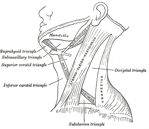
The anterior jugular vein is a vein in the neck. Structure The anterior jugular vein lies lateral to the cricothyroid ligament. It begins near the hyoid bone by the confluence of several superficial veins from the submandibular region. Its tributaries are some laryngeal veins, and occasionally a small thyroid vein. It descends between the median line and the anterior border of the sternocleidomastoid muscle, and, at the lower part of the neck, passes beneath that muscle to open into the termination of the external jugular vein, or, in some instances, into the subclavian vein. Just above the sternum the two anterior jugular veins communicate by a transverse trunk, the venous jugular arch, which receive tributaries from the inferior thyroid veins; each also communicates with the internal jugular. There are no valves in this vein. The pretracheal lymph nodes follow the anterior jugular vein on each side of the midline. Variation The anterior jugular vein varies considerably ... [...More Info...] [...Related Items...] OR: [Wikipedia] [Google] [Baidu] |
External Jugular Vein
The external jugular vein receives the greater part of the blood from the exterior of the cranium and the deep parts of the face, being formed by the junction of the posterior division of the retromandibular vein with the posterior auricular vein. Structure It commences in the substance of the parotid gland, on a level with the angle of the mandible, and runs perpendicularly down the neck, in the direction of a line drawn from the angle of the mandible to the middle of the clavicle superficial to the sternocleidomastoideus. In its course it crosses the sternocleidomastoideus obliquely, and in the subclavian triangle perforates the deep fascia, and ends in the subclavian vein lateral to or in front of the scalenus anterior, piercing the roof of the posterior triangle. It is separated from the sternocleidomastoideus by the investing layer of the deep cervical fascia, and is covered by the platysma, the superficial fascia, and the integument; it crosses the cutaneous cervical nerv ... [...More Info...] [...Related Items...] OR: [Wikipedia] [Google] [Baidu] |
Venous Jugular Arch
Just above the sternum the two anterior jugular veins communicate by a transverse trunk, the jugular venous arch (or venous jugular arch), which receive tributaries from the inferior thyroid veins The inferior thyroid veins appear two, frequently three or four, in number, and arise in the venous plexus on the thyroid gland, communicating with the middle and superior thyroid veins. While the superior and middle thyroid veins serve as direc ...; each also communicates with the internal jugular. There are no valves in this vein. References Veins of the head and neck {{circulatory-stub ... [...More Info...] [...Related Items...] OR: [Wikipedia] [Google] [Baidu] |
Cervical Vertebra 6
In tetrapods, cervical vertebrae (singular: vertebra) are the vertebrae of the neck, immediately below the skull. Truncal vertebrae (divided into thoracic and lumbar vertebrae in mammals) lie caudal (toward the tail) of cervical vertebrae. In sauropsid species, the cervical vertebrae bear cervical ribs. In lizards and saurischian dinosaurs, the cervical ribs are large; in birds, they are small and completely fused to the vertebrae. The vertebral transverse processes of mammals are homologous to the cervical ribs of other amniotes. Most mammals have seven cervical vertebrae, with the only three known exceptions being the manatee with six, the two-toed sloth with five or six, and the three-toed sloth with nine. In humans, cervical vertebrae are the smallest of the true vertebrae and can be readily distinguished from those of the thoracic or lumbar regions by the presence of a foramen (hole) in each transverse process, through which the vertebral artery, vertebral veins, and in ... [...More Info...] [...Related Items...] OR: [Wikipedia] [Google] [Baidu] |
Bleeding
Bleeding, hemorrhage, haemorrhage or blood loss, is blood escaping from the circulatory system from damaged blood vessels. Bleeding can occur internally, or externally either through a natural opening such as the mouth, nose, ear, urethra, vagina or anus, or through a puncture in the skin. Hypovolemia is a massive decrease in blood volume, and death by excessive loss of blood is referred to as exsanguination. Typically, a healthy person can endure a loss of 10–15% of the total blood volume without serious medical difficulties (by comparison, blood donation typically takes 8–10% of the donor's blood volume). The stopping or controlling of bleeding is called hemostasis and is an important part of both first aid and surgery. Types * Upper head ** Intracranial hemorrhage – bleeding in the skull. ** Cerebral hemorrhage – a type of intracranial hemorrhage, bleeding within the brain tissue itself. ** Intracerebral hemorrhage – bleeding in the brain caused by the ruptur ... [...More Info...] [...Related Items...] OR: [Wikipedia] [Google] [Baidu] |
Tracheotomy
Tracheotomy (, ), or tracheostomy, is a surgical airway management procedure which consists of making an incision (cut) on the anterior aspect (front) of the neck and opening a direct airway through an incision in the trachea (windpipe). The resulting stoma (hole) can serve independently as an airway or as a site for a tracheal tube or tracheostomy tube to be inserted; this tube allows a person to breathe without the use of the nose or mouth. Etymology and terminology The etymology of the word ''tracheotomy'' comes from two Greek words: the root ''tom-'' (from Greek τομή ''tomḗ'') meaning "to cut", and the word ''trachea'' (from Greek τραχεία ''tracheía''). The word ''tracheostomy'', including the root ''stom-'' (from Greek στόμα ''stóma'') meaning "mouth," refers to the making of a semi-permanent or permanent opening, and to the opening itself. Some sources offer different definitions of the above terms. Part of the ambiguity is due to the uncertainty of ... [...More Info...] [...Related Items...] OR: [Wikipedia] [Google] [Baidu] |
Ultrasound
Ultrasound is sound waves with frequency, frequencies higher than the upper audible limit of human hearing range, hearing. Ultrasound is not different from "normal" (audible) sound in its physical properties, except that humans cannot hear it. This limit varies from person to person and is approximately 20 Hertz, kilohertz (20,000 hertz) in healthy young adults. Ultrasound devices operate with frequencies from 20 kHz up to several gigahertz. Ultrasound is used in many different fields. Ultrasonic devices are used to detect objects and measure distances. Ultrasound imaging or sonography is often used in medicine. In the nondestructive testing of products and structures, ultrasound is used to detect invisible flaws. Industrially, ultrasound is used for cleaning, mixing, and accelerating chemical processes. Animals such as bats and porpoises use ultrasound for locating Predation, prey and obstacles. History Acoustics, the science of sound, starts as far back as Pyth ... [...More Info...] [...Related Items...] OR: [Wikipedia] [Google] [Baidu] |
Pretracheal Lymph Nodes
The pretracheal lymph nodes are lymph nodes located anterior to the trachea in the neck. Structure The pretracheal lymph nodes lie anterior to the trachea. They follow the anterior jugular veins either side of the midline. They drain into the deep cervical lymph nodes on the right and the thoracic duct In human anatomy, the thoracic duct is the larger of the two lymph ducts of the lymphatic system. It is also known as the ''left lymphatic duct'', ''alimentary duct'', ''chyliferous duct'', and ''Van Hoorne's canal''. The other duct is the right ... on the left. References External links * http://www.emedicine.com/ent/topic306.htm#section~anatomy_of_the_cervical_lymphatics {{lymphatic-stub Lymphatics of the head and neck ... [...More Info...] [...Related Items...] OR: [Wikipedia] [Google] [Baidu] |
Inferior Thyroid Veins
The inferior thyroid veins appear two, frequently three or four, in number, and arise in the venous plexus on the thyroid gland, communicating with the middle and superior thyroid veins. While the superior and middle thyroid veins serve as direct tributaries to the internal jugular vein, the inferior thyroid veins drain directly to the brachiocephalic veins. The inferior thyroid veins form a plexus in front of the trachea, behind the sternothyroid muscle. From this plexus, the left vein descends and joins the left brachiocephalic vein, and the right vein passes obliquely downward and to the right across the brachiocephalic artery to open into the right brachiocephalic vein, just at its junction with the superior vena cava; sometimes the right and left veins open by a common trunk in the latter situation. The inferior thyroid veins receive esophageal, tracheal, and inferior laryngeal veins, and are provided with valves at their terminations in the brachiocephalic veins The le ... [...More Info...] [...Related Items...] OR: [Wikipedia] [Google] [Baidu] |
Subclavian Vein
The subclavian vein is a paired large vein, one on either side of the body, that is responsible for draining blood from the upper extremities, allowing this blood to return to the heart. The left subclavian vein plays a key role in the absorption of lipids, by allowing products that have been carried by lymph in the thoracic duct to enter the bloodstream. The diameter of the subclavian veins is approximately 1–2 cm, depending on the individual. Structure Each subclavian vein is a continuation of the axillary vein and runs from the outer border of the first rib to the medial border of anterior scalene muscle. From here it joins with the internal jugular vein to form the brachiocephalic vein (also known as "innominate vein"). The angle of union is termed the venous angle. The subclavian vein follows the subclavian artery and is separated from the subclavian artery by the insertion of anterior scalene. Thus, the subclavian vein lies anterior to the anterior scalene while the su ... [...More Info...] [...Related Items...] OR: [Wikipedia] [Google] [Baidu] |
Vein
Veins are blood vessels in humans and most other animals that carry blood towards the heart. Most veins carry deoxygenated blood from the tissues back to the heart; exceptions are the pulmonary and umbilical veins, both of which carry oxygenated blood to the heart. In contrast to veins, arteries carry blood away from the heart. Veins are less muscular than arteries and are often closer to the skin. There are valves (called ''pocket valves'') in most veins to prevent backflow. Structure Veins are present throughout the body as tubes that carry blood back to the heart. Veins are classified in a number of ways, including superficial vs. deep, pulmonary vs. systemic, and large vs. small. * Superficial veins are those closer to the surface of the body, and have no corresponding arteries. *Deep veins are deeper in the body and have corresponding arteries. *Perforator veins drain from the superficial to the deep veins. These are usually referred to in the lower limbs and feet. *Communic ... [...More Info...] [...Related Items...] OR: [Wikipedia] [Google] [Baidu] |
External Jugular Vein
The external jugular vein receives the greater part of the blood from the exterior of the cranium and the deep parts of the face, being formed by the junction of the posterior division of the retromandibular vein with the posterior auricular vein. Structure It commences in the substance of the parotid gland, on a level with the angle of the mandible, and runs perpendicularly down the neck, in the direction of a line drawn from the angle of the mandible to the middle of the clavicle superficial to the sternocleidomastoideus. In its course it crosses the sternocleidomastoideus obliquely, and in the subclavian triangle perforates the deep fascia, and ends in the subclavian vein lateral to or in front of the scalenus anterior, piercing the roof of the posterior triangle. It is separated from the sternocleidomastoideus by the investing layer of the deep cervical fascia, and is covered by the platysma, the superficial fascia, and the integument; it crosses the cutaneous cervical nerv ... [...More Info...] [...Related Items...] OR: [Wikipedia] [Google] [Baidu] |
Sternocleidomastoid Muscle
The sternocleidomastoid muscle is one of the largest and most superficial cervical muscles. The primary actions of the muscle are rotation of the head to the opposite side and flexion of the neck. The sternocleidomastoid is innervated by the accessory nerve. Etymology and location It is given the name ''sternocleidomastoid'' because it originates at the manubrium of the sternum (''sterno-'') and the clavicle (''cleido-'') and has an insertion at the mastoid process of the temporal bone of the skull. Structure The sternocleidomastoid muscle originates from two locations: the manubrium of the sternum and the clavicle. It travels obliquely across the side of the neck and inserts at the mastoid process of the temporal bone of the skull by a thin aponeurosis. The sternocleidomastoid is thick and narrow at its centre, and broader and thinner at either end. The sternal head is a round fasciculus, tendinous in front, fleshy behind, arising from the upper part of the front of the manubriu ... [...More Info...] [...Related Items...] OR: [Wikipedia] [Google] [Baidu] |


