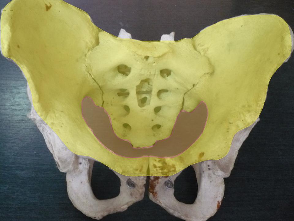
|
Anterior Branch Of The Obturator Artery
The obturator artery is a branch of the internal iliac artery that passes antero-inferiorly (forwards and downwards) on the lateral wall of the pelvis, to the upper part of the obturator foramen, and, escaping from the pelvic cavity through the obturator canal, it divides into both an anterior and a posterior branch. Structure In the pelvic cavity this vessel is in relation, laterally, with the obturator fascia; medially, with the ureter, ductus deferens, and peritoneum; while a little below it is the obturator nerve. The obturator artery usually arises from the internal iliac artery. Inside the pelvis the obturator artery gives off iliac branches to the iliac fossa, which supply the bone and the Iliacus, and anastomose with the ilio-lumbar artery; a vesical branch, which runs backward to supply the bladder; and a pubic branch, which is given off from the vessel just before it leaves the pelvic cavity. The pubic branch ascends upon the back of the pubis, communicating with the co ... [...More Info...] [...Related Items...] OR: [Wikipedia] [Google] [Baidu] |
Femoral Ring
The femoral ring is the opening at the proximal, abdominal end of the femoral canal, and represents the (superiorly directed/oriented) base of the conically-shaped femoral canal. The femoral ring is oval-shaped, with its long diameter being directed transversely and measuring about 1.25 cm.'''' The opening of the femoral ring is filled in by extraperitoneal fat, forming the femoral septum. Part of the intestine can sometimes pass through the femoral ring into the femoral canal causing a femoral hernia. Boundaries The femoral ring is bounded as follows:'''' * anteriorly by the inguinal ligament. * posteriorly by the pectineal ligament. * medially by the crescentic base of the lacunar ligament. * laterally by the fibrous septum on the medial side of the femoral vein. Additional images File:Gray1227.png, Front of abdomen, showing surface markings for arteries and inguinal canal. See also * Femoral canal * Femoral hernia * Inguinal canal The inguinal canals are the tw ... [...More Info...] [...Related Items...] OR: [Wikipedia] [Google] [Baidu] |
Peritoneum
The peritoneum is the serous membrane forming the lining of the abdominal cavity or coelom in amniotes and some invertebrates, such as annelids. It covers most of the intra-abdominal (or coelomic) organs, and is composed of a layer of mesothelium supported by a thin layer of connective tissue. This peritoneal lining of the cavity supports many of the abdominal organs and serves as a conduit for their blood vessels, lymphatic vessels, and nerves. The abdominal cavity (the space bounded by the vertebrae, abdominal muscles, diaphragm, and pelvic floor) is different from the intraperitoneal space (located within the abdominal cavity but wrapped in peritoneum). The structures within the intraperitoneal space are called "intraperitoneal" (e.g., the stomach and intestines), the structures in the abdominal cavity that are located behind the intraperitoneal space are called "retroperitoneal" (e.g., the kidneys), and those structures below the intraperitoneal space are called "subp ... [...More Info...] [...Related Items...] OR: [Wikipedia] [Google] [Baidu] |
Gracilis Muscle
The gracilis muscle (; Latin for "slender") is the most superficial muscle on the medial side of the thigh. It is thin and flattened, broad above, narrow and tapering below. Structure It arises by a thin aponeurosis from the anterior margins of the lower half of the symphysis pubis and the upper half of the pubic arch. The muscle's fibers run vertically downward, ending in a rounded tendon. This tendon passes behind the medial condyle of the femur, curves around the medial condyle of the tibia where it becomes flattened, and inserts into the upper part of the medial surface of the body of the tibia, below the condyle. For this reason, the muscle is a lower limb adductor. At its insertion the tendon is situated immediately above that of the semitendinosus muscle, and its upper edge is overlapped by the tendon of the sartorius muscle, which it joins to form the pes anserinus. The pes anserinus is separated from the medial collateral ligament of the knee-joint by a bursa. A ... [...More Info...] [...Related Items...] OR: [Wikipedia] [Google] [Baidu] |
Adductor Muscles Of The Hip
The adductor muscles of the hip are a group of muscles mostly used for bringing the thighs together (called adduction). Structure The adductor group is made up of: *Adductor brevis *Adductor longus *Adductor magnus * Adductor minimus This is often considered to be a part of adductor magnus. * pectineus * gracilis *Obturator externusPlatzer, Werner (2004), Color Atlas of Human Anatomy, Vol. 1, Locomotor System', Thieme, 5th ed, p 240 and are also part of the medial compartment of thigh The adductors originate on the pubis and ischium bones and insert mainly on the medial posterior surface of the femur. Nerve supply The pectineus is the only adductor muscle that is innervated by the femoral nerve. The other adductor muscles are innervated by the obturator nerve with the exception of a small part of the adductor magnus which is innervated by the tibial nerve. Variation In 33% of people a supernumerary muscle is found between the adductor brevis and adductor minimus. When ... [...More Info...] [...Related Items...] OR: [Wikipedia] [Google] [Baidu] |
Pectineus
The pectineus muscle (, from the Latin word ''pecten'', meaning comb) is a flat, quadrangular muscle, situated at the anterior (front) part of the upper and medial (inner) aspect of the thigh. The pectineus muscle is the most anterior adductor of the hip. The muscle does adduct and internally rotate the thigh but its primary function is hip flexion. It can be classified in the medial compartment of thigh (when the function is emphasized) or the anterior compartment of thigh (when the nerve is emphasized). Structure The pectineus muscle arises from the pectineal line of the pubis and to a slight extent from the surface of bone in front of it, between the iliopectineal eminence and pubic tubercle, and from the fascia covering the anterior surface of the muscle; the fibers pass downward, backward, and lateral, to be inserted into the pectineal line of the femur which leads from the lesser trochanter to the linea aspera. Relations The pectineus is in relation by its anterior sur ... [...More Info...] [...Related Items...] OR: [Wikipedia] [Google] [Baidu] |
Obturator Membrane , i. e., within the margin.
Both obturator muscles are connected with this membrane ...
The obturator membrane is a thin fibrous sheet, which almost completely closes the obturator foramen. Its fibers are arranged in interlacing bundles mainly transverse in direction; the uppermost bundle is attached to the obturator tubercles and completes the obturator canal for the passage of the obturator vessels and nerve. The membrane is attached to the sharp margin of the obturator foramen except at its lower lateral angle, where it is fixed to the pelvic surface of the inferior ramus of the ischium The ischium () form ... [...More Info...] [...Related Items...] OR: [Wikipedia] [Google] [Baidu] |
Artery
An artery (plural arteries) () is a blood vessel in humans and most animals that takes blood away from the heart to one or more parts of the body (tissues, lungs, brain etc.). Most arteries carry oxygenated blood; the two exceptions are the pulmonary and the umbilical arteries, which carry deoxygenated blood to the organs that oxygenate it (lungs and placenta, respectively). The effective arterial blood volume is that extracellular fluid which fills the arterial system. The arteries are part of the circulatory system, that is responsible for the delivery of oxygen and nutrients to all cells, as well as the removal of carbon dioxide and waste products, the maintenance of optimum blood pH, and the circulation of proteins and cells of the immune system. Arteries contrast with veins, which carry blood back towards the heart. Structure The anatomy of arteries can be separated into gross anatomy, at the macroscopic level, and microanatomy, which must be studied with a microscop ... [...More Info...] [...Related Items...] OR: [Wikipedia] [Google] [Baidu] |
Obturator Externus
The external obturator muscle, obturator externus muscle (; OE) is a flat, triangular muscle, which covers the outer surface of the anterior wall of the pelvis. It is sometimes considered part of the medial compartment of thigh, and sometimes considered part of the gluteal region. Structure It arises from the margin of bone immediately around the medial side of the obturator membrane and surrounding bone, viz., from the inferior pubic ramus, and the ramus of the ischium; it also arises from the medial two-thirds of the outer surface of the obturator membrane, and from the tendinous arch which completes the canal for the passage of the obturator vessels and nerves. The fibers springing from the pubic arch extend on to the inner surface of the bone, where they obtain a narrow origin between the margin of the foramen and the attachment of the obturator membrane. The fibers converge and pass posterolateral and upward, and end in a tendon which runs across the back of the neck of ... [...More Info...] [...Related Items...] OR: [Wikipedia] [Google] [Baidu] |
Inferior Epigastric Artery
In human anatomy, inferior epigastric artery refers to the artery that arises from the external iliac artery. It anastomoses with the superior epigastric artery. Along its course, it is accompanied by a similarly named vein, the inferior epigastric vein. These epigastric vessels form the lateral border of Hesselbach's triangle, which outlines the area through which direct inguinal hernias protrude. Structure Origin The inferior epigastric artery arises from the external iliac artery, immediately superior to the inguinal ligament. Course and relations It curves forward in the subperitoneal tissue, and then ascends obliquely along the medial margin of the abdominal inguinal ring; continuing its course upward, it pierces the transversalis fascia, and, passing in front of the linea semicircularis, ascends between the rectus abdominis muscle and the posterior lamella of its sheath. It finally divides into numerous branches, which anastomose, above the umbilicus, with the sup ... [...More Info...] [...Related Items...] OR: [Wikipedia] [Google] [Baidu] |
Pelvic Cavity
The pelvic cavity is a body cavity that is bounded by the bones of the pelvis. Its oblique roof is the pelvic inlet (the superior opening of the pelvis). Its lower boundary is the pelvic floor. The pelvic cavity primarily contains the reproductive organs, urinary bladder, distal ureters, proximal urethra, terminal sigmoid colon, rectum, and anal canal. In females, the uterus, Fallopian tubes, ovaries and upper vagina occupy the area between the other viscera. The rectum is located at the back of the pelvis, in the curve of the sacrum and coccyx; the bladder is in front, behind the pubic symphysis. The pelvic cavity also contains major arteries, veins, muscles, and nerves. These structures coexist in a crowded space, and disorders of one pelvic component may impact upon another; for example, constipation may overload the rectum and compress the urinary bladder, or childbirth might damage the pudendal nerves and later lead to anal weakness. Structure The pelvis has an antero ... [...More Info...] [...Related Items...] OR: [Wikipedia] [Google] [Baidu] |
Urinary Bladder
The urinary bladder, or simply bladder, is a hollow organ in humans and other vertebrates that stores urine from the kidneys before disposal by urination. In humans the bladder is a distensible organ that sits on the pelvic floor. Urine enters the bladder via the ureters and exits via the urethra. The typical adult human bladder will hold between 300 and (10.14 and ) before the urge to empty occurs, but can hold considerably more. The Latin phrase for "urinary bladder" is ''vesica urinaria'', and the term ''vesical'' or prefix ''vesico -'' appear in connection with associated structures such as vesical veins. The modern Latin word for "bladder" – ''cystis'' – appears in associated terms such as cystitis (inflammation of the bladder). Structure In humans, the bladder is a hollow muscular organ situated at the base of the pelvis. In gross anatomy, the bladder can be divided into a broad , a body, an apex, and a neck. The apex (also called the vertex) is directed forward ... [...More Info...] [...Related Items...] OR: [Wikipedia] [Google] [Baidu] |
Ilio-lumbar Artery
The iliolumbar artery is the first branch of the posterior trunk of the internal iliac artery. Structure The iliolumbar artery is the first branch of the posterior trunk of the internal iliac artery. It turns upward behind the obturator nerve and the external iliac artery and vein, to the medial border of the psoas major muscle, behind which it divides into: * Lumbar branch of iliolumbar artery * Iliac branch of iliolumbar artery The iliac branch of the iliolumbar artery (ramus iliacus) descends to supply the iliacus muscle; some offsets, running between the muscle and the bone, anastomose with the iliac branches of the obturator artery; one of these enters an oblique canal ... Anastomoses *1. Last lumbar→iliolumbar *2. Lateral sacral↔lateral sacral *3. Middle sacral→lateral sacral *4. Superior hemorrhoidal→middle hemorrhoidal *5. Medial femoral circumflex→inferior gluteal *6. Medial femoral circumflex↔obturator *7. Lateral femoral circumflex→superior gluteal *8. Dee ... [...More Info...] [...Related Items...] OR: [Wikipedia] [Google] [Baidu] |


