|
Anoplosaurus
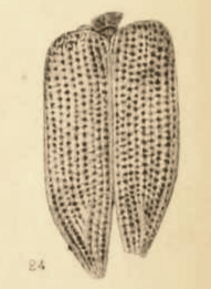
''Anoplosaurus'' (meaning "unarmored or unarmed lizard") is an extinct genus of herbivorous nodosaurid dinosaur, from the late Albian-age Lower Cretaceous Cambridge Greensand of Cambridgeshire, England. It has in the past been classified with either the armored dinosaurs or the ornithopods, but current thought has been in agreement with the "armored dinosaur" interpretation, placing it in the Ankylosauria. History Harry Govier Seeley named this genus in 1879 for a disarticulated partial postcranial skeleton that had been uncovered at Reach, Cambridgeshire, composed of a left dentary fragment, numerous vertebrae from the neck, back, and sacrum, parts of the pectoral girdle, humerus fragments, part of the left femur, left tibia, foot bones, ribs, and other fragments. He regarded it as possibly juvenile, due to its small size, with a length of about five feet. The type species is ''Anoplosaurus curtonotus''. The generic name, derived from the Greek ''hoplo''~, a word element ... [...More Info...] [...Related Items...] OR: [Wikipedia] [Google] [Baidu] |
Cambridge Greensand
The Cambridge Greensand is a geological unit in England whose strata are earliest Cenomanian in age. It lies above the erosive contact between the Gault Formation and the Chalk Group in the vicinity of Cambridgeshire, and technically forms the lowest member bed of the West Melbury Marly Chalk Formation.Cambridge Greensand at BGS It is a remanié deposit, containing reworked fossils of late age, including those of dinosaurs and pterosaurs. Description The lithology is made out of |
Nodosaurid
Nodosauridae is a family of ankylosaurian dinosaurs, from the Late Jurassic to the Late Cretaceous period in what is now North America, South America, Europe, and Asia. Description Nodosaurids, like their close relatives the ankylosaurids, were heavily armored dinosaurs adorned with rows of bony armor nodules and spines (osteoderms), which were covered in keratin sheaths. All nodosaurids, like other ankylosaurians, were medium-sized to large, heavily built, quadrupedal, herbivorous dinosaurs, possessing small, leaf-shaped teeth. Unlike ankylosaurids, nodosaurids lacked mace-like tail clubs, instead having flexible tail tips. Many nodosaurids had spikes projecting outward from their shoulders. One particularly well-preserved nodosaurid "mummy", known as the Suncor nodosaur (''Borealopelta markmitchelli''), preserved a nearly complete set of armor in life position, as well as the keratin covering and mineralized remains of the underlying skin, which indicate reddish dorsal pigmen ... [...More Info...] [...Related Items...] OR: [Wikipedia] [Google] [Baidu] |
1879 In Paleontology
Plants Ferns Arthropods Insects Ichthyosauromorpha Ichthyosaurs Archosauromorphs Newly named non-avian dinosaurs Synapsids "Pelycosaurians" See also References {{Reflist 1870s in paleontology Paleontology, 1879 In ... [...More Info...] [...Related Items...] OR: [Wikipedia] [Google] [Baidu] |
Foot
The foot ( : feet) is an anatomical structure found in many vertebrates. It is the terminal portion of a limb which bears weight and allows locomotion. In many animals with feet, the foot is a separate organ at the terminal part of the leg made up of one or more segments or bones, generally including claws or nails. Etymology The word "foot", in the sense of meaning the "terminal part of the leg of a vertebrate animal" comes from "Old English fot "foot," from Proto-Germanic *fot (source also of Old Frisian fot, Old Saxon fot, Old Norse fotr, Danish fod, Swedish fot, Dutch voet, Old High German fuoz, German Fuß, Gothic fotus "foot"), from PIE root *ped- "foot". The "plural form feet is an instance of i-mutation." Structure The human foot is a strong and complex mechanical structure containing 26 bones, 33 joints (20 of which are actively articulated), and more than a hundred muscles, tendons, and ligaments.Podiatry Channel, ''Anatomy of the foot and ankle'' The joints of the ... [...More Info...] [...Related Items...] OR: [Wikipedia] [Google] [Baidu] |
Sacrum
The sacrum (plural: ''sacra'' or ''sacrums''), in human anatomy, is a large, triangular bone at the base of the spine that forms by the fusing of the sacral vertebrae (S1S5) between ages 18 and 30. The sacrum situates at the upper, back part of the pelvic cavity, between the two wings of the pelvis. It forms joints with four other bones. The two projections at the sides of the sacrum are called the alae (wings), and articulate with the ilium at the L-shaped sacroiliac joints. The upper part of the sacrum connects with the last lumbar vertebra (L5), and its lower part with the coccyx (tailbone) via the sacral and coccygeal cornua. The sacrum has three different surfaces which are shaped to accommodate surrounding pelvic structures. Overall it is concave (curved upon itself). The base of the sacrum, the broadest and uppermost part, is tilted forward as the sacral promontory internally. The central part is curved outward toward the posterior, allowing greater room for the pel ... [...More Info...] [...Related Items...] OR: [Wikipedia] [Google] [Baidu] |
Pectoral Girdle
The shoulder girdle or pectoral girdle is the set of bones in the appendicular skeleton which connects to the arm on each side. In humans it consists of the clavicle and scapula; in those species with three bones in the shoulder, it consists of the clavicle, scapula, and coracoid. Some mammalian species (such as the dog and the horse) have only the scapula. The pectoral girdles are to the upper limbs as the pelvic girdle is to the lower limbs; the girdles are the parts of the appendicular skeleton that anchor the appendages to the axial skeleton. In humans, the only true anatomical joints between the shoulder girdle and the axial skeleton are the sternoclavicular joints on each side. No anatomical joint exists between each scapula and the rib cage; instead the muscular connection or physiological joint between the two permits great mobility of the shoulder girdle compared to the compact pelvic girdle; because the upper limb is not usually involved in weight bearing, its stabilit ... [...More Info...] [...Related Items...] OR: [Wikipedia] [Google] [Baidu] |
Humerus
The humerus (; ) is a long bone in the arm that runs from the shoulder to the elbow. It connects the scapula and the two bones of the lower arm, the radius and ulna, and consists of three sections. The humeral upper extremity consists of a rounded head, a narrow neck, and two short processes (tubercles, sometimes called tuberosities). The body is cylindrical in its upper portion, and more prismatic below. The lower extremity consists of 2 epicondyles, 2 processes (trochlea & capitulum), and 3 fossae (radial fossa, coronoid fossa, and olecranon fossa). As well as its true anatomical neck, the constriction below the greater and lesser tubercles of the humerus is referred to as its surgical neck due to its tendency to fracture, thus often becoming the focus of surgeons. Etymology The word "humerus" is derived from la, humerus, umerus meaning upper arm, shoulder, and is linguistically related to Gothic ''ams'' shoulder and Greek ''ōmos''. Structure Upper extremity The upper or pr ... [...More Info...] [...Related Items...] OR: [Wikipedia] [Google] [Baidu] |
Femur
The femur (; ), or thigh bone, is the proximal bone of the hindlimb in tetrapod vertebrates. The head of the femur articulates with the acetabulum in the pelvic bone forming the hip joint, while the distal part of the femur articulates with the tibia (shinbone) and patella (kneecap), forming the knee joint. By most measures the two (left and right) femurs are the strongest bones of the body, and in humans, the largest and thickest. Structure The femur is the only bone in the upper leg. The two femurs converge medially toward the knees, where they articulate with the proximal ends of the tibiae. The angle of convergence of the femora is a major factor in determining the femoral-tibial angle. Human females have thicker pelvic bones, causing their femora to converge more than in males. In the condition ''genu valgum'' (knock knee) the femurs converge so much that the knees touch one another. The opposite extreme is ''genu varum'' (bow-leggedness). In the general populatio ... [...More Info...] [...Related Items...] OR: [Wikipedia] [Google] [Baidu] |
Tibia
The tibia (; ), also known as the shinbone or shankbone, is the larger, stronger, and anterior (frontal) of the two bones in the leg below the knee in vertebrates (the other being the fibula, behind and to the outside of the tibia); it connects the knee with the ankle. The tibia is found on the medial side of the leg next to the fibula and closer to the median plane. The tibia is connected to the fibula by the interosseous membrane of leg, forming a type of fibrous joint called a syndesmosis with very little movement. The tibia is named for the flute ''tibia''. It is the second largest bone in the human body, after the femur. The leg bones are the strongest long bones as they support the rest of the body. Structure In human anatomy, the tibia is the second largest bone next to the femur. As in other vertebrates the tibia is one of two bones in the lower leg, the other being the fibula, and is a component of the knee and ankle joints. The ossification or formation of the bone ... [...More Info...] [...Related Items...] OR: [Wikipedia] [Google] [Baidu] |
Albian
The Albian is both an age of the geologic timescale and a stage in the stratigraphic column. It is the youngest or uppermost subdivision of the Early/Lower Cretaceous Epoch/Series. Its approximate time range is 113.0 ± 1.0 Ma to 100.5 ± 0.9 Ma (million years ago). The Albian is preceded by the Aptian and followed by the Cenomanian. Stratigraphic definitions The Albian Stage was first proposed in 1842 by Alcide d'Orbigny. It was named after Alba, the Latin name for River Aube in France. A Global Boundary Stratotype Section and Point (GSSP), ratified by the IUGS in 2016, defines the base of the Albian as the first occurrence of the planktonic foraminiferan '' Microhedbergella renilaevis'' at the Col de Pré-Guittard section, Arnayon, Drôme, France. The top of the Albian Stage (the base of the Cenomanian Stage and Upper Cretaceous Series) is defined as the place where the foram species '' Rotalipora globotruncanoides'' first appears in the stratigraphic column. The Albia ... [...More Info...] [...Related Items...] OR: [Wikipedia] [Google] [Baidu] |
Juvenile (organism)
A juvenile is an individual organism that has not yet reached its adult form, sexual maturity or size. Juveniles can look very different from the adult form, particularly in colour, and may not fill the same niche as the adult form. In many organisms the juvenile has a different name from the adult (see List of animal names). Some organisms reach sexual maturity in a short metamorphosis, such as eclosion in many insects. For others, the transition from juvenile to fully mature is a more prolonged process—puberty in humans and other species, for example. In such cases, juveniles during this transformation are sometimes called subadults. Many invertebrates, on reaching the adult stage, are fully mature and their development and growth stops. Their juveniles are larvae or nymphs. In vertebrates and some invertebrates (e.g. spiders), larval forms (e.g. tadpoles) are usually considered a development stage of their own, and "juvenile" refers to a post-larval stage that is not full ... [...More Info...] [...Related Items...] OR: [Wikipedia] [Google] [Baidu] |
Neck
The neck is the part of the body on many vertebrates that connects the head with the torso. The neck supports the weight of the head and protects the nerves that carry sensory and motor information from the brain down to the rest of the body. In addition, the neck is highly flexible and allows the head to turn and flex in all directions. The structures of the human neck are anatomically grouped into four compartments; vertebral, visceral and two vascular compartments. Within these compartments, the neck houses the cervical vertebrae and cervical part of the spinal cord, upper parts of the respiratory and digestive tracts, endocrine glands, nerves, arteries and veins. Muscles of the neck are described separately from the compartments. They bound the neck triangles. In anatomy, the neck is also called by its Latin names, or , although when used alone, in context, the word ''cervix'' more often refers to the uterine cervix, the neck of the uterus. Thus the adjective ''cervical'' ma ... [...More Info...] [...Related Items...] OR: [Wikipedia] [Google] [Baidu] |


