|
Annular Ligaments Of Fingers
In human anatomy, the annular ligaments of the fingers, often referred to as A pulleys, are the :wikt:annular, annular part of the fibrous sheathes of the fingers. Four or five such annular pulleys, together with three Cruciate ligaments of fingers, cruciate pulleys, form a fibro-osseous tunnel on the palmar aspect of the hand through which passes the Flexor digitorum profundis muscle, deep and Flexor digitorum superficialis muscle, superficial flexor tendons. The annular and cruciate ligaments serve to govern the flexor mechanism of the hand and wrist, providing critical constraints to the flexor tendons to prevent bowstringing upon contraction and excursion of extrinsic flexor musculo-tendinous units. The first annular pulley (A1 pulley), near the head of the metacarpal bone, lies in the flexor groove in the deep transverse metacarpal ligament. As a general rule, the A1, A3, and A5 pulleys in the fingers are "joint pulleys" that originate from the Palmar plate, volar plate on th ... [...More Info...] [...Related Items...] OR: [Wikipedia] [Google] [Baidu] |
Phalanx Bones
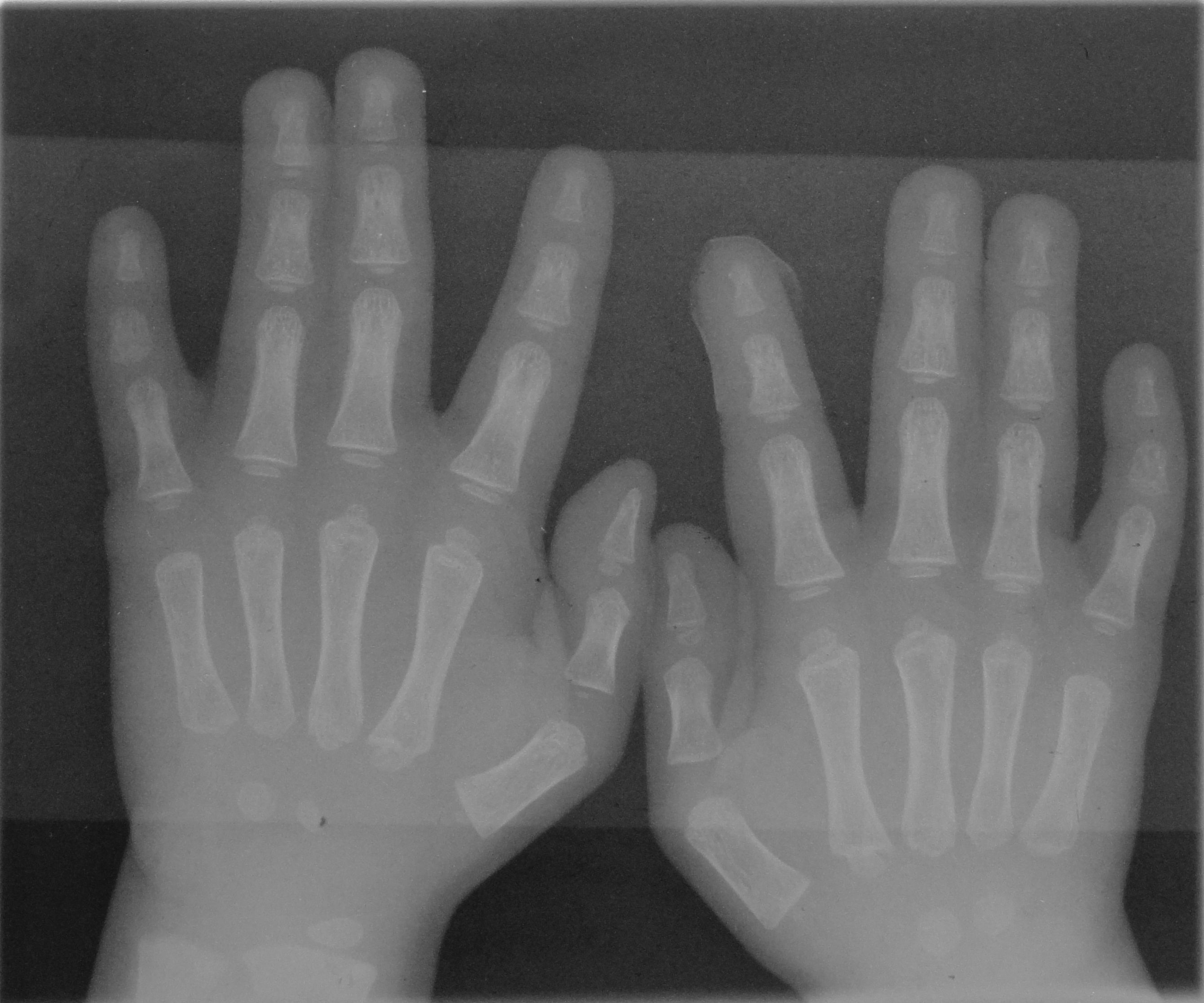
The phalanges (singular: ''phalanx'' ) are digital bones in the hands and feet of most vertebrates. In primates, the thumbs and big toes have two phalanges while the other digits have three phalanges. The phalanges are classed as long bones. Structure The phalanges are the bones that make up the fingers of the hand and the toes of the foot. There are 56 phalanges in the human body, with fourteen on each hand and foot. Three phalanges are present on each finger and toe, with the exception of the thumb and large toe, which possess only two. The middle and far phalanges of the fifth toes are often fused together (symphalangism). The phalanges of the hand are commonly known as the finger bones. The phalanges of the foot differ from the hand in that they are often shorter and more compressed, especially in the proximal phalanges, those closest to the torso. A phalanx is named according to whether it is proximal, middle, or distal and its associated finger or toe. The proximal ... [...More Info...] [...Related Items...] OR: [Wikipedia] [Google] [Baidu] |
Human Anatomy
The human body is the structure of a human being. It is composed of many different types of cells that together create tissues and subsequently organ systems. They ensure homeostasis and the viability of the human body. It comprises a head, hair, neck, trunk (which includes the thorax and abdomen), arms and hands, legs and feet. The study of the human body involves anatomy, physiology, histology and embryology. The body varies anatomically in known ways. Physiology focuses on the systems and organs of the human body and their functions. Many systems and mechanisms interact in order to maintain homeostasis, with safe levels of substances such as sugar and oxygen in the blood. The body is studied by health professionals, physiologists, anatomists, and by artists to assist them in their work. Composition The human body is composed of elements including hydrogen, oxygen, carbon, calcium and phosphorus. These elements reside in trillions of cells and non-cellula ... [...More Info...] [...Related Items...] OR: [Wikipedia] [Google] [Baidu] |
Finger
A finger is a limb of the body and a type of digit, an organ of manipulation and sensation found in the hands of most of the Tetrapods, so also with humans and other primates. Most land vertebrates have five fingers ( Pentadactyly). Chambers 1998 p. 603 Oxford Illustrated pp. 311, 380 Land vertebrate fingers The five-rayed anterior limbs of terrestrial vertebrates can be derived phylogenetically from the pectoral fins of fish. Within the taxa of the terrestrial vertebrates, the basic pentadactyl plan, and thus also the fingers and phalanges, undergo many variations. Morphologically the different fingers of terrestrial vertebrates are homolog. The wings of birds and those of bats are not homologous, they are analogue flight organs. However, the phalanges within them are homologous. Chimpanzees have lower limbs that are specialized for manipulation, and (arguably) have fingers on their lower limbs as well. In the case of Primates in general, the digits of the hand a ... [...More Info...] [...Related Items...] OR: [Wikipedia] [Google] [Baidu] |
Cruciate Ligaments Of Fingers
{{disambig ...
Cruciate, and similar words, can mean: *The cruciate ligaments in the knee *For a magic spell in the Harry Potter scenario, see crucio *Latin and early-English word for crusade The Crusades were a series of religious wars initiated, supported, and sometimes directed by the Latin Church in the medieval period. The best known of these Crusades are those to the Holy Land in the period between 1095 and 1291 that were i ... [...More Info...] [...Related Items...] OR: [Wikipedia] [Google] [Baidu] |
Flexor Digitorum Profundis Muscle
The flexor digitorum profundus is a muscle in the forearm of humans that flexes the fingers (also known as digits). It is considered an extrinsic hand muscle because it acts on the hand while its muscle belly is located in the forearm. Together the flexor pollicis longus, pronator quadratus, and flexor digitorum profundus form the deep layer of ventral forearm muscles.Platzer 2004, p 162 The muscle is named . Structure Flexor digitorum profundus originates in the upper 3/4 of the anterior and medial surfaces of the ulna, interosseous membrane and deep fascia of the forearm. The muscle fans out into four tendons (one to each of the second to fifth fingers) to the palmar base of the distal phalanx. Along with the flexor digitorum superficialis, it has long tendons that run down the arm and through the carpal tunnel and attach to the palmar side of the phalanges of the fingers. Flexor digitorum profundus lies deep to the superficialis, but it attaches more distally. Therefore, p ... [...More Info...] [...Related Items...] OR: [Wikipedia] [Google] [Baidu] |
Flexor Digitorum Superficialis Muscle
Flexor digitorum superficialis (''flexor digitorum sublimis'') is an extrinsic flexor muscle of the fingers at the proximal interphalangeal joints. It is in the anterior compartment of the forearm. It is sometimes considered to be the deepest part of the superficial layer of this compartment, and sometimes considered to be a distinct, "intermediate layer" of this compartment. It is relatively common for the Flexor digitorum superficialis to be missing from the little finger, bilaterally and unilaterally, which can cause problems when diagnosing a little finger injury. Structure The muscle has two classically described heads – the humeroulnar and radial – and it is between these heads that the median nerve and ulnar artery pass. The ulnar collateral ligament of elbow joint gives its origin to part of this muscle. Four long tendons come off this muscle near the wrist and travel through the carpal tunnel formed by the flexor retinaculum. These tendons, along with those of flex ... [...More Info...] [...Related Items...] OR: [Wikipedia] [Google] [Baidu] |
Metacarpal Bone
In human anatomy, the metacarpal bones or metacarpus form the intermediate part of the skeletal hand located between the phalanges of the fingers and the carpal bones of the wrist, which forms the connection to the forearm. The metacarpal bones are analogous to the metatarsal bones in the foot. Structure The metacarpals form a transverse arch to which the rigid row of distal carpal bones are fixed. The peripheral metacarpals (those of the thumb and little finger) form the sides of the cup of the palmar gutter and as they are brought together they deepen this concavity. The index metacarpal is the most firmly fixed, while the thumb metacarpal articulates with the trapezium and acts independently from the others. The middle metacarpals are tightly united to the carpus by intrinsic interlocking bone elements at their bases. The ring metacarpal is somewhat more mobile while the fifth metacarpal is semi-independent.Tubiana ''et al'' 1998, p 11 Each metacarpal bone consists of a body o ... [...More Info...] [...Related Items...] OR: [Wikipedia] [Google] [Baidu] |
Deep Transverse Metacarpal Ligament
The deep transverse metacarpal ligament (also called the deep transverse palmar ligament) is a narrow fibrous band which runs across the palmar surfaces of the heads of the second, third, fourth and fifth metacarpal bones, connecting them together. Structure The deep transverse metacarpal ligament connects the palmar surfaces of the heads of the second, third, fourth, and fifth metacarpal bones. It is blended with the palmar metacarpophalangeal ligaments. Its palmar surface is concave where the flexor tendons pass over it. Behind it, the tendons of the interosseous muscles of the hand The interosseous muscles of the hand are muscles found near the metacarpal bones that help to control the fingers. They are considered voluntary muscles. They are generally divided into two sets: * 4 Dorsal interossei - Abduct the digits away f ... pass to their insertions. Clinical significance Rarely, the deep transverse metacarpal ligament may rupture. Additional Images File:Dissecti ... [...More Info...] [...Related Items...] OR: [Wikipedia] [Google] [Baidu] |
Palmar Plate
In the human hand, palmar or volar plates (also referred to as palmar or volar ligaments) are found in the metacarpophalangeal (MCP) and interphalangeal (IP) joints, where they reinforce the joint capsules, enhance joint stability, and limit hyperextension. The plates of the MCP and IP joints are structurally and functionally similar, except that in the MCP joints they are interconnected by a deep transverse ligament. In the MCP joints, they also indirectly provide stability to the longitudinal palmar arches of the hand. (MCP joints) (IP joints) The volar plate of the thumb MCP joint has a transverse longitudinal rectangular shape, shorter than those in the fingers. Structure This fibrocartilaginous structure is attached to the volar base of the phalanx distal to the joint. From there, it forms a palmar continuation of the articular surface of the phalanx bone and its inner surface thus adds to the articular surface during extension. In its proximal end, the volar plat ... [...More Info...] [...Related Items...] OR: [Wikipedia] [Google] [Baidu] |
Metacarpophalangeal Joint
The metacarpophalangeal joints (MCP) are situated between the metacarpal bones and the proximal phalanges of the fingers. These joints are of the condyloid kind, formed by the reception of the rounded heads of the metacarpal bones into shallow cavities on the proximal ends of the proximal phalanges. Being condyloid, they allow the movements of flexion, extension, abduction, adduction and circumduction at the joint. Structure Ligaments Each joint has: * palmar ligaments of metacarpophalangeal articulations * collateral ligaments of metacarpophalangeal articulations Dorsal surfaces The dorsal surfaces of these joints are covered by the expansions of the Extensor tendons, together with some loose areolar tissue which connects the deep surfaces of the tendons to the bones. Function The movements which occur in these joints are flexion, extension, adduction, abduction, and circumduction; the movements of abduction and adduction are very limited, and cannot be performed while th ... [...More Info...] [...Related Items...] OR: [Wikipedia] [Google] [Baidu] |
Interphalangeal Articulations Of Hand
The interphalangeal joints of the hand are the hinge joints between the phalanges of the fingers that provide flexion towards the palm of the hand. There are two sets in each finger (except in the thumb, which has only one joint): * "proximal interphalangeal joints" (PIJ or PIP), those between the first (also called proximal) and second (intermediate) phalanges * "distal interphalangeal joints" (DIJ or DIP), those between the second (intermediate) and third (distal) phalanges Anatomically, the proximal and distal interphalangeal joints are very similar. There are some minor differences in how the palmar plates are attached proximally and in the segmentation of the flexor tendon sheath, but the major differences are the smaller dimension and reduced mobility of the distal joint. Joint structure The PIP joint exhibits great lateral stability. Its transverse diameter is greater than its antero-posterior diameter and its thick collateral ligaments are tight in all positions duri ... [...More Info...] [...Related Items...] OR: [Wikipedia] [Google] [Baidu] |


_dorsal_view.png)
