|
Anatomical Snuff Box
The anatomical snuff box or snuffbox or foveola radialis is a triangular deepening on the radial, dorsal aspect of the hand—at the level of the carpal bones, specifically, the scaphoid and trapezium bones forming the floor. The name originates from the use of this surface for placing and then sniffing powdered tobacco, or " snuff." It is sometimes referred to by its French name ''tabatière''. Structure Boundaries * The medial border (ulnar side) of the snuffbox is the tendon of the extensor pollicis longus. * The lateral border (radial side) is a pair of parallel and intimate tendons, of the extensor pollicis brevis and the abductor pollicis longus. (Accordingly, the anatomical snuffbox is most visible, having a more pronounced concavity, during thumb extension.) * The proximal border is formed by the styloid process of the radius * The distal border is formed by the approximate apex of the schematic snuffbox isosceles triangle. * The floor of the snuffbox varies de ... [...More Info...] [...Related Items...] OR: [Wikipedia] [Google] [Baidu] |
Radial Artery
In human anatomy, the radial artery is the main artery of the lateral aspect of the forearm. Structure The radial artery arises from the bifurcation of the brachial artery in the antecubital fossa. It runs distally on the anterior part of the forearm. There, it serves as a landmark for the division between the anterior and posterior compartments of the forearm, with the posterior compartment beginning just lateral to the artery. The artery winds laterally around the wrist, passing through the anatomical snuff box and between the heads of the first dorsal interosseous muscle. It passes anteriorly between the heads of the adductor pollicis, and becomes the deep palmar arch, which joins with the deep branch of the ulnar artery. Along its course, it is accompanied by a similarly named vein, the radial vein. Branches The named branches of the radial artery may be divided into three groups, corresponding with the three regions in which the vessel is situated. In the for ... [...More Info...] [...Related Items...] OR: [Wikipedia] [Google] [Baidu] |
Extension (kinesiology)
Motion, the process of movement, is described using specific anatomical terms. Motion includes movement of organs, joints, limbs, and specific sections of the body. The terminology used describes this motion according to its direction relative to the anatomical position of the body parts involved. Anatomists and others use a unified set of terms to describe most of the movements, although other, more specialized terms are necessary for describing unique movements such as those of the hands, feet, and eyes. In general, motion is classified according to the anatomical plane it occurs in. ''Flexion'' and ''extension'' are examples of ''angular'' motions, in which two axes of a joint are brought closer together or moved further apart. ''Rotational'' motion may occur at other joints, for example the shoulder, and are described as ''internal'' or ''external''. Other terms, such as ''elevation'' and ''depression'', describe movement above or below the horizontal plane. Many anato ... [...More Info...] [...Related Items...] OR: [Wikipedia] [Google] [Baidu] |
Anatomical Terms Of Bone
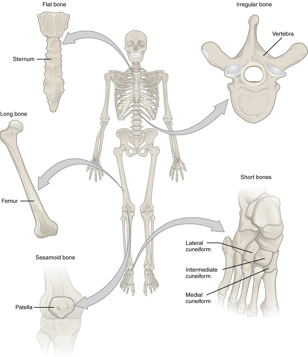
Many anatomical terms descriptive of bone are defined in anatomical terminology, and are often derived from Greek and Latin. Bone in the human body is categorized into long bone, short bone, flat bone, irregular bone and sesamoid bone. Types of bone Long bones A long bone is one that is cylindrical in shape, being longer than it is wide. However, the term describes the shape of a bone, not its size, which is relative. Long bones are found in the arms (humerus, ulna, radius) and legs (femur, tibia, fibula), as well as in the fingers (metacarpals, phalanges) and toes ( metatarsals, phalanges). Long bones function as levers; they move when muscles contract. They are responsible for the body's height. Short bones A short bone is one that is cube-like in shape, being approximately equal in length, width, and thickness. The only short bones in the human skeleton are in the carpals of the wrists and the tarsals of the ankles. Short bones provide stability and support as well as som ... [...More Info...] [...Related Items...] OR: [Wikipedia] [Google] [Baidu] |
Anatomical Terminology
Anatomical terminology is a form of scientific terminology used by anatomists, zoologists, and health professionals such as doctors. Anatomical terminology uses many unique terms, suffixes, and prefixes deriving from Ancient Greek and Latin. These terms can be confusing to those unfamiliar with them, but can be more precise, reducing ambiguity and errors. Also, since these anatomical terms are not used in everyday conversation, their meanings are less likely to change, and less likely to be misinterpreted. To illustrate how inexact day-to-day language can be: a scar "above the wrist" could be located on the forearm two or three inches away from the hand or at the base of the hand; and could be on the palm-side or back-side of the arm. By using precise anatomical terminology such ambiguity is eliminated. An international standard for anatomical terminology, ''Terminologia Anatomica'' has been created. Word formation Anatomical terminology has quite regular morphology: the s ... [...More Info...] [...Related Items...] OR: [Wikipedia] [Google] [Baidu] |
Necrosis
Necrosis () is a form of cell injury which results in the premature death of cells in living tissue by autolysis. Necrosis is caused by factors external to the cell or tissue, such as infection, or trauma which result in the unregulated digestion of cell components. In contrast, apoptosis is a naturally occurring programmed and targeted cause of cellular death. While apoptosis often provides beneficial effects to the organism, necrosis is almost always detrimental and can be fatal. Cellular death due to necrosis does not follow the apoptotic signal transduction pathway, but rather various receptors are activated and result in the loss of cell membrane integrity and an uncontrolled release of products of cell death into the extracellular space. This initiates in the surrounding tissue an inflammatory response, which attracts leukocytes and nearby phagocytes which eliminate the dead cells by phagocytosis. However, microbial damaging substances released by leukocytes woul ... [...More Info...] [...Related Items...] OR: [Wikipedia] [Google] [Baidu] |
Scaphoid Fracture
A scaphoid fracture is a break of the scaphoid bone in the wrist. Symptoms generally includes pain at the base of the thumb which is worse with use of the hand. The anatomic snuffbox is generally tender and swelling may occur. Complications may include nonunion of the fracture, avascular necrosis of the proximal part of the bone, and arthritis. Scaphoid fractures are most commonly caused by a fall on an outstretched hand. Diagnosis is generally based on a combination of clinical examination and medical imaging. Some fractures may not be visible on plain X-rays. In such cases the affected area may be immobilised in a splint or cast and reviewed with repeat X-rays in two weeks, or alternatively an MRI or bone scan may be performed. The fracture may be preventable by using wrist guards during certain activities. In those in whom the fracture remains well aligned a cast is generally sufficient. If the fracture is displaced then surgery is generally recommended. Healing may take ... [...More Info...] [...Related Items...] OR: [Wikipedia] [Google] [Baidu] |
Tenderness (medicine)
In medicine, tenderness is pain or discomfort when an affected area is touched. It should not be confused with the pain that a patient perceives without touching. Pain is patient's perception, while tenderness is a sign that a clinician elicits. See also * Rebound tenderness, an indication of peritonitis Peritonitis is inflammation of the localized or generalized peritoneum, the lining of the inner wall of the abdomen and cover of the abdominal organs. Symptoms may include severe pain, swelling of the abdomen, fever, or weight loss. One part or .... References Pain {{Med-sign-stub ... [...More Info...] [...Related Items...] OR: [Wikipedia] [Google] [Baidu] |
Superficial Branch Of Radial Nerve
The superficial branch of the radial nerve passes along the front of the radial side of the forearm to the commencement of its lower third. It is a sensory nerve. It lies at first slightly lateral to the radial artery, concealed beneath the Brachioradialis. In the middle third of the forearm, it lies behind the same muscle, close to the lateral side of the artery. It quits the artery about 7 cm. above the wrist, passes beneath the tendon of the Brachioradialis, and, piercing the deep fascia, divides into two branches: lateral and medial. Structure Lateral branch The ''lateral branch'', the smaller, supplies the radial side of the thumb (by a digital nerve), the skin of the radial side and ball of the thumb, joining with the volar branch of the lateral antebrachial cutaneous nerve. Medial branch The ''medial branch'' communicates, above the wrist, with the dorsal branch of the lateral antebrachial cutaneous, and, on the back of the hand, with the dorsal branch of the u ... [...More Info...] [...Related Items...] OR: [Wikipedia] [Google] [Baidu] |
Lateral Cutaneous Nerve Of Forearm
The lateral antebrachial cutaneous nerve (or lateral cutaneous nerve of forearm) (branch of musculocutaneous nerve, also sometimes spelled "antebrachial") passes behind the cephalic vein, and divides, opposite the elbow-joint, into a volar and a dorsal branch. Volar branch The volar branch (ramus volaris; anterior branch) descends along the radial border of the forearm to the wrist, and supplies the skin over the lateral half of its volar surface. At the wrist-joint it is placed in front of the radial artery, and some filaments, piercing the deep fascia, accompany that vessel to the dorsal surface of the carpus. The nerve then passes downward to the ball of the thumb, where it ends in cutaneous filaments. It communicates with the superficial branch of the radial nerve, and with the palmar cutaneous branch of the median nerve. Dorsal branch The dorsal branch (ramus dorsalis; posterior branch) descends, along the dorsal surface of the radial side of the forearm to the wrist ... [...More Info...] [...Related Items...] OR: [Wikipedia] [Google] [Baidu] |
Superficial Branch Of Radial Nerve
The superficial branch of the radial nerve passes along the front of the radial side of the forearm to the commencement of its lower third. It is a sensory nerve. It lies at first slightly lateral to the radial artery, concealed beneath the Brachioradialis. In the middle third of the forearm, it lies behind the same muscle, close to the lateral side of the artery. It quits the artery about 7 cm. above the wrist, passes beneath the tendon of the Brachioradialis, and, piercing the deep fascia, divides into two branches: lateral and medial. Structure Lateral branch The ''lateral branch'', the smaller, supplies the radial side of the thumb (by a digital nerve), the skin of the radial side and ball of the thumb, joining with the volar branch of the lateral antebrachial cutaneous nerve. Medial branch The ''medial branch'' communicates, above the wrist, with the dorsal branch of the lateral antebrachial cutaneous, and, on the back of the hand, with the dorsal branch of the u ... [...More Info...] [...Related Items...] OR: [Wikipedia] [Google] [Baidu] |
Metacarpals
In human anatomy, the metacarpal bones or metacarpus form the intermediate part of the skeletal hand located between the phalanges of the fingers and the carpal bones of the wrist, which forms the connection to the forearm. The metacarpal bones are analogous to the metatarsal bones in the foot. Structure The metacarpals form a transverse arch to which the rigid row of distal carpal bones are fixed. The peripheral metacarpals (those of the thumb and little finger) form the sides of the cup of the palmar gutter and as they are brought together they deepen this concavity. The index metacarpal is the most firmly fixed, while the thumb metacarpal articulates with the trapezium and acts independently from the others. The middle metacarpals are tightly united to the carpus by intrinsic interlocking bone elements at their bases. The ring metacarpal is somewhat more mobile while the fifth metacarpal is semi-independent.Tubiana ''et al'' 1998, p 11 Each metacarpal bone consists of a body ... [...More Info...] [...Related Items...] OR: [Wikipedia] [Google] [Baidu] |
Scaphoid
The scaphoid bone is one of the carpal bones of the wrist. It is situated between the hand and forearm on the thumb side of the wrist (also called the lateral or radial side). It forms the radial border of the carpal tunnel. The scaphoid bone is the largest bone of the proximal row of wrist bones, its long axis being from above downward, lateralward, and forward. It is approximately the size and shape of a medium cashew. Structure The scaphoid is situated between the proximal and distal rows of carpal bones. It is located on the radial side of the wrist, and articulates with the radius, lunate, trapezoid, trapezium, and capitate. Over 80% of the bone is covered in articular cartilage. Bone The palmar surface of the scaphoid is concave, and forming a distal tubercle, giving attachment to the transverse carpal ligament. The proximal surface is triangular, smooth and convex. The lateral surface is narrow and gives attachment to the radial collateral ligament. The medial sur ... [...More Info...] [...Related Items...] OR: [Wikipedia] [Google] [Baidu] |


_dorsal_view.png)