|
Amanita Atkinsoniana
''Amanita atkinsoniana'', also known as the Atkinson's amanita, is a species of fungus in the family Amanitaceae. It is found in the northeastern, southeastern, and southern United States as well as southern Canada, where it grows solitarily or in small groups on the ground in mixed woods. The fruit body is white to brownish, with caps that measure up to in diameter, and stems up to long and thick. The surface of the cap is covered with reddish-brown to grayish-brown conical warts. The stem has a bulbous base covered with grayish-brown scales. The fruit bodies smell faintly like bleaching powder. Although not known to be poisonous, the mushroom is not recommended for consumption. Taxonomy The species was first described by American botanist William Chambers Coker in 1917, in his monograph of Amanitas of the eastern United States. Coker's description was based on several specimens he had collected from various locations in North Carolina in September and October 1914. The spe ... [...More Info...] [...Related Items...] OR: [Wikipedia] [Google] [Baidu] |
William Chambers Coker
William Chambers Coker (October 24, 1872 – June 26, 1953) was an American botanist and mycologist. Biography He was born at Hartsville, South Carolina on October 24, 1872. He graduated from South Carolina College in 1894 and took postgraduate courses at Johns Hopkins University and in Germany. He taught for several years in the summer schools of the Brooklyn Institute of Arts and Sciences, at Cold Spring Harbor, L. I., and in 1902 became associate professor of botany at the University of North Carolina. He established the Coker Arboretum in 1903. He was made professor in 1907 and Kenan professor of botany in 1920. In 1903, he was chief of the botanic staff of the Bahama Expedition of the Geographical Society of Baltimore. Professor Coker was a member of many scientific societies and the author of ''The Plant Life of Hartsville, S. C.'' (1912); ''The Trees of North Carolina'' (with Henry Roland Totten) (1916); and ''The Saprolegniaceae of the United States'' (1921). ... [...More Info...] [...Related Items...] OR: [Wikipedia] [Google] [Baidu] |
Biological Classification
In biology, taxonomy () is the scientific study of naming, defining ( circumscribing) and classifying groups of biological organisms based on shared characteristics. Organisms are grouped into taxa (singular: taxon) and these groups are given a taxonomic rank; groups of a given rank can be aggregated to form a more inclusive group of higher rank, thus creating a taxonomic hierarchy. The principal ranks in modern use are domain, kingdom, phylum (''division'' is sometimes used in botany in place of ''phylum''), class, order, family, genus, and species. The Swedish botanist Carl Linnaeus is regarded as the founder of the current system of taxonomy, as he developed a ranked system known as Linnaean taxonomy for categorizing organisms and binomial nomenclature for naming organisms. With advances in the theory, data and analytical technology of biological systematics, the Linnaean system has transformed into a system of modern biological classification intended to reflect the evolut ... [...More Info...] [...Related Items...] OR: [Wikipedia] [Google] [Baidu] |
Fusiform
Fusiform means having a spindle-like shape that is wide in the middle and tapers at both ends. It is similar to the lemon-shape, but often implies a focal broadening of a structure that continues from one or both ends, such as an aneurysm on a blood vessel. Examples * Fusiform, a body shape common to many aquatic animals, characterized by being tapered at both the head and the tail * Fusiform, a classification of aneurysm * Fusiform bacteria (spindled rods, that is, fusiform bacilli), such as the Fusobacteriota * Fusiform cell (biology) * Fusiform face area, a part of the human visual system which seems to specialize in facial recognition * Fusiform gyrus, part of the temporal lobe of the brain * Fusiform muscle, where the fibres run parallel along the length of the muscle * Fusiform neuron, a spindle-shaped neuron A neuron, neurone, or nerve cell is an electrically excitable cell that communicates with other cells via specialized connections called synapses. The neuron i ... [...More Info...] [...Related Items...] OR: [Wikipedia] [Google] [Baidu] |
Ventricose
Ventricose is an adjective describing the condition of a mushroom, gastropod or plant that it is "swollen, distended, or inflated especially on one side". Mycology In mycology, ventricose is a condition in which the cystidia, lamella or stipe of a mushroom A mushroom or toadstool is the fleshy, spore-bearing fruiting body of a fungus, typically produced above ground, on soil, or on its food source. ''Toadstool'' generally denotes one poisonous to humans. The standard for the name "mushroom" is t ... is swollen in the middle. Gastropods In gastropods, if the shell of a snail is ventricose or subventricose, it means the whorl of the shell is swollen. References Mycology Fungal morphology and anatomy {{Basidiomycota-stub ... [...More Info...] [...Related Items...] OR: [Wikipedia] [Google] [Baidu] |
Floccose
{{Short pages monitor ... [...More Info...] [...Related Items...] OR: [Wikipedia] [Google] [Baidu] |
Lamella (mycology)
In mycology, a lamella, or gill, is a papery hymenophore rib under the cap of some mushroom species, most often agarics. The gills are used by the mushrooms as a means of spore dispersal, and are important for species identification. The attachment of the gills to the stem is classified based on the shape of the gills when viewed from the side, while color, crowding and the shape of individual gills can also be important features. Additionally, gills can have distinctive microscopic or macroscopic features. For instance, ''Lactarius'' species typically seep latex from their gills. It was originally believed that all gilled fungi were Agaricales, but as fungi were studied in more detail, some gilled species were demonstrated not to be. It is now clear that this is a case of convergent evolution (i.e. gill-like structures evolved separately) rather than being an anatomic feature that evolved only once. The apparent reason that various basidiomycetes have evolved gills is that ... [...More Info...] [...Related Items...] OR: [Wikipedia] [Google] [Baidu] |
Partial Veil
In mycology, a partial veil (also called an inner veil, to differentiate it from the "outer", or universal veil) is a temporary structure of tissue found on the fruiting bodies of some basidiomycete fungi, typically agarics. Its role is to isolate and protect the developing spore-producing surface, represented by gills or tubes, found on the lower surface of the cap. A partial veil, in contrast to a universal veil, extends from the stem surface to the cap edge. The partial veil later disintegrates, once the fruiting body has matured and the spores are ready for dispersal. It might then give rise to a stem ring, or fragments attached to the stem or cap edge. In some mushrooms, both a partial veil and a universal veil may be present. Structure In the immature fruit bodies of some basidiomycete fungi, the partial veil extends from the stem surface to the cap margin and shields the gills during development, and later breaks to expose the mature gills. The presence, absence, or struct ... [...More Info...] [...Related Items...] OR: [Wikipedia] [Google] [Baidu] |
Universal Veil
In mycology, a universal veil is a temporary membranous tissue that fully envelops immature fruiting bodies of certain gilled mushrooms. The developing Caesar's mushroom (''Amanita caesarea''), for example, which may resemble a small white sphere at this point, is protected by this structure. The veil will eventually rupture and disintegrate by the force of the expanding and maturing mushroom, but will usually leave evidence of its former shape with remnants. These remnants include the volva, or cup-like structure at the base of the stipe, and patches or "warts" on top of the cap. This macrofeature is useful in wild mushroom identification because it is an easily observed, taxonomically significant feature. It is a character present among species of basidiomycete fungi belonging to the genera ''Amanita'' and ''Volvariella''. This has particular importance due to the disproportionately high number of potentially lethal species contained within the former genus. A membrane envel ... [...More Info...] [...Related Items...] OR: [Wikipedia] [Google] [Baidu] |
Pileus (mycology)
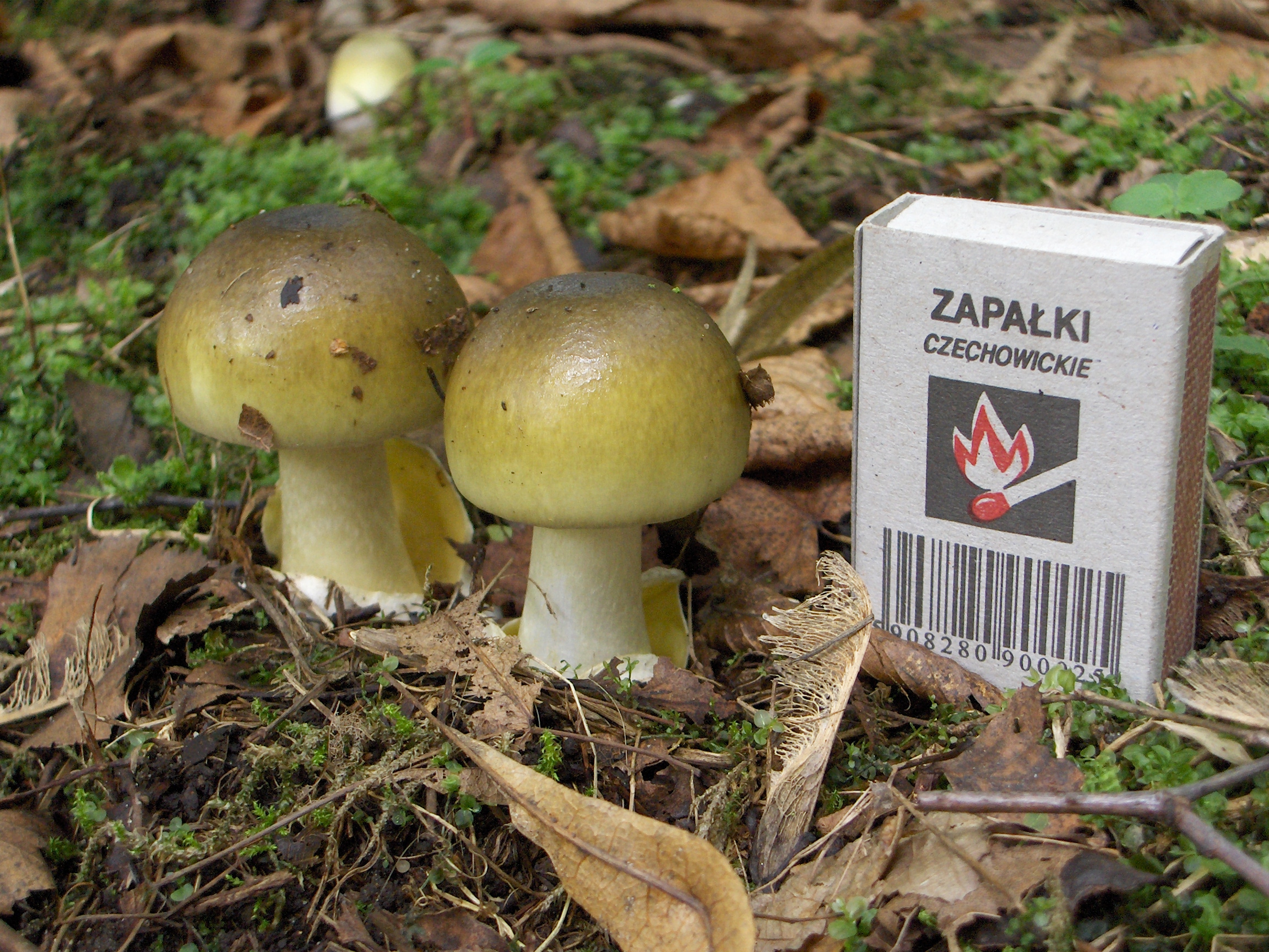
The pileus is the technical name for the cap, or cap-like part, of a basidiocarp or ascocarp (fungal fruiting body) that supports a spore-bearing surface, the hymenium.Moore-Landecker, E: "Fundamentals of the Fungi", page 560. Prentice Hall, 1972. The hymenium (hymenophore) may consist of lamellae, tubes, or teeth, on the underside of the pileus. A pileus is characteristic of agarics, boletes, some polypores, tooth fungi, and some ascomycetes. Classification Pilei can be formed in various shapes, and the shapes can change over the course of the developmental cycle of a fungus. The most familiar pileus shape is hemispherical or ''convex.'' Convex pilei often continue to expand as they mature until they become flat. Many well-known species have a convex pileus, including the button mushroom, various ''Amanita'' species and boletes. Some, such as the parasol mushroom, have distinct bosses or umbos and are described as ''umbonate''. An umbo is a knobby protrusion at the center of th ... [...More Info...] [...Related Items...] OR: [Wikipedia] [Google] [Baidu] |
Amanita Atkinsoniana 72050 Crop
The genus ''Amanita'' contains about 600 species of agarics, including some of the most toxic known mushrooms found worldwide, as well as some well-regarded edible species. This genus is responsible for approximately 95% of the fatalities resulting from mushroom poisoning, with the death cap accounting for about 50% on its own. The most potent toxin present in these mushrooms is α-Amanitin. The genus also contains many edible mushrooms, but mycologists discourage mushroom hunters, other than experts, from selecting any of these for human consumption. Nonetheless, in some cultures, the larger local edible species of ''Amanita'' are mainstays of the markets in the local growing season. Samples of this are ''Amanita zambiana'' and other fleshy species in central Africa, '' A. basii'' and similar species in Mexico, '' A. caesarea'' and the "Blusher" ''Amanita rubescens'' in Europe, and '' A. chepangiana'' in South-East Asia. Other species are used for colouring sauces, such as the ... [...More Info...] [...Related Items...] OR: [Wikipedia] [Google] [Baidu] |
Hypha
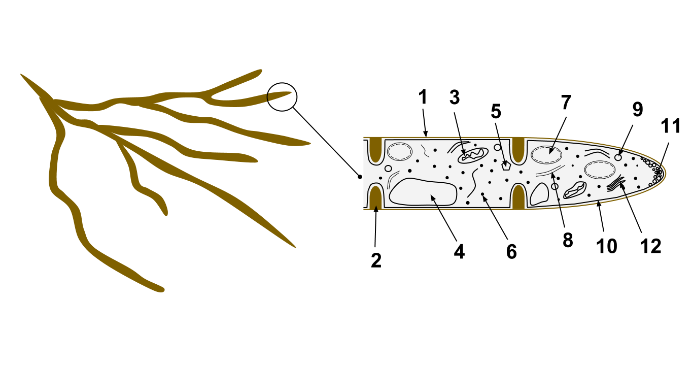
A hypha (; ) is a long, branching, filamentous structure of a fungus, oomycete, or actinobacterium. In most fungi, hyphae are the main mode of vegetative growth, and are collectively called a mycelium. Structure A hypha consists of one or more cells surrounded by a tubular cell wall. In most fungi, hyphae are divided into cells by internal cross-walls called "septa" (singular septum). Septa are usually perforated by pores large enough for ribosomes, mitochondria, and sometimes nuclei to flow between cells. The major structural polymer in fungal cell walls is typically chitin, in contrast to plants and oomycetes that have cellulosic cell walls. Some fungi have aseptate hyphae, meaning their hyphae are not partitioned by septa. Hyphae have an average diameter of 4–6 µm. Growth Hyphae grow at their tips. During tip growth, cell walls are extended by the external assembly and polymerization of cell wall components, and the internal production of new cell membrane. The S ... [...More Info...] [...Related Items...] OR: [Wikipedia] [Google] [Baidu] |
Volva (mycology)
In mycology, a volva is a cup-like structure at the base of a mushroom that is a remnant of the universal veil, or the remains of the peridium that encloses the immature fruit bodies of gasteroid fungi. This macrofeature is important in wild mushroom identification because it is an easily observed, taxonomically significant feature that frequently signifies a member of Amanitaceae. This has particular importance due to the disproportionately high number of deadly poisonous species contained within that family. A mushroom's volva is often partially or completely buried in the ground, and therefore care must be taken to check for its presence when identifying mushrooms. Cutting or pulling mushrooms and attempting to identify them later without having noted this feature could be a fatal error. References {{Reflist, refs= {{cite book , vauthors=Kirk PM, Cannon PF, Minter DW, Stalpers JA , title=Dictionary of the Fungi , edition=10th , publisher=CAB International , location=Wallin ... [...More Info...] [...Related Items...] OR: [Wikipedia] [Google] [Baidu] |


