|
Alcohol-related Brain Damage
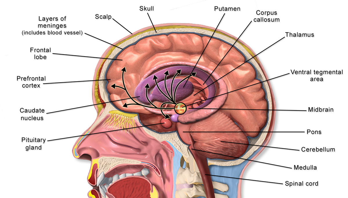
Alcohol-related brain damage alters both the structure and function of the brain as a result of the direct Neurotoxicity, neurotoxic effects of Alcoholic drink, alcohol intoxication or acute alcohol withdrawal. Increased alcohol intake is associated with damage to brain regions including the frontal lobe, limbic system, and cerebellum, with widespread cerebral atrophy, or brain shrinkage caused by Neurodegeneration, neuron degeneration. This damage can be seen on neuroimaging scans. Frontal lobe damage becomes the most prominent as alcoholics age and can lead to impaired neuropsychological performance in areas such as problem solving, good judgement, and goal-directed behaviors. Impaired emotional processing results from damage to the limbic system. This may lead to troubles recognizing emotional facial expressions and "interpreting nonverbal emotional cues". Binge drinking, or heavy episodic drinking, can lead to damage in the limbic system that occurs after a relatively short pe ... [...More Info...] [...Related Items...] OR: [Wikipedia] [Google] [Baidu] |
Neurotoxicity
Neurotoxicity is a form of toxicity in which a biological, chemical, or physical agent produces an adverse effect on the structure or function of the central and/or peripheral nervous system. It occurs when exposure to a substance – specifically, a neurotoxin or neurotoxicant– alters the normal activity of the nervous system in such a way as to cause permanent or reversible damage to nervous tissue. This can eventually disrupt or even kill neurons, which are cells that transmit and process signals in the brain and other parts of the nervous system. Neurotoxicity can result from organ transplants, radiation treatment, certain drug therapies, recreational drug use, and exposure to heavy metals, bites from certain species of venomous snakes, pesticides, certain industrial cleaning solvents, fuels and certain naturally occurring substances. Symptoms may appear immediately after exposure or be delayed. They may include limb weakness or numbness, loss of memory, vision, and/or ... [...More Info...] [...Related Items...] OR: [Wikipedia] [Google] [Baidu] |
White Matter
White matter refers to areas of the central nervous system (CNS) that are mainly made up of myelinated axons, also called tracts. Long thought to be passive tissue, white matter affects learning and brain functions, modulating the distribution of action potentials, acting as a relay and coordinating communication between different brain regions. White matter is named for its relatively light appearance resulting from the lipid content of myelin. However, the tissue of the freshly cut brain appears pinkish-white to the naked eye because myelin is composed largely of lipid tissue veined with capillaries. Its white color in prepared specimens is due to its usual preservation in formaldehyde. Structure White matter White matter is composed of bundles, which connect various grey matter areas (the locations of nerve cell bodies) of the brain to each other, and carry nerve impulses between neurons. Myelin acts as an insulator, which allows electrical signals to jump, rather than c ... [...More Info...] [...Related Items...] OR: [Wikipedia] [Google] [Baidu] |
Cerebellar Vermis
The cerebellar vermis (from Latin ''vermis,'' "worm") is located in the medial, cortico-nuclear zone of the cerebellum, which is in the posterior fossa of the cranium. The primary fissure in the vermis curves ventrolaterally to the superior surface of the cerebellum, dividing it into anterior and posterior lobes. Functionally, the vermis is associated with bodily posture and locomotion. The vermis is included within the spinocerebellum and receives somatic sensory input from the head and proximal body parts via ascending spinal pathways. The cerebellum develops in a rostro-caudal manner, with rostral regions in the midline giving rise to the vermis, and caudal regions developing into the cerebellar hemispheres. By 4 months of prenatal development, the vermis becomes fully foliated, while development of the hemispheres lags by 30–60 days. Postnatally, proliferation and organization of the cellular components of the cerebellum continues, with completion of the f ... [...More Info...] [...Related Items...] OR: [Wikipedia] [Google] [Baidu] |
Purkinje Cell
Purkinje cells, or Purkinje neurons, are a class of GABAergic inhibitory neurons located in the cerebellum. They are named after their discoverer, Czech people, Czech anatomist Jan Evangelista Purkyně, who characterized the cells in 1839. Structure These Cell (biology), cells are some of the largest neurons in the human brain (Betz cells being the largest), with an intricately elaborate dendrite, dendritic arbor, characterized by a large number of dendritic spines. Purkinje cells are found within the Cerebellum#Microanatomy, Purkinje layer in the cerebellum. Purkinje cells are aligned like dominos stacked one in front of the other. Their large dendritic arbors form nearly two-dimensional layers through which parallel fibers from the deeper-layers pass. These parallel fibers make relatively weaker excitatory synapse, excitatory (glutamatergic) synapses to spines in the Purkinje cell dendrite, whereas climbing fibers originating from the inferior olivary nucleus in the medull ... [...More Info...] [...Related Items...] OR: [Wikipedia] [Google] [Baidu] |
Alzheimer's Disease
Alzheimer's disease (AD) is a neurodegeneration, neurodegenerative disease that usually starts slowly and progressively worsens. It is the cause of 60–70% of cases of dementia. The most common early symptom is difficulty in short-term memory, remembering recent events. As the disease advances, symptoms can include primary progressive aphasia, problems with language, Orientation (mental), disorientation (including easily getting lost), mood swings, loss of motivation, self-neglect, and challenging behaviour, behavioral issues. As a person's condition declines, they often withdraw from family and society. Gradually, bodily functions are lost, ultimately leading to death. Although the speed of progression can vary, the typical life expectancy following diagnosis is three to nine years. The cause of Alzheimer's disease is poorly understood. There are many environmental and genetic risk factors associated with its development. The strongest genetic risk factor is from an alle ... [...More Info...] [...Related Items...] OR: [Wikipedia] [Google] [Baidu] |
Soma (biology)
The soma (pl. ''somata'' or ''somas''), perikaryon (pl. ''perikarya''), neurocyton, or cell body is the bulbous, non-process portion of a neuron or other brain cell type, containing the cell nucleus. The word 'soma' comes from the Greek '' σῶμα'', meaning 'body'. Although it is often used to refer to neurons, it can also refer to other cell types as well, including astrocytes, oligodendrocytes, and microglia. There are many different specialized types of neurons, and their sizes vary from as small as about 5 micrometres to over 10 millimetres for some of the smallest and largest neurons of invertebrates, respectively. The soma of a neuron (i.e., the main part of the neuron in which the dendrites branch off of) contains many organelles, including granules called Nissl granules, which are composed largely of rough endoplasmic reticulum and free polyribosomes. The cell nucleus is a key feature of the soma. The nucleus is the source of most of the RNA that is produced in neuro ... [...More Info...] [...Related Items...] OR: [Wikipedia] [Google] [Baidu] |
Corpus Callosum
The corpus callosum (Latin for "tough body"), also callosal commissure, is a wide, thick nerve tract, consisting of a flat bundle of commissural fibers, beneath the cerebral cortex in the brain. The corpus callosum is only found in placental mammals. It spans part of the longitudinal fissure, connecting the left and right cerebral hemispheres, enabling communication between them. It is the largest white matter structure in the human brain, about in length and consisting of 200–300 million axonal projections. A number of separate nerve tracts, classed as subregions of the corpus callosum, connect different parts of the hemispheres. The main ones are known as the genu, the rostrum, the trunk or body, and the splenium. Structure The corpus callosum forms the floor of the longitudinal fissure that separates the two cerebral hemispheres. Part of the corpus callosum forms the roof of the lateral ventricles. The corpus callosum has four main parts – individual nerve tracts ... [...More Info...] [...Related Items...] OR: [Wikipedia] [Google] [Baidu] |
N-Acetylaspartic Acid
''N''-Acetylaspartic acid, or ''N''-acetylaspartate (NAA), is a derivative of aspartic acid with a formula of C6H9NO5 and a molecular weight of 175.139. NAA is the second-most-concentrated molecule in the brain after the amino acid glutamate. It is detected in the adult brain in neurons, oligodendrocytes and myelin and is synthesized in the mitochondria from the amino acid aspartic acid and acetyl-coenzyme A. Function The various functions served by NAA are under investigation, but the primary proposed functions include: * Neuronal osmolyte that is involved in fluid balance in the brain * Source of acetate for lipid and myelin synthesis in oligodendrocytes, the glial cells that myelinate neuronal axons * Precursor for the synthesis of the neuronal dipeptide N-Acetylaspartylglutamate * Contributor to energy production from the amino acid glutamate in neuronal mitochondria In the brain, NAA was thought to be present predominantly in neuronal cell bodies, where it acts as a ... [...More Info...] [...Related Items...] OR: [Wikipedia] [Google] [Baidu] |
MRI Brain Scan
Magnetic resonance imaging of the brain uses magnetic resonance imaging (MRI) to produce high quality two-dimensional or three-dimensional images of the brain and brainstem as well as the cerebellum without the use of ionizing radiation (X-rays) or radioactive tracers. History The first MR images of a human brain were obtained in 1978 by two groups of researchers at EMI Laboratories led by Ian Robert Young and Hugh Clow. In 1986, Charles L. Dumoulin and Howard R. Hart at General Electric developed MR angiography, and Denis Le Bihan obtained the first images and later patented diffusion MRI. In 1988, Arno Villringer and colleagues demonstrated that susceptibility contrast agents may be employed in perfusion MRI. In 1990, Seiji Ogawa at AT&T Bell labs recognized that oxygen-depleted blood with dHb was attracted to a magnetic field, and discovered the technique that underlies Functional Magnetic Resonance Imaging (fMRI). In the early 1980s to the early 1990s, 'Jedi' helmets, inspire ... [...More Info...] [...Related Items...] OR: [Wikipedia] [Google] [Baidu] |
Dentate Gyrus
The dentate gyrus (DG) is part of the hippocampal formation in the temporal lobe of the brain, which also includes the hippocampus and the subiculum. The dentate gyrus is part of the hippocampal trisynaptic circuit and is thought to contribute to the formation of new episodic memories, the spontaneous exploration of novel environments and other functions. It is notable as being one of a select few brain structures known to have significant rates of adult neurogenesis in many species of mammals, from rodents to primates. Other sites of adult neurogenesis include the subventricular zone, the striatum and the cerebellum. However, whether significant neurogenesis exists in the adult human dentate gyrus has been a matter of debate. 2019 evidence has shown that adult neurogenesis does take place in the subventricular zone and in the subgranular zone of the dentate gyrus. Structure The dentate gyrus, like the hippocampus, consists of three distinct layers: an outer molecular layer ... [...More Info...] [...Related Items...] OR: [Wikipedia] [Google] [Baidu] |
Entorhinal Cortex
The entorhinal cortex (EC) is an area of the brain's allocortex, located in the medial temporal lobe, whose functions include being a widespread network hub for memory, navigation, and the perception of time.Integrating time from experience in the lateral entorhinal cortex Albert Tsao, Jørgen Sugar, Li Lu, Cheng Wang, James J. Knierim, May-Britt Moser & Edvard I. Moser Naturevolume 561, pages57–62 (2018) The EC is the main interface between the hippocampus and neocortex. The EC-hippocampus system plays an important role in declarative (autobiographical/episodic/semantic) memories and in particular spatial memories including memory formation, memory consolidation, and memory optimization in sleep. The EC is also responsible for the pre-processing (familiarity) of the input signals in the reflex nictitating membrane response of classical trace conditioning; the association of impulses from the eye and the ear occurs in the entorhinal cortex. Structure In rodents, the EC ... [...More Info...] [...Related Items...] OR: [Wikipedia] [Google] [Baidu] |
Perirhinal Cortex
The perirhinal cortex is a cortical region in the medial temporal lobe that is made up of Brodmann areas 35 and 36. It receives highly processed sensory information from all sensory regions, and is generally accepted to be an important region for memory. It is bordered caudally by postrhinal cortex or parahippocampal cortex (homologous regions in rodents and primates, respectively) and ventrally and medially by entorhinal cortex. Structure The perirhinal cortex is composed of two regions: areas 36 and 35. Area 36 is sometimes divided into three subdivisions: 36d is the most rostral and dorsal, 36r ventral and caudal, and 36c the most caudal. Area 35 can be divided in the same manner, into 35d and 35v (for dorsal and ventral, respectively). Area 36 is six-layered, dysgranular, meaning that its layer IV is relatively sparse. Area 35 is agranular cortex (lacking any cells in layer IV). Function The perirhinal cortex is involved in both visual perception and memory; it fac ... [...More Info...] [...Related Items...] OR: [Wikipedia] [Google] [Baidu] |


.jpg)


