|
Adductor Minimus Muscle
In human anatomy, the adductor minimus (adductor femoris minimus or adductor quartus) is a small and flat skeletal muscle in the thigh which constitutes the upper, lateral part of the adductor magnus muscle.Bergman, Ronald A.; Afifi, Adel K.; Miyauchi, Ryosuke (2010)''Adductor Minimus (Henle, Günther)'' Anatomy Atlases It adducts and laterally rotates the femur. Structure The adductor minimus originates on the pelvis at the inferior ramus of the pubis as the anterior-most part of the adductor magnus. It is inserted on the back of the femur at the medial lip of the ''linea aspera'' and thus crosses the proximal part of the true adductor magnus.Platzer, Werner (2004), Color Atlas of Human Anatomy, Vol. 1, Locomotor System', Thieme, 5th ed, p 242 The adductor minimus and the adductor magnus are frequently separated by a branch of the superior perforating branch of the profunda femoris artery and the former muscle is considered independent from the latter because it is primarily ... [...More Info...] [...Related Items...] OR: [Wikipedia] [Google] [Baidu] |
Inferior Pubic Ramus
In vertebrates, the pubic region ( la, pubis) is the most forward-facing (ventral and anterior) of the three main regions making up the coxal bone. The left and right pubic regions are each made up of three sections, a superior ramus, inferior ramus, and a body. Structure The pubic region is made up of a ''body'', ''superior ramus'', and ''inferior ramus'' (). The left and right coxal bones join at the pubic symphysis. It is covered by a layer of fat, which is covered by the mons pubis. The pubis is the lower limit of the suprapubic region. In the female, the pubic region is anterior to the urethral sponge. Body The body forms the wide, strong, middle and flat part of the pubic region. The bodies of the left and right pubic regions join at the pubic symphysis. The rough upper edge is the pubic crest, ending laterally in the pubic tubercle. This tubercle, found roughly 3 cm from the pubic symphysis, is a distinctive feature on the lower part of the abdominal wall; important ... [...More Info...] [...Related Items...] OR: [Wikipedia] [Google] [Baidu] |
Linea Aspera
The linea aspera ( la, rough line) is a ridge of roughened surface on the posterior surface of the shaft of the femur. It is the site of attachments of muscles and the intermuscular septum. Its margins diverge above and below. The linea aspera is a prominent longitudinal ridge or crest, on the middle third of the bone, presenting a medial and a lateral lip, and a narrow rough, intermediate line. It is an important insertion point for the adductors and the lateral and medial intermuscular septa that divides the thigh into three compartments. The tension generated by muscle attached to the bones is responsible for the formation of the ridges. Structure Above Above, the linea aspera is prolonged by three ridges. * The lateral ridge is very rough, and runs almost vertically upward to the base of the greater trochanter. It is termed the gluteal tuberosity, and gives attachment to part of the gluteus maximus: its upper part is often elongated into a roughened crest, on which a more ... [...More Info...] [...Related Items...] OR: [Wikipedia] [Google] [Baidu] |
Hip Adductors
The adductor muscles of the hip are a group of muscles mostly used for bringing the thighs together (called adduction). Structure The adductor group is made up of: *Adductor brevis * Adductor longus *Adductor magnus * Adductor minimus This is often considered to be a part of adductor magnus. *pectineus * gracilis *Obturator externusPlatzer, Werner (2004), Color Atlas of Human Anatomy, Vol. 1, Locomotor System', Thieme, 5th ed, p 240 and are also part of the medial compartment of thigh The adductors originate on the pubis and ischium bones and insert mainly on the medial posterior surface of the femur. Nerve supply The pectineus is the only adductor muscle that is innervated by the femoral nerve. The other adductor muscles are innervated by the obturator nerve with the exception of a small part of the adductor magnus which is innervated by the tibial nerve. Variation In 33% of people a supernumerary muscle is found between the adductor brevis and adductor minimus. When p ... [...More Info...] [...Related Items...] OR: [Wikipedia] [Google] [Baidu] |
Ontogeny
Ontogeny (also ontogenesis) is the origination and development of an organism (both physical and psychological, e.g., moral development), usually from the time of fertilization of the egg to adult. The term can also be used to refer to the study of the entirety of an organism's lifespan. Ontogeny is the developmental history of an organism within its own lifetime, as distinct from phylogeny, which refers to the evolutionary history of a species. Another way to think of ontogeny is that it is the process of an organism going through all of the developmental stages over its lifetime. The developmental history includes all the developmental events that occur during the existence of an organism, beginning with the changes in the egg at the time of fertilization and events from the time of birth or hatching and afterward (i.e., growth, remolding of body shape, development of secondary sexual characteristics, etc.). While developmental (i.e., ontogenetic) processes can influence ... [...More Info...] [...Related Items...] OR: [Wikipedia] [Google] [Baidu] |
Obturator Externus Muscle
The external obturator muscle, obturator externus muscle (; OE) is a flat, triangular muscle, which covers the outer surface of the anterior wall of the pelvis. It is sometimes considered part of the medial compartment of thigh, and sometimes considered part of the gluteal region. Structure It arises from the margin of bone immediately around the medial side of the obturator membrane and surrounding bone, viz., from the inferior pubic ramus, and the ramus of the ischium; it also arises from the medial two-thirds of the outer surface of the obturator membrane, and from the tendinous arch which completes the canal for the passage of the obturator vessels and nerves. The fibers springing from the pubic arch extend on to the inner surface of the bone, where they obtain a narrow origin between the margin of the foramen and the attachment of the obturator membrane. The fibers converge and pass posterolateral and upward, and end in a tendon which runs across the back of the neck of t ... [...More Info...] [...Related Items...] OR: [Wikipedia] [Google] [Baidu] |
Lesser Trochanter
The lesser trochanter is a conical posteromedial bony projection of the femoral shaft. it serves as the principal insertion site of the iliopsoas muscle. Structure The lesser trochanter is a conical posteromedial projection of the shaft of the femur, projecting from the posteroinferior aspect of its junction with the femoral neck. The summit and anterior surface of the lesser trochanter are rough, whereas its posterior surface is smooth. From its apex three well-marked borders extend: * two of these are above ** a medial continuous with the lower border of the femur neck ** a lateral with the intertrochanteric crest * the inferior border is continuous with the middle division of the linea aspera Attachments The summit of the lesser trochanter gives insertion to the tendon of the psoas major muscle and the iliacus muscle; the lesser trochanter represents the principal attachment of the iliopsoas. Anatomical relations The intertrochanteric crest (which demarcates the j ... [...More Info...] [...Related Items...] OR: [Wikipedia] [Google] [Baidu] |
Pectineal Line (femur)
On the posterior surface of the femur, the intermediate ridge or pectineal line is continued to the base of the lesser trochanter and gives attachment to the pectineus muscle The pectineus muscle (, from the Latin word ''pecten'', meaning comb) is a flat, quadrangular muscle, situated at the anterior (front) part of the upper and medial (inner) aspect of the thigh. The pectineus muscle is the most anterior adductor o .... References Bones of the lower limb Femur {{musculoskeletal-stub ... [...More Info...] [...Related Items...] OR: [Wikipedia] [Google] [Baidu] |
Aponeurosis
An aponeurosis (; plural: ''aponeuroses'') is a type or a variant of the deep fascia, in the form of a sheet of pearly-white fibrous tissue that attaches sheet-like muscles needing a wide area of attachment. Their primary function is to join muscles and the body parts they act upon, whether bone or other muscles. They have a shiny, whitish-silvery color, are histologically similar to tendons, and are very sparingly supplied with blood vessels and nerves. When dissected, aponeuroses are papery and peel off by sections. The primary regions with thick aponeuroses are in the ventral abdominal region, the dorsal lumbar region, the ventriculus in birds, and the palmar (palms) and plantar (soles) regions. Anatomy Anterior abdominal aponeuroses The anterior abdominal aponeuroses are located just superficial to the rectus abdominis muscle. It has for its borders the external oblique, pectoralis muscles, and the latissimus dorsi. Posterior lumbar aponeuroses The posterior lumbar aponeu ... [...More Info...] [...Related Items...] OR: [Wikipedia] [Google] [Baidu] |
Supernumerary Body Part
Supernumerary body parts are most commonly a congenital disorder involving the growth of an additional part of the body and a deviation from the body plan. Body parts may be easily visible or hidden away, such as internal organs. Many additional body parts form by the same process as conjoined twins: the zygote begins to split but fails to completely separate. This condition may also be a symptom of repeated occurrences of continuous inbreeding in a genetic line. Specific types of occurrence Specific types of additional body parts include: *Accessory breast – one or more additional breastsOnline Mendelian Inheritance in Man. Nipples, Supernumerary. Accessed 10 July 2006. *Accessory spleen – one or more additional spleens *Cervical rib – an additional rib *Diphallia – having two penes/penises *Hermaphroditism – having both male and female sex organs *Hyperdontia – additional teeth * Pelvic digit – a bony growth in the soft tissue of the pelvic region * Polycephaly ... [...More Info...] [...Related Items...] OR: [Wikipedia] [Google] [Baidu] |
Sciatic Nerve
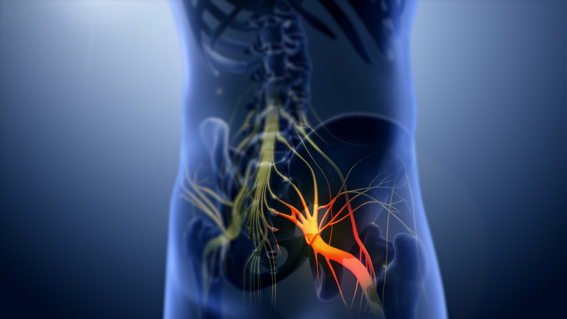
The sciatic nerve, also called the ischiadic nerve, is a large nerve in humans and other vertebrate animals which is the largest branch of the sacral plexus and runs alongside the hip joint and down the lower limb. It is the longest and widest single nerve in the human body, going from the top of the leg to the foot on the posterior aspect. The sciatic nerve has no cutaneous branches for the thigh. This nerve provides the connection to the nervous system for the skin of the lateral leg and the whole foot, the muscles of the back of the thigh, and those of the leg and foot. It is derived from spinal nerves L4 to S3. It contains fibers from both the anterior and posterior divisions of the lumbosacral plexus. Structure In humans, the sciatic nerve is formed from the L4 to S3 segments of the sacral plexus, a collection of nerve fibres that emerge from the sacral part of the spinal cord. The lumbosacral trunk from the L4 and L5 roots descends between the sacral promontory and al ... [...More Info...] [...Related Items...] OR: [Wikipedia] [Google] [Baidu] |
Obturator Nerve
The obturator nerve in human anatomy arises from the ventral divisions of the second, third, and fourth lumbar nerves in the lumbar plexus; the branch from the third is the largest, while that from the second is often very small. Structure The obturator nerve originates from the anterior divisions of the L2, L3, and L4 spinal nerve roots. It descends through the fibers of the psoas major, and emerges from its medial border near the brim of the pelvis. It then passes behind the common iliac arteries, and on the lateral side of the internal iliac artery and vein, and runs along the lateral wall of the lesser pelvis, above and in front of the obturator vessels, to the upper part of the obturator foramen. Here it enters the thigh, through the obturator canal, and divides into an anterior and a posterior branch, which are separated at first by some of the fibers of the obturator externus, and lower down by the adductor brevis. An accessory obturator nerve may be present in app ... [...More Info...] [...Related Items...] OR: [Wikipedia] [Google] [Baidu] |
Profunda Femoris Artery
The deep artery of the thigh, (profunda femoris artery or deep femoral artery) is a large branch of the femoral artery. It travels more deeply (posteriorly) than the rest of the femoral artery. Structure The deep artery of the thigh branches off the posterolateral side of the femoral artery soon after its origin. It travels down the thigh closer to the femur than the femoral artery. It runs between the pectineus muscle and the adductor longus muscle. It runs on the posterior side of adductor longus muscle. It pierces the adductor magnus muscle, and may be known as the Perforating arteries, fourth perforating artery as it continues. The deep femoral artery does not leave the thigh. Branches The deep artery of the thigh gives off the following branches: * Lateral circumflex femoral artery. * Medial circumflex femoral artery. * 3 Perforating arteries - perforate the adductor magnus muscle to the posterior and medial fascial compartment, compartments of the thigh to connect with the ... [...More Info...] [...Related Items...] OR: [Wikipedia] [Google] [Baidu] |
