|
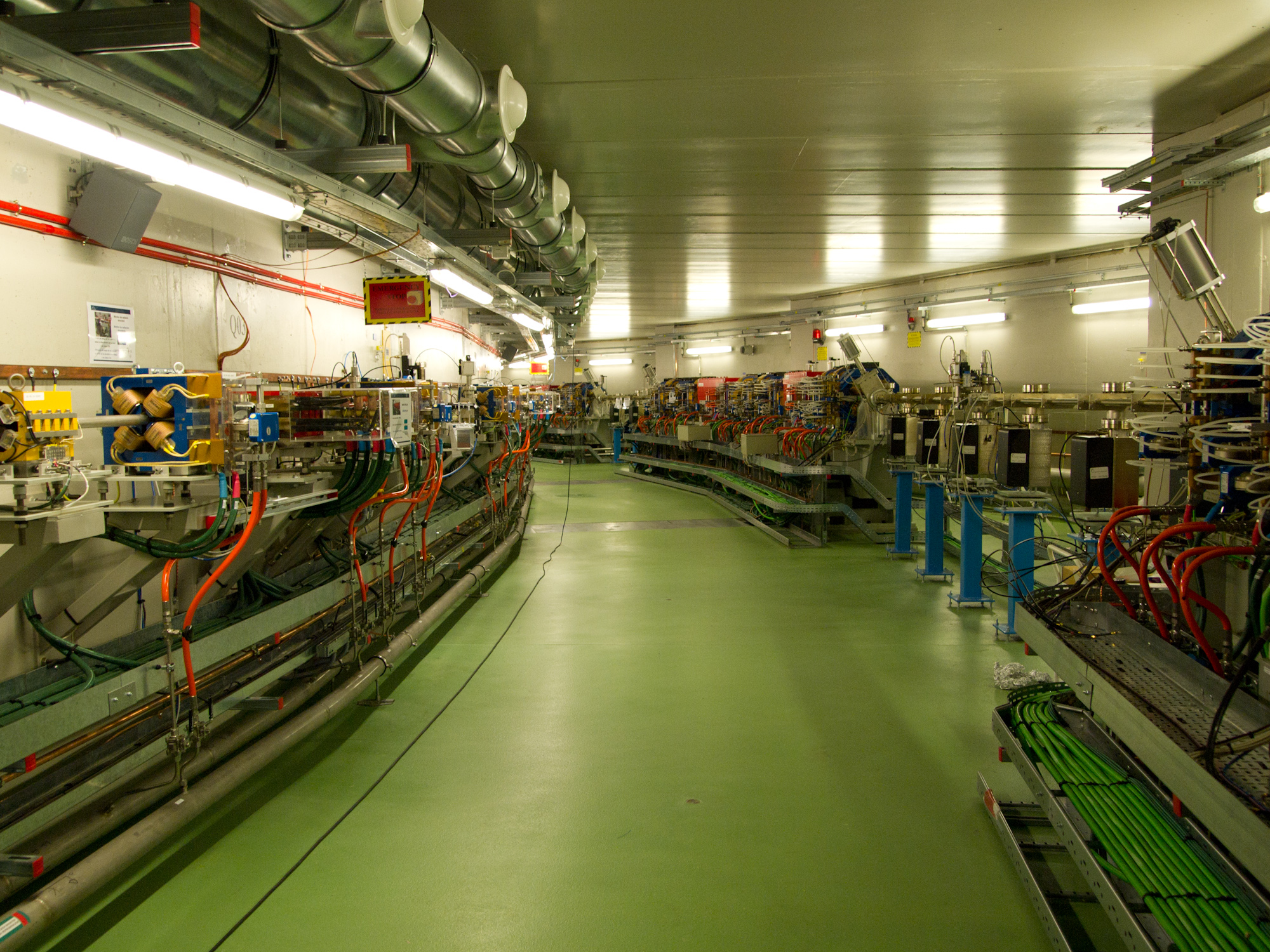
ALBA (synchrotron)
ALBA (meaning "Sunrise" in Catalan and in Spanish) is a third-generation synchrotron light source facility located in the Barcelona Synchrotron Park in Cerdanyola del Vallès near Barcelona, in Catalonia (Spain). It was constructed and is operated by CELLS (sp: ''Consorcio para la Construcción, Equipamiento y Explotación del Laboratorio de Luz de Sincrotrón'', the Consortium for the Exploitation of the Synchrotron Light Laboratory), and co-financed by the Spanish central administration and regional Catalan Government. After nearly ten years of planning and design work by the Spanish scientific community, the project was approved in 2002 by the Spanish and the regional Catalan governments. After scientific workshops and meetings with prospective users, the facility was redesigned in 2004, and in 2006 construction started. The laboratory was officially opened for experiments on seven beamlines in March 2010. History The project was launched in 1994, the construction began in ... [...More Info...] [...Related Items...] OR: [Wikipedia] [Google] [Baidu] |
Angle Of Incidence (optics)
The angle of incidence, in geometric optics, is the angle between a ray incident on a surface and the line perpendicular (at 90 degree angle) to the surface at the point of incidence, called the normal. The ray can be formed by any waves, such as optical, acoustic, microwave, and X-ray. In the figure below, the line representing a ray makes an angle θ with the normal (dotted line). The angle of incidence at which light is first totally internally reflected is known as the critical angle. The angle of reflection and angle of refraction are other angles related to beams. In computer graphics and geography, the angle of incidence is also known as the illumination angle of a surface with a light source, such as the Earth's surface and the Sun. It can also be equivalently described as the angle between the tangent plane of the surface and another plane at right angles to the light rays. This means that the illumination angle of a certain point on Earth's surface is 0° if the Sun ... [...More Info...] [...Related Items...] OR: [Wikipedia] [Google] [Baidu] |
Wide-angle X-ray Scattering
In X-ray crystallography, wide-angle X-ray scattering (WAXS) or wide-angle X-ray diffraction (WAXD) is the analysis of Bragg peaks scattered to wide angles, which (by Bragg's law) are caused by sub-nanometer-sized structures. It is an X-ray-diffraction method and commonly used to determine a range of information about crystalline materials. The term WAXS is commonly used in polymer sciences to differentiate it from SAXS but many scientists doing "WAXS" would describe the measurements as Bragg/X-ray/powder diffraction or crystallography. Wide-angle X-ray scattering is similar to small-angle X-ray scattering (SAXS) but the increasing angle between the sample and detector is probing smaller length scales. This requires samples to be more ordered/crystalline for information to be extracted. In a dedicated SAXS instrument the distance from sample to the detector is longer to increase angular resolution. Most diffractometers can be used to perform both WAXS and limited SAXS in a single ... [...More Info...] [...Related Items...] OR: [Wikipedia] [Google] [Baidu] |
Small-angle X-ray Scattering
Small-angle X-ray scattering (SAXS) is a small-angle scattering technique by which nanoscale density differences in a sample can be quantified. This means that it can determine nanoparticle size distributions, resolve the size and shape of (monodisperse) macromolecules, determine pore sizes, characteristic distances of partially ordered materials, and much more. This is achieved by analyzing the elastic scattering behaviour of X-rays when travelling through the material, recording their scattering at small angles (typically 0.1 – 10°, hence the "Small-angle" in its name). It belongs to the family of small-angle scattering (SAS) techniques along with small-angle neutron scattering, and is typically done using hard X-rays with a wavelength of 0.07 – 0.2 nm.. Depending on the angular range in which a clear scattering signal can be recorded, SAXS is capable of delivering structural information of dimensions between 1 and 100 nm, and of repeat distances in partially ordered s ... [...More Info...] [...Related Items...] OR: [Wikipedia] [Google] [Baidu] |
Zernike Phase Contrast Microscopy
__NOTOC__ Phase-contrast microscopy (PCM) is an optical microscopy technique that converts phase shifts in light passing through a transparent specimen to brightness changes in the image. Phase shifts themselves are invisible, but become visible when shown as brightness variations. When light waves travel through a medium other than a vacuum, interaction with the medium causes the wave amplitude and phase to change in a manner dependent on properties of the medium. Changes in amplitude (brightness) arise from the scattering and absorption of light, which is often wavelength-dependent and may give rise to colors. Photographic equipment and the human eye are only sensitive to amplitude variations. Without special arrangements, phase changes are therefore invisible. Yet, phase changes often convey important information. Phase-contrast microscopy is particularly important in biology. It reveals many cellular structures that are invisible with a bright-field microscope, as exemplif ... [...More Info...] [...Related Items...] OR: [Wikipedia] [Google] [Baidu] |
Spatial Resolution
In physics and geosciences, the term spatial resolution refers to distance between independent measurements, or the physical dimension that represents a pixel of the image. While in some instruments, like cameras and telescopes, spatial resolution is directly connected to angular resolution, other instruments, like synthetic aperture radar or a network of weather stations, produce data whose spatial sampling layout is more related to the Earth's surface, such as in remote sensing and satellite imagery. See also * Image resolution * Ground sample distance * Level of detail * Resel In image analysis, a resel (from ''res''olution ''el''ement) represents the actual spatial resolution in an image or a volumetric dataset. The number of resels in the image may be lower or equal to the number of pixel/voxels in the image. In an act ... References Accuracy and precision {{physics-stub ... [...More Info...] [...Related Items...] OR: [Wikipedia] [Google] [Baidu] |
CCD Camera
A charge-coupled device (CCD) is an integrated circuit containing an array of linked, or coupled, capacitors. Under the control of an external circuit, each capacitor can transfer its electric charge to a neighboring capacitor. CCD sensors are a major technology used in digital imaging. In a CCD image sensor, pixels are represented by p-doped metal–oxide–semiconductor (MOS) capacitors. These MOS capacitors, the basic building blocks of a CCD, are biased above the threshold for inversion when image acquisition begins, allowing the conversion of incoming photons into electron charges at the semiconductor-oxide interface; the CCD is then used to read out these charges. Although CCDs are not the only technology to allow for light detection, CCD image sensors are widely used in professional, medical, and scientific applications where high-quality image data are required. In applications with less exacting quality demands, such as consumer and professional digital cameras, active ... [...More Info...] [...Related Items...] OR: [Wikipedia] [Google] [Baidu] |
Fresnel Zone Plate
A zone plate is a device used to focus light or other things exhibiting wave character.G. W. Webb, I. V. Minin and O. V. Minin, “Variable Reference Phase in Diffractive Antennas”, ''IEEE Antennas and Propagation Magazine'', vol. 53, no. 2, April. 2011, pp. 77-94. Unlike lenses or curved mirrors, zone plates use diffraction instead of refraction or reflection. Based on analysis by French physicist Augustin-Jean Fresnel, they are sometimes called Fresnel zone plates in his honor. The zone plate's focusing ability is an extension of the Arago spot phenomenon caused by diffraction from an opaque disc. A zone plate consists of a set of concentric rings, known as Fresnel zones, which alternate between being opaque and transparent. Light hitting the zone plate will diffract around the opaque zones. The zones can be spaced so that the diffracted light constructively interferes at the desired focus, creating an image there. Design and manufacture To get constructive interference ... [...More Info...] [...Related Items...] OR: [Wikipedia] [Google] [Baidu] |
Monochromatic Light
{{Short description, Electromagnetic radiation with a single constant frequency In physics, monochromatic radiation is electromagnetic radiation with a single constant frequency. When that frequency is part of the visible spectrum (or near it) the term monochromatic light is often used. Monochromatic light is perceived by the human eye as a spectral color. When monochromatic radiation propagates through vacuum or a homogeneous transparent medium, it has a single constant wavelength. Practical monochromaticity No radiation can be totally monochromatic, since that would require a wave of infinite duration as a consequence of the Fourier transform's localization property (cf. spectral coherence). In practice, "monochromatic" radiation — even from lasers or spectral lines — always consists of components with a range of frequencies of non-zero width. Generation Monochromatic radiation can be produced by a number of methods. Isaac Newton observed that a beam of light from th ... [...More Info...] [...Related Items...] OR: [Wikipedia] [Google] [Baidu] |
Absorption Edge
An absorption edge, absorption discontinuity or absorption limit is a sharp discontinuity in the absorption spectrum of a substance. These discontinuities occur at wavelengths where the energy of an absorbed photon corresponds to an electronic transition or ionization potential. When the quantum energy of the incident radiation becomes smaller than the work required to eject an electron from one or other quantum states in the constituent absorbing atom, the incident radiation ceases to be absorbed by that state. For example, incident radiation on an atom of a wavelength that has a corresponding energy just below the binding energy of the K-shell electron in that atom cannot eject the K-shell electron."The Penguin Dictionary of Physics", 3rd ed., Longman Group Ltd. (2000), p. 3. Siegbahn notation is used for notating absorption edges. In compound semiconductors, the bonding between atoms of different species forms a set of dipoles. These dipoles can absorb energy from an electromag ... [...More Info...] [...Related Items...] OR: [Wikipedia] [Google] [Baidu] |
Water Window
The water window is a region of the electromagnetic spectrum in which water is transparent to soft x-rays. The window extends from the K-absorption edge of carbon at 282 eV (68 PHz, 4.40 nm wavelength) to the K-edge of oxygen at 533 eV (129 PHz, 2.33 nm wavelength). Water is transparent to these X-rays, but carbon (and thus, organic molecules) is absorbing. These wavelengths could be used in an x-ray microscope An X-ray microscope uses electromagnetic radiation in the soft X-ray band to produce magnified images of objects. Since X-rays penetrate most objects, there is no need to specially prepare them for X-ray microscopy observations. Unlike visible li ... for viewing living specimens. References * {{X-ray science X-rays ... [...More Info...] [...Related Items...] OR: [Wikipedia] [Google] [Baidu] |
Nanotomography
Nanotomography, much like its related modalities tomography and microtomography, uses x-rays to create cross-sections from a 3D-object that later can be used to recreate a virtual model without destroying the original model, applying Nondestructive testing. The term nano is used to indicate that the pixel sizes of the cross-sections are in the nanometer range Nano-CT beamlines have been built at 3rd generation synchrotron radiation facilities, including the Advanced Photon Source of Argonne National Laboratory, SPring-8, and ESRF from early 2000s. They have been applied to wide variety of three-dimensional visualization studies, such as those of comet samples returned by the Startdust mission, mechanical degradation in lithium-ion batteries, and neuron deformation in schizophrenic brains. Although a lot of research is done to create nano-CT scanners, currently there are only a few available commercially. The SkyScan-201has a range of about 150 to 250 nanometers per pixel with a ... [...More Info...] [...Related Items...] OR: [Wikipedia] [Google] [Baidu] |


