|
AII Amacrine Cells
AII amacrine cells are a subtype of amacrine cells present in the retina of mammals. AII amacrine cell serve the critical role of transferring light signals from rod photoreceptors to the retinal ganglion cells (which contain the axons of the optic nerve) The Classical Rod Pathway described the role of AII amacrine cells in the mammalian retina. This can be summarised as follows: * In scotopic conditions, if a rod photoreceptor receives light in scotopic (dark) conditions, it will hyperpolarise. * The rod photoreceptor synapses with the rod bipolar cell.There is only one type of rod bipolar cell: an ON-bipolar cellThis is a 'sign-inverting' synapse. * This rod bipolar cell will directly (exclusively) synapse with an AII amacrine cell in sublamina B (within the inner plexiform layer)This is a 'sign-conserving' synapse. * The AII amacrine cells becomes activated (i.e., it depolarises) when light stimulates a rod. (Once activated, the AII amacrine cell then modulates the con ... [...More Info...] [...Related Items...] OR: [Wikipedia] [Google] [Baidu] |
Amacrine Cells
Amacrine cells are interneurons in the retina. They are named from the Greek roots ''a–'' ("non"), ''makr–'' ("long") and ''in–'' ("fiber"), because of their short neuronal processes. Amacrine cells are inhibitory neurons, and they project their dendritic arbors onto the inner plexiform layer (IPL), they interact with retinal ganglion cells and/or bipolar cells. Structure Amacrine cells operate at inner plexiform layer (IPL), the second synaptic retinal layer where bipolar cells and retinal ganglion cells form synapses. There are at least 33 different subtypes of amacrine cells based just on their dendrite morphology and stratification. Like horizontal cells, amacrine cells work laterally, but whereas horizontal cells are connected to the output of rod and cone cells, amacrine cells affect the output from bipolar cells, and are often more specialized. Each type of amacrine cell releases one or several neurotransmitters where it connects with other cells. They are often ... [...More Info...] [...Related Items...] OR: [Wikipedia] [Google] [Baidu] |
Inner Plexiform Layer
The inner plexiform layer is an area of the retina that is made up of a dense reticulum of fibrils formed by interlaced dendrites of retinal ganglion cells and cells of the inner nuclear layer The inner nuclear layer or layer of inner granules, of the retina, is made up of a number of closely packed cells, of which there are three varieties, viz.: bipolar cells, horizontal cells, and amacrine cells. Bipolar cells The bipolar cells, by .... Within this reticulum a few branched spongioblasts are sometimes embedded. References External links Overviewat utah.edu * Human eye anatomy {{eye-stub ... [...More Info...] [...Related Items...] OR: [Wikipedia] [Google] [Baidu] |
Synapses
In the nervous system, a synapse is a structure that permits a neuron (or nerve cell) to pass an electrical or chemical signal to another neuron or to the target effector cell. Synapses are essential to the transmission of nervous impulses from one neuron to another. Neurons are specialized to pass signals to individual target cells, and synapses are the means by which they do so. At a synapse, the plasma membrane of the signal-passing neuron (the ''presynaptic'' neuron) comes into close apposition with the membrane of the target (''postsynaptic'') cell. Both the presynaptic and postsynaptic sites contain extensive arrays of molecular machinery that link the two membranes together and carry out the signaling process. In many synapses, the presynaptic part is located on an axon and the postsynaptic part is located on a dendrite or soma. Astrocytes also exchange information with the synaptic neurons, responding to synaptic activity and, in turn, regulating neurotransmission. Syna ... [...More Info...] [...Related Items...] OR: [Wikipedia] [Google] [Baidu] |
Glycine
Glycine (symbol Gly or G; ) is an amino acid that has a single hydrogen atom as its side chain. It is the simplest stable amino acid (carbamic acid is unstable), with the chemical formula NH2‐ CH2‐ COOH. Glycine is one of the proteinogenic amino acids. It is encoded by all the codons starting with GG (GGU, GGC, GGA, GGG). Glycine is integral to the formation of alpha-helices in secondary protein structure due to its compact form. For the same reason, it is the most abundant amino acid in collagen triple-helices. Glycine is also an inhibitory neurotransmitter – interference with its release within the spinal cord (such as during a ''Clostridium tetani'' infection) can cause spastic paralysis due to uninhibited muscle contraction. It is the only achiral proteinogenic amino acid. It can fit into hydrophilic or hydrophobic environments, due to its minimal side chain of only one hydrogen atom. History and etymology Glycine was discovered in 1820 by the French chemist He ... [...More Info...] [...Related Items...] OR: [Wikipedia] [Google] [Baidu] |
Bipolar Cells
A bipolar neuron, or bipolar cell, is a type of neuron that has two extensions (one axon and one dendrite). Many bipolar cells are specialized sensory neurons for the transmission of sense. As such, they are part of the sensory pathways for smell, sight, taste, hearing, touch, balance and proprioception. The other shape classifications of neurons include unipolar, pseudounipolar and multipolar. During embryonic development, pseudounipolar neurons begin as bipolar in shape but become pseudounipolar as they mature. Common examples are the retina bipolar cell, the ganglia of the vestibulocochlear nerve, the extensive use of bipolar cells to transmit efferent (motor) signals to control muscles, olfactory receptor neurons in the olfactory epithelium for smell (axons form the olfactory nerve), and neurons in the spiral ganglion for hearing (CN VIII). In the retina Often found in the retina, bipolar cells are crucial as they serve as both direct and indirect cell pathways. The sp ... [...More Info...] [...Related Items...] OR: [Wikipedia] [Google] [Baidu] |
Cone
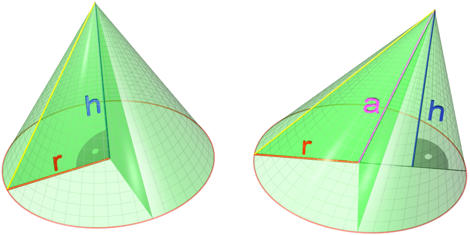
A cone is a three-dimensional geometric shape that tapers smoothly from a flat base (frequently, though not necessarily, circular) to a point called the apex or vertex. A cone is formed by a set of line segments, half-lines, or lines connecting a common point, the apex, to all of the points on a base that is in a plane that does not contain the apex. Depending on the author, the base may be restricted to be a circle, any one-dimensional quadratic form in the plane, any closed one-dimensional figure, or any of the above plus all the enclosed points. If the enclosed points are included in the base, the cone is a solid object; otherwise it is a two-dimensional object in three-dimensional space. In the case of a solid object, the boundary formed by these lines or partial lines is called the ''lateral surface''; if the lateral surface is unbounded, it is a conical surface. In the case of line segments, the cone does not extend beyond the base, while in the case of half-lin ... [...More Info...] [...Related Items...] OR: [Wikipedia] [Google] [Baidu] |
Gap Junctions
Gap junctions are specialized intercellular connections between a multitude of animal cell-types. They directly connect the cytoplasm of two cells, which allows various molecules, ions and electrical impulses to directly pass through a regulated gate between cells. One gap junction channel is composed of two protein hexamers (or hemichannels) called connexons in vertebrates and innexons in invertebrates. The hemichannel pair connect across the intercellular space bridging the gap between two cells. Gap junctions are analogous to the plasmodesmata that join plant cells. Gap junctions occur in virtually all tissues of the body, with the exception of adult fully developed skeletal muscle and mobile cell types such as sperm or erythrocytes. Gap junctions are not found in simpler organisms such as sponges and slime molds. A gap junction may also be called a ''nexus'' or ''macula communicans''. While an ephapse has some similarities to a gap junction, by modern definition the tw ... [...More Info...] [...Related Items...] OR: [Wikipedia] [Google] [Baidu] |
Dendrites
Dendrites (from Greek δένδρον ''déndron'', "tree"), also dendrons, are branched protoplasmic extensions of a nerve cell that propagate the electrochemical stimulation received from other neural cells to the cell body, or soma, of the neuron from which the dendrites project. Electrical stimulation is transmitted onto dendrites by upstream neurons (usually via their axons) via synapses which are located at various points throughout the dendritic tree. Dendrites play a critical role in integrating these synaptic inputs and in determining the extent to which action potentials are produced by the neuron. Dendritic arborization, also known as dendritic branching, is a multi-step biological process by which neurons form new dendritic trees and branches to create new synapses. The morphology of dendrites such as branch density and grouping patterns are highly correlated to the function of the neuron. Malformation of dendrites is also tightly correlated to impaired nervous syste ... [...More Info...] [...Related Items...] OR: [Wikipedia] [Google] [Baidu] |
Retina
The retina (from la, rete "net") is the innermost, light-sensitive layer of tissue of the eye of most vertebrates and some molluscs. The optics of the eye create a focused two-dimensional image of the visual world on the retina, which then processes that image within the retina and sends nerve impulses along the optic nerve to the visual cortex to create visual perception. The retina serves a function which is in many ways analogous to that of the film or image sensor in a camera. The neural retina consists of several layers of neurons interconnected by synapses and is supported by an outer layer of pigmented epithelial cells. The primary light-sensing cells in the retina are the photoreceptor cells, which are of two types: rods and cones. Rods function mainly in dim light and provide monochromatic vision. Cones function in well-lit conditions and are responsible for the perception of colour through the use of a range of opsins, as well as high-acuity vision used for task ... [...More Info...] [...Related Items...] OR: [Wikipedia] [Google] [Baidu] |


