|
6TMS Neutral Amino Acid Transporter Family
The 6TMS Neutral Amino Acid Transporter (NAAT) FamilyTC# 2.A.95 is a family of transporters belonging to the Lysine Exporter (LysE) Superfamily. Homologues are found in numerous Gram-negative and Gram-positive bacteria including many human pathogens. Several archaea also encode MarC (see below) homologues. Some of these organisms have 2 or more paralogues. Most of these proteins are of about the same size (180-230 aas) although a few are larger. They exhibit 6 (or in some cases, possibly 5) putative TMSs. A representative list of members belonging to the NAAT family can be found in thTransporter Classification Database SnatA A gene encoding a small neutral amino acid transporter was cloned from the genome of the hyperthermophilic archaeon ''Thermococcus'' sp. KS-1. The cloned gene, , encodes a protein of 216 amino acid residues, SnatATC# 2.A.95.1.4, with six membrane-spanning segments (TMSs). Competition studies indicated that SnatA transports various L-type neutral amino acids ... [...More Info...] [...Related Items...] OR: [Wikipedia] [Google] [Baidu] |
Homology (biology)
In biology, homology is similarity due to shared ancestry between a pair of structures or genes in different taxa. A common example of homologous structures is the forelimbs of vertebrates, where the wings of bats and birds, the arms of primates, the front flippers of whales and the forelegs of four-legged vertebrates like dogs and crocodiles are all derived from the same ancestral tetrapod structure. Evolutionary biology explains homologous structures adapted to different purposes as the result of descent with modification from a common ancestor. The term was first applied to biology in a non-evolutionary context by the anatomist Richard Owen in 1843. Homology was later explained by Charles Darwin's theory of evolution in 1859, but had been observed before this, from Aristotle onwards, and it was explicitly analysed by Pierre Belon in 1555. In developmental biology, organs that developed in the embryo in the same manner and from similar origins, such as from matching p ... [...More Info...] [...Related Items...] OR: [Wikipedia] [Google] [Baidu] |
Gram-negative Bacteria
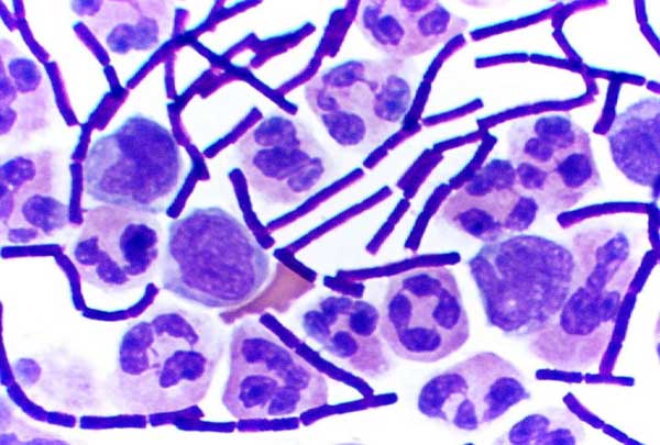
Gram-negative bacteria are bacteria that do not retain the crystal violet stain used in the Gram staining method of bacterial differentiation. They are characterized by their cell envelopes, which are composed of a thin peptidoglycan cell wall sandwiched between an inner cytoplasmic cell membrane and a bacterial outer membrane. Gram-negative bacteria are found in virtually all environments on Earth that support life. The gram-negative bacteria include the model organism ''Escherichia coli'', as well as many pathogenic bacteria, such as ''Pseudomonas aeruginosa'', '' Chlamydia trachomatis'', and ''Yersinia pestis''. They are a significant medical challenge as their outer membrane protects them from many antibiotics (including penicillin), detergents that would normally damage the inner cell membrane, and lysozyme, an antimicrobial enzyme produced by animals that forms part of the innate immune system. Additionally, the outer leaflet of this membrane comprises a complex lipopol ... [...More Info...] [...Related Items...] OR: [Wikipedia] [Google] [Baidu] |
Gram-positive Bacteria
In bacteriology, gram-positive bacteria are bacteria that give a positive result in the Gram stain test, which is traditionally used to quickly classify bacteria into two broad categories according to their type of cell wall. Gram-positive bacteria take up the crystal violet stain used in the test, and then appear to be purple-coloured when seen through an optical microscope. This is because the thick peptidoglycan layer in the bacterial cell wall retains the stain after it is washed away from the rest of the sample, in the decolorization stage of the test. Conversely, gram-negative bacteria cannot retain the violet stain after the decolorization step; alcohol used in this stage degrades the outer membrane of gram-negative cells, making the cell wall more porous and incapable of retaining the crystal violet stain. Their peptidoglycan layer is much thinner and sandwiched between an inner cell membrane and a bacterial outer membrane, causing them to take up the counterstain (sa ... [...More Info...] [...Related Items...] OR: [Wikipedia] [Google] [Baidu] |
Archaea
Archaea ( ; singular archaeon ) is a domain of single-celled organisms. These microorganisms lack cell nuclei and are therefore prokaryotes. Archaea were initially classified as bacteria, receiving the name archaebacteria (in the Archaebacteria kingdom), but this term has fallen out of use. Archaeal cells have unique properties separating them from the other two domains, Bacteria and Eukaryota. Archaea are further divided into multiple recognized phyla. Classification is difficult because most have not been isolated in a laboratory and have been detected only by their gene sequences in environmental samples. Archaea and bacteria are generally similar in size and shape, although a few archaea have very different shapes, such as the flat, square cells of ''Haloquadratum walsbyi''. Despite this morphological similarity to bacteria, archaea possess genes and several metabolic pathways that are more closely related to those of eukaryotes, notably for the enzymes involved ... [...More Info...] [...Related Items...] OR: [Wikipedia] [Google] [Baidu] |
Hyperthermophile
A hyperthermophile is an organism that thrives in extremely hot environments—from 60 °C (140 °F) upwards. An optimal temperature for the existence of hyperthermophiles is often above 80 °C (176 °F). Hyperthermophiles are often within the domain Archaea, although some bacteria are also able to tolerate extreme temperatures. Some of these bacteria are able to live at temperatures greater than 100 °C, deep in the ocean where high pressures increase the boiling point of water. Many hyperthermophiles are also able to withstand other environmental extremes, such as high acidity or high radiation levels. Hyperthermophiles are a subset of extremophiles. Their existence may support the possibility of extraterrestrial life, showing that life can thrive in environmental extremes. History Hyperthermophiles isolated from hot springs in Yellowstone National Park were first reported by Thomas D. Brock in 1965. Since then, more than 70 species have been established. The most extreme hypert ... [...More Info...] [...Related Items...] OR: [Wikipedia] [Google] [Baidu] |
Protonophore
A protonophore, also known as a proton translocator, is an ionophore that moves protons across lipid bilayers or other type of membranes. This would otherwise not occur as protons cations (H+) have positive charge and hydrophilic properties, making them unable to cross without a channel or transporter in the form of a protonophore. Protonophores are generally aromatic compounds with a negative charge, that are both hydrophobic and capable of distributing the negative charge over a number of atoms by π- orbitals which delocalize a proton's charge when it attaches to the molecule. Both the neutral and the charged protonophore can diffuse across the lipid bilayer by passive diffusion and simultaneously facilitate proton transport. Protonophores uncouple oxidative phosphorylation via a decrease in the membrane potential of the inner membrane of mitochondria. They stimulate mitochondria respiration and heat production. Protonophores (uncouplers) are often used in biochemistry research t ... [...More Info...] [...Related Items...] OR: [Wikipedia] [Google] [Baidu] |
Valinomycin
Valinomycin is a naturally occurring dodecadepsipeptide used in the transport of potassium and as an antibiotic. Valinomycin is obtained from the cells of several '' Streptomyces'' species, '' S. fulvissimus'' being a notable one. It is a member of the group of natural neutral ionophores because it does not have a residual charge. It consists of enantiomers D- and L-valine (Val), D- alpha-hydroxyisovaleric acid, and L-lactic acid. Structures are alternately bound via amide and ester bridges. Valinomycin is highly selective for potassium ions over sodium ions within the cell membrane. It functions as a potassium-specific transporter and facilitates the movement of potassium ions through lipid membranes "down" the electrochemical potential gradient. The stability constant K for the potassium-valinomycin complex is nearly 100,000 times larger than that of the sodium-valinomycin complex. This difference is important for maintaining the selectivity of valinomycin for the transport of ... [...More Info...] [...Related Items...] OR: [Wikipedia] [Google] [Baidu] |
Nigericin
Nigericin is an antibiotic derived from '' Streptomyces hygroscopicus''. Its isolation was described in the 1950s, and in 1968 the structure could be elucidated by X-ray crystallography. The structure and properties of nigericin are similar to the antibiotic monensin. Commercially it is obtained as a byproduct, or contaminant, at the fermentation of Geldanamycin. It is also called Polyetherin A, Azalomycin M, Helixin C, Antibiotic K178, Antibiotic X-464. Nigericin acts as an H+, K+, Pb2+ ionophore. Most commonly it is an antiporter of H+ and K+. In the past nigericin was used as an antibiotic active against gram positive bacteria. It inhibits the Golgi functions in Eukaryotic cells. Its ability to induce K+ efflux also makes it a potent activator of the NLRP3 inflammasome Inflammasomes are cytosolic multiprotein oligomers of the innate immune system responsible for the activation of inflammatory responses. Activation and assembly of the inflammasome promotes proteolytic ... [...More Info...] [...Related Items...] OR: [Wikipedia] [Google] [Baidu] |
Murein
Peptidoglycan or murein is a unique large macromolecule, a polysaccharide, consisting of sugars and amino acids that forms a mesh-like peptidoglycan layer outside the plasma membrane, the rigid cell wall (murein sacculus) characteristic of most bacteria (domain ''Bacteria''). The sugar component consists of alternating residues of β-(1,4) linked ''N''-acetylglucosamine (NAG) and ''N''-acetylmuramic acid (NAM). Attached to the ''N''-acetylmuramic acid is a oligopeptide chain made of three to five amino acids. The peptide chain can be cross-linked to the peptide chain of another strand forming the 3D mesh-like layer. Peptidoglycan serves a structural role in the bacterial cell wall, giving structural strength, as well as counteracting the osmotic pressure of the cytoplasm. This repetitive linking results in a dense peptidoglycan layer which is critical for maintaining cell form and withstanding high osmotic pressures, and it is regularly replaced by peptidoglycan production. Peptid ... [...More Info...] [...Related Items...] OR: [Wikipedia] [Google] [Baidu] |
Pleiotropic-drug-resistance Protein
Steroid-transporting ATPase (, ''pleiotropic-drug-resistance protein'', ''PDR protein'') is an enzyme with systematic name ''ATP phosphohydrolase (steroid-exporting)''. This enzyme catalyses the following chemical reaction : ATP + H2O + steroidin \rightleftharpoons ADP + phosphate In chemistry, a phosphate is an anion, salt, functional group or ester derived from a phosphoric acid. It most commonly means orthophosphate, a derivative of orthophosphoric acid . The phosphate or orthophosphate ion is derived from phospho ... + steroidout This enzyme has two similar ATP-binding domains. References External links * EC 3.6.3 {{3.6-enzyme-stub ... [...More Info...] [...Related Items...] OR: [Wikipedia] [Google] [Baidu] |
Protein Families
A protein family is a group of evolutionarily related proteins. In many cases, a protein family has a corresponding gene family, in which each gene encodes a corresponding protein with a 1:1 relationship. The term "protein family" should not be confused with family as it is used in taxonomy. Proteins in a family descend from a common ancestor and typically have similar three-dimensional structures, functions, and significant sequence similarity. The most important of these is sequence similarity (usually amino-acid sequence), since it is the strictest indicator of homology and therefore the clearest indicator of common ancestry. A fairly well developed framework exists for evaluating the significance of similarity between a group of sequences using sequence alignment methods. Proteins that do not share a common ancestor are very unlikely to show statistically significant sequence similarity, making sequence alignment a powerful tool for identifying the members of protein familie ... [...More Info...] [...Related Items...] OR: [Wikipedia] [Google] [Baidu] |


.jpg)
