|
4Pi Microscopy
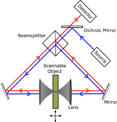
A 4Pi microscope is a laser scanning fluorescence microscope with an improved axial resolution. With it the typical range of the axial resolution of 500–700 nm can be improved to 100–150 nm, which corresponds to an almost spherical focal spot with 5–7 times less volume than that of standard confocal microscopy. Working principle The improvement in resolution is achieved by using two opposing objective lenses, which both are focused to the same geometrical location. Also the difference in optical path length through each of the two objective lenses is carefully aligned to be minimal. By this method, molecules residing in the common focal area of both objectives can be illuminated coherently from both sides and the reflected or emitted light can also be collected coherently, i.e. coherent superposition of emitted light on the detector is possible. The solid angle \Omega that is used for illumination and detection is increased and approaches its maximum. In this case t ... [...More Info...] [...Related Items...] OR: [Wikipedia] [Google] [Baidu] |
Fluorescence Microscope
A fluorescence microscope is an optical microscope that uses fluorescence instead of, or in addition to, scattering, reflection, and attenuation or absorption, to study the properties of organic or inorganic substances. "Fluorescence microscope" refers to any microscope that uses fluorescence to generate an image, whether it is a simple set up like an epifluorescence microscope or a more complicated design such as a confocal microscope, which uses optical sectioning to get better resolution of the fluorescence image. Principle The specimen is illuminated with light of a specific wavelength (or wavelengths) which is absorbed by the fluorophores, causing them to emit light of longer wavelengths (i.e., of a different color than the absorbed light). The illumination light is separated from the much weaker emitted fluorescence through the use of a spectral emission filter. Typical components of a fluorescence microscope are a light source (xenon arc lamp or mercury-vapor lamp are ... [...More Info...] [...Related Items...] OR: [Wikipedia] [Google] [Baidu] |
Hologram
Holography is a technique that enables a wavefront to be recorded and later re-constructed. Holography is best known as a method of generating real three-dimensional images, but it also has a wide range of other Holography#Applications, applications. In principle, it is possible to make a hologram for any type of Holography#Non-optical holography, wave. A hologram is made by superimposing a second wavefront (normally called the reference beam) on the wavefront of interest, thereby generating an interference pattern which is recorded on a physical medium. When only the second wavefront illuminates the interference pattern, it is diffracted to recreate the original wavefront. Holograms can also be Computer-generated holography, computer-generated by modelling the two wavefronts and adding them together digitally. The resulting digital image is then printed onto a suitable mask or film and illuminated by a suitable source to reconstruct the wavefront of interest. Overview and ... [...More Info...] [...Related Items...] OR: [Wikipedia] [Google] [Baidu] |
Laboratory Equipment
A laboratory (; ; colloquially lab) is a facility that provides controlled conditions in which scientific or technological research, experiments, and measurement may be performed. Laboratory services are provided in a variety of settings: physicians' offices, clinics, hospitals, and regional and national referral centers. Overview The organisation and contents of laboratories are determined by the differing requirements of the specialists working within. A physics laboratory might contain a particle accelerator or vacuum chamber, while a metallurgy laboratory could have apparatus for casting or refining metals or for testing their strength. A chemist or biologist might use a wet laboratory, while a psychologist's laboratory might be a room with one-way mirrors and hidden cameras in which to observe behavior. In some laboratories, such as those commonly used by computer scientists, computers (sometimes supercomputers) are used for either simulations or the analysis of data. Scien ... [...More Info...] [...Related Items...] OR: [Wikipedia] [Google] [Baidu] |
Cell Imaging
Cell most often refers to: * Cell (biology), the functional basic unit of life Cell may also refer to: Locations * Monastic cell, a small room, hut, or cave in which a religious recluse lives, alternatively the small precursor of a monastery with only a few monks or nuns * Prison cell, a room used to hold people in prisons Groups of people * Cell, a group of people in a cell group, a form of Christian church organization * Cell, a unit of a clandestine cell system, a penetration-resistant form of a secret or outlawed organization * Cellular organizational structure, such as in business management Science, mathematics, and technology Computing and telecommunications * Cell (EDA), a term used in an electronic circuit design schematics * Cell (microprocessor), a microprocessor architecture developed by Sony, Toshiba, and IBM * Memory cell (computing) The memory cell is the fundamental building block of computer memory. The memory cell is an electronic circuit that stores on ... [...More Info...] [...Related Items...] OR: [Wikipedia] [Google] [Baidu] |
Fluorescence
Fluorescence is the emission of light by a substance that has absorbed light or other electromagnetic radiation. It is a form of luminescence. In most cases, the emitted light has a longer wavelength, and therefore a lower photon energy, than the absorbed radiation. A perceptible example of fluorescence occurs when the absorbed radiation is in the ultraviolet region of the electromagnetic spectrum (invisible to the human eye), while the emitted light is in the visible region; this gives the fluorescent substance a distinct color that can only be seen when the substance has been exposed to UV light. Fluorescent materials cease to glow nearly immediately when the radiation source stops, unlike phosphorescent materials, which continue to emit light for some time after. Fluorescence has many practical applications, including mineralogy, gemology, medicine, chemical sensors (fluorescence spectroscopy), fluorescent labelling, dyes, biological detectors, cosmic-ray detection, vacu ... [...More Info...] [...Related Items...] OR: [Wikipedia] [Google] [Baidu] |
Microscopes
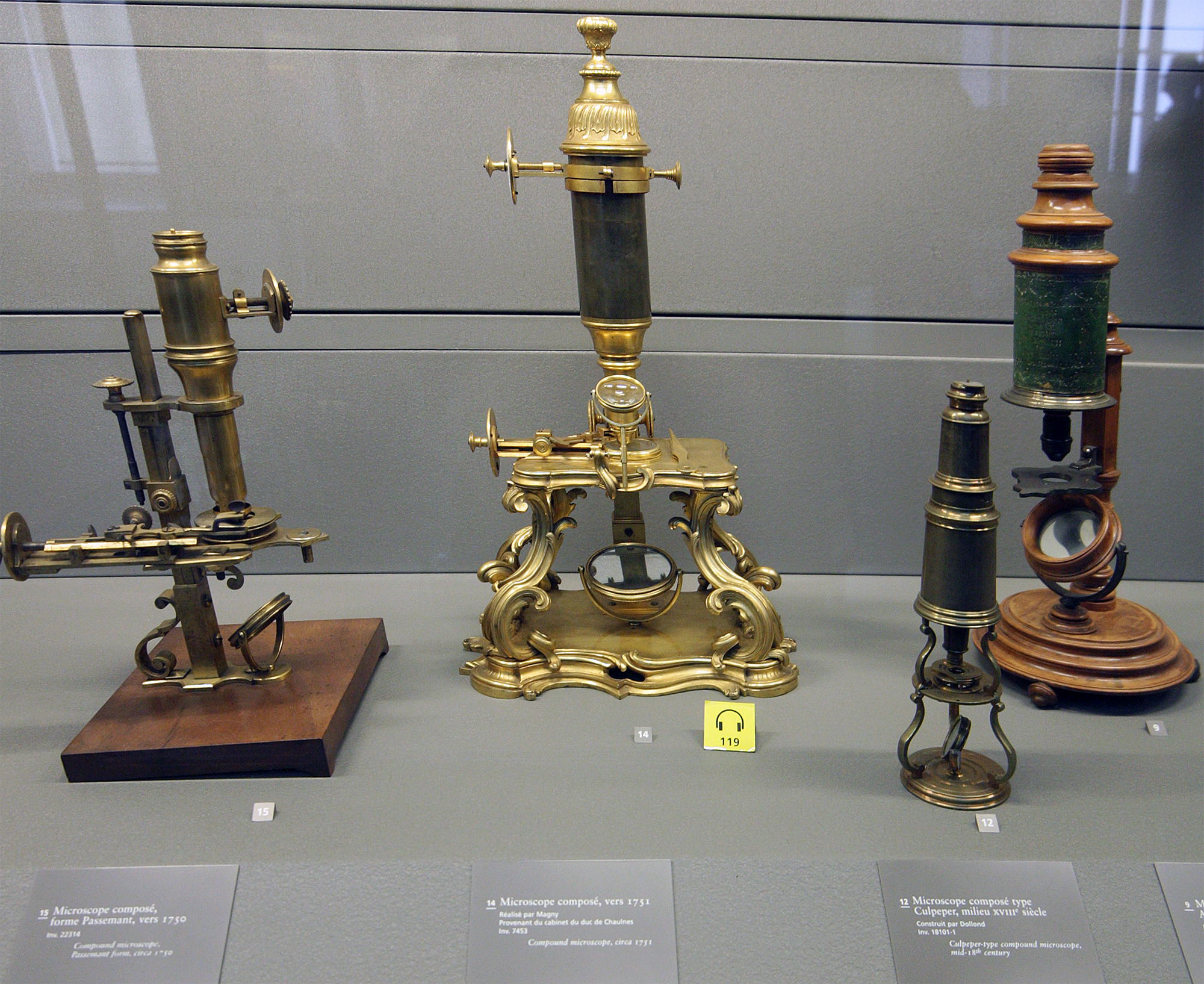
A microscope () is a laboratory instrument used to examine objects that are too small to be seen by the naked eye. Microscopy is the science of investigating small objects and structures using a microscope. Microscopic means being invisible to the eye unless aided by a microscope. There are many types of microscopes, and they may be grouped in different ways. One way is to describe the method an instrument uses to interact with a sample and produce images, either by sending a beam of light or electrons through a sample in its optical path, by detecting photon emissions from a sample, or by scanning across and a short distance from the surface of a sample using a probe. The most common microscope (and the first to be invented) is the optical microscope, which uses lenses to refract visible light that passed through a thinly sectioned sample to produce an observable image. Other major types of microscopes are the fluorescence microscope, electron microscope (both the transmi ... [...More Info...] [...Related Items...] OR: [Wikipedia] [Google] [Baidu] |
Multifocal Plane Microscopy
Multifocal plane microscopy (MUM) or multiplane microscopy or multifocus microscopy is a form of light microscopy that allows the tracking of the 3D dynamics in live cells at high temporal and spatial resolution by simultaneously imaging different focal planes within the specimen. In this methodology, the light collected from the sample by an infinity-corrected objective lens is split into two paths. In each path the split light is focused onto a detector which is placed at a specific calibrated distance from the tube lens. In this way, each detector images a distinct plane within the sample. The first developed MUM setup was capable of imaging two distinct planes within the sample. However, the setup can be modified to image more than two planes by further splitting the light in each light path and focusing it onto detectors placed at specific calibrated distances. It has later been improved for imaging up to four distinct planes. To image a greater number of focal planes, simple ... [...More Info...] [...Related Items...] OR: [Wikipedia] [Google] [Baidu] |
Stimulated Emission Depletion Microscope
Stimulated emission depletion (STED) microscopy is one of the techniques that make up super-resolution microscopy. It creates super-resolution images by the selective deactivation of fluorophores, minimizing the area of illumination at the focal point, and thus enhancing the achievable resolution for a given system. It was developed by Stefan W. Hell and Jan Wichmann in 1994, and was first experimentally demonstrated by Hell and Thomas Klar in 1999. Hell was awarded the Nobel Prize in Chemistry in 2014 for its development. In 1986, V.A. Okhonin (Institute of Biophysics, USSR Academy of Sciences, Siberian Branch, Krasnoyarsk) had patented the STED idea. This patent was unknown to Hell and Wichmann in 1994. STED microscopy is one of several types of super resolution microscopy techniques that have recently been developed to bypass the diffraction limit of light microscopy to increase resolution. STED is a deterministic functional technique that exploits the non-linear response of f ... [...More Info...] [...Related Items...] OR: [Wikipedia] [Google] [Baidu] |
RESOLFT
RESOLFT, an acronym for REversible Saturable OpticaL Fluorescence Transitions, denotes a group of optical fluorescence microscopy techniques with very high resolution. Using standard far field visible light optics a resolution far below the diffraction limit down to molecular scales can be obtained. With conventional microscopy techniques, it is not possible to distinguish features that are located at distances less than about half the wavelength used (i.e. about 200 nm for visible light). This diffraction limit is based on the wave nature of light. In conventional microscopes the limit is determined by the used wavelength and the numerical aperture of the optical system. The RESOLFT concept surmounts this limit by temporarily switching the molecules to a state in which they cannot send a (fluorescence-) signal upon illumination. This concept is different from for example electron microscopy where instead the used wavelength is much smaller. Working principle RESOLFT micro ... [...More Info...] [...Related Items...] OR: [Wikipedia] [Google] [Baidu] |
Stimulated Emission Depletion Microscope
Stimulated emission depletion (STED) microscopy is one of the techniques that make up super-resolution microscopy. It creates super-resolution images by the selective deactivation of fluorophores, minimizing the area of illumination at the focal point, and thus enhancing the achievable resolution for a given system. It was developed by Stefan W. Hell and Jan Wichmann in 1994, and was first experimentally demonstrated by Hell and Thomas Klar in 1999. Hell was awarded the Nobel Prize in Chemistry in 2014 for its development. In 1986, V.A. Okhonin (Institute of Biophysics, USSR Academy of Sciences, Siberian Branch, Krasnoyarsk) had patented the STED idea. This patent was unknown to Hell and Wichmann in 1994. STED microscopy is one of several types of super resolution microscopy techniques that have recently been developed to bypass the diffraction limit of light microscopy to increase resolution. STED is a deterministic functional technique that exploits the non-linear response of f ... [...More Info...] [...Related Items...] OR: [Wikipedia] [Google] [Baidu] |
Leica Microsystems
Leica Microsystems GmbH is a German microscope manufacturing company. It is a manufacturer of optical microscopes, equipment for the preparation of microscopic specimens and related products. There are ten plants in eight countries with distribution partners in over 100 countries. Leica Microsystems emerged in 1997 out of a 1990 merger between Wild-Leitz, headquartered in Heerbrugg Switzerland, and Cambridge Instruments of Cambridge England. The merger of those two umbrella companies created an alliance of the following 8 individual manufacturers of scientific instruments. American Optical Scientific Products, Carl Reichert Optische Werke AG, R.Jung, Bausch and Lomb Optical Scientific Products Division, Cambridge Instruments, E.Leitz Wetzlar, Kern & Co., and Wild Heerbrugg AG, bringing much-needed modernization and a broader degree of expertise to the newly created entity called Leica Holding B.V. group. In 1997 the name was changed to Leica Microsystems and is a wholly-owned ent ... [...More Info...] [...Related Items...] OR: [Wikipedia] [Google] [Baidu] |
Mitochondria
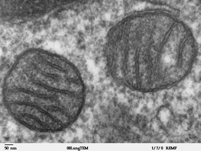
A mitochondrion (; ) is an organelle found in the Cell (biology), cells of most Eukaryotes, such as animals, plants and Fungus, fungi. Mitochondria have a double lipid bilayer, membrane structure and use aerobic respiration to generate adenosine triphosphate (ATP), which is used throughout the cell as a source of chemical energy. They were discovered by Albert von Kölliker in 1857 in the voluntary muscles of insects. The term ''mitochondrion'' was coined by Carl Benda in 1898. The mitochondrion is popularly nicknamed the "powerhouse of the cell", a phrase coined by Philip Siekevitz in a 1957 article of the same name. Some cells in some multicellular organisms lack mitochondria (for example, mature mammalian red blood cells). A large number of unicellular organisms, such as microsporidia, parabasalids and diplomonads, have reduced or transformed their mitochondria into mitosome, other structures. One eukaryote, ''Monocercomonoides'', is known to have completely lost its mitocho ... [...More Info...] [...Related Items...] OR: [Wikipedia] [Google] [Baidu] |


