Upper Extremity Of Radius on:
[Wikipedia]
[Google]
[Amazon]
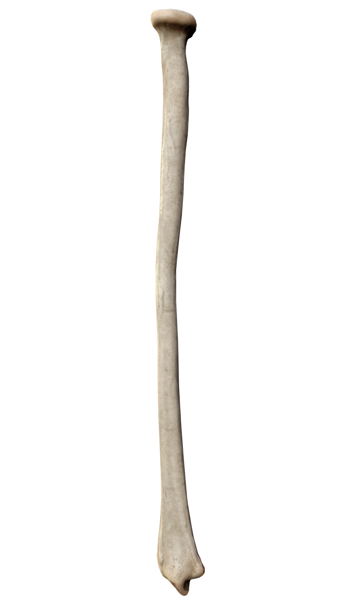
The radius or radial bone is one of the two large


 The long narrow medullary cavity is enclosed in a strong wall of compact bone. It is thickest along the interosseous border and thinnest at the extremities, same over the cup-shaped articular surface (fovea) of the head.
The trabeculae of the spongy tissue are somewhat arched at the upper end and pass upward from the compact layer of the shaft to the ''fovea capituli'' (the
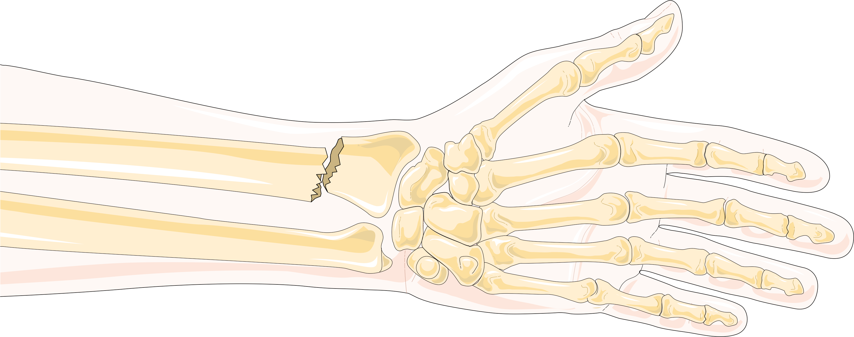
 Specific fracture types of the radius include:
*Proximal radius fracture. A fracture within the capsule of the
  ** Essex-Lopresti fracture – a fracture of the
at Wheeless' Textbook of Orthopaedics online *Radial shaft fracture * Distal radius fracture ** Galeazzi fracture – a fracture of the radius with dislocation of the
bone
A bone is a rigid organ that constitutes part of the skeleton in most vertebrate animals. Bones protect the various other organs of the body, produce red and white blood cells, store minerals, provide structure and support for the body, an ...
s of the forearm
The forearm is the region of the upper limb between the elbow and the wrist. The term forearm is used in anatomy to distinguish it from the arm, a word which is most often used to describe the entire appendage of the upper limb, but which in ...
, the other being the ulna
The ulna (''pl''. ulnae or ulnas) is a long bone found in the forearm that stretches from the elbow to the smallest finger, and when in anatomical position, is found on the medial side of the forearm. That is, the ulna is on the same side of t ...
. It extends from the lateral
Lateral is a geometric term of location which may refer to:
Healthcare
*Lateral (anatomy), an anatomical direction
* Lateral cricoarytenoid muscle
* Lateral release (surgery), a surgical procedure on the side of a kneecap
Phonetics
*Lateral co ...
side of the elbow
The elbow is the region between the arm and the forearm that surrounds the elbow joint. The elbow includes prominent landmarks such as the olecranon, the cubital fossa (also called the chelidon, or the elbow pit), and the lateral and the m ...
to the thumb
The thumb is the first digit of the hand, next to the index finger. When a person is standing in the medical anatomical position (where the palm is facing to the front), the thumb is the outermost digit. The Medical Latin English noun for thu ...
side of the wrist
In human anatomy, the wrist is variously defined as (1) the carpus or carpal bones, the complex of eight bones forming the proximal skeletal segment of the hand; "The wrist contains eight bones, roughly aligned in two rows, known as the carpal ...
and runs parallel to the ulna. The ulna is usually slightly longer than the radius, but the radius is thicker. Therefore the radius is considered to be the larger of the two. It is a long bone, prism
Prism usually refers to:
* Prism (optics), a transparent optical component with flat surfaces that refract light
* Prism (geometry), a kind of polyhedron
Prism may also refer to:
Science and mathematics
* Prism (geology), a type of sedimentary ...
-shaped and slightly curved longitudinally.
The radius is part of two joints
A joint or articulation (or articular surface) is the connection made between bones, ossicles, or other hard structures in the body which link an animal's skeletal system into a functional whole.Saladin, Ken. Anatomy & Physiology. 7th ed. McGraw- ...
: the elbow
The elbow is the region between the arm and the forearm that surrounds the elbow joint. The elbow includes prominent landmarks such as the olecranon, the cubital fossa (also called the chelidon, or the elbow pit), and the lateral and the m ...
and the wrist
In human anatomy, the wrist is variously defined as (1) the carpus or carpal bones, the complex of eight bones forming the proximal skeletal segment of the hand; "The wrist contains eight bones, roughly aligned in two rows, known as the carpal ...
. At the elbow, it joins with the capitulum of the humerus
In human anatomy of the arm, the capitulum of the humerus is a smooth, rounded eminence on the lateral portion of the distal articular surface of the humerus. It articulates with the cupshaped depression on the head of the radius, and is limite ...
, and in a separate region, with the ulna at the radial notch. At the wrist, the radius forms a joint with the ulna bone.
The corresponding bone in the lower leg
The human leg, in the general word sense, is the entire lower limb of the human body, including the foot, thigh or sometimes even the hip or gluteal region. However, the definition in human anatomy refers only to the section of the lower limb ext ...
is the fibula
The fibula or calf bone is a human leg, leg bone on the Lateral (anatomy), lateral side of the tibia, to which it is connected above and below. It is the smaller of the two bones and, in proportion to its length, the most slender of all the long ...
.
Structure


 The long narrow medullary cavity is enclosed in a strong wall of compact bone. It is thickest along the interosseous border and thinnest at the extremities, same over the cup-shaped articular surface (fovea) of the head.
The trabeculae of the spongy tissue are somewhat arched at the upper end and pass upward from the compact layer of the shaft to the ''fovea capituli'' (the
 humerus
The humerus (; ) is a long bone in the arm that runs from the shoulder to the elbow. It connects the scapula and the two bones of the lower arm, the radius and ulna, and consists of three sections. The humeral upper extremity consists of a roun ...
's cup-shaped articulatory notch); they are crossed by others parallel to the surface of the fovea. The arrangement at the lower end is somewhat similar. It is missing in radial aplasia.
The radius has a body and two extremities. The upper extremity of the radius consists of a somewhat cylindrical head articulating with the ulna and the humerus, a neck, and a radial tuberosity
Beneath the neck of the radius, on the medial side, is an eminence, the radial tuberosity; its surface is divided into:
* a ''posterior, rough portion'', for the insertion of the tendon of the biceps brachii.
* an ''anterior, smooth portion'', on ...
. The body of the radius is self-explanatory, and the lower extremity of the radius is roughly quadrilateral in shape, with articular surfaces for the ulna
The ulna (''pl''. ulnae or ulnas) is a long bone found in the forearm that stretches from the elbow to the smallest finger, and when in anatomical position, is found on the medial side of the forearm. That is, the ulna is on the same side of t ...
, scaphoid
The scaphoid bone is one of the carpal bones of the wrist. It is situated between the hand and forearm on the thumb side of the wrist (also called the lateral or radial side). It forms the radial border of the carpal tunnel. The scaphoid bone ...
and lunate bone
The lunate bone (semilunar bone) is a carpal bone in the human hand. It is distinguished by its deep concavity and crescentic outline. It is situated in the center of the proximal row carpal bones, which lie between the ulna and radius and the han ...
s. The distal end of the radius forms two palpable points, radially the styloid process and Lister's tubercle on the ulnar side. Along with the proximal
Standard anatomical terms of location are used to unambiguously describe the anatomy of animals, including humans. The terms, typically derived from Latin or Greek roots, describe something in its standard anatomical position. This position ...
and distal radioulnar articulation
The distal radioulnar articulation (also known as the distal radioulnar joint, or inferior radioulnar joint) is a synovial pivot joint between the two bones in the forearm; the radius and ulna. It is one of two joints between the radius and ulna, ...
s, an interosseous membrane
An interosseous membrane is a thick dense fibrous sheet of connective tissue that spans the space between two bones, forming a type of syndesmosis joint.
Interosseous membranes in the human body:
* Interosseous membrane of forearm
The interosse ...
originates medially along the length of the body of the radius to attach the radius to the ulna.
Near the wrist
The distal end of the radius is large and of quadrilateral form. ;Joint surfaces It is provided with two articular surfaces – one below, for the carpus, and another at the medial side, for theulna
The ulna (''pl''. ulnae or ulnas) is a long bone found in the forearm that stretches from the elbow to the smallest finger, and when in anatomical position, is found on the medial side of the forearm. That is, the ulna is on the same side of t ...
.
* The ''carpal'' articular surface is triangular, concave, smooth, and divided by a slight antero-posterior ridge into two parts. Of these, the lateral, triangular, articulates with the scaphoid bone; the medial, quadrilateral, with the lunate bone
The lunate bone (semilunar bone) is a carpal bone in the human hand. It is distinguished by its deep concavity and crescentic outline. It is situated in the center of the proximal row carpal bones, which lie between the ulna and radius and the han ...
.
* The articular surface for the ''ulna'' is called the ulnar notch (''sigmoid cavity'') of the radius; it is narrow, concave, smooth, and articulates with the head of the ulna.
These two articular surfaces are separated by a prominent ridge, to which the base of the triangular articular disk is attached; this disk separates the wrist-joint from the distal radioulnar articulation.
;Other surfaces
This end of the bone has three non-articular surfaces – volar, dorsal, and lateral.
* The ''volar surface'', rough and irregular, affords attachment to the volar radiocarpal ligament
The palmar radiocarpal ligament (anterior ligament, volar radiocarpal ligament) is a broad membranous band, attached above to the distal end of the radius, and passing downward to the scaphoid, lunate, triquetrum and capitate of the carpal bones ...
.
* The ''dorsal surface'' is convex, affords attachment to the dorsal radiocarpal ligament, and is marked by three grooves. Enumerated from the lateral side:
** The ''first'' groove is broad, but shallow, and subdivided into two by a slight ridge: the lateral of these two, transmits the tendon of the extensor carpi radialis longus muscle; the medial, the tendon of the extensor carpi radialis brevis muscle
In human anatomy, extensor carpi radialis brevis is a muscle in the forearm that acts to extend and abduct the wrist. It is shorter and thicker than its namesake extensor carpi radialis longus which can be found above the proximal end of the exten ...
.
** The ''second'' is deep but narrow, and bounded laterally by a sharply defined ridge; it is directed obliquely from above downward and lateralward, and transmits the tendon of the extensor pollicis longus muscle
In human anatomy, the extensor pollicis longus muscle (EPL) is a skeletal muscle located dorsally on the forearm. It is much larger than the extensor pollicis brevis, the origin of which it partly covers and acts to stretch the thumb together wi ...
.
** The ''third'' is broad, for the passage of the tendons of the extensor indicis proprius and extensor digitorum communis
The extensor digitorum muscle (also known as extensor digitorum communis) is a muscle of the posterior forearm present in humans and other animals. It extends the medial four digits of the hand. Extensor digitorum is innervated by the posterior int ...
.
* The ''lateral surface'' is prolonged obliquely downward into a strong, conical projection, the styloid process, which gives attachment by its base to the tendon of the brachioradialis, and by its apex to the radial collateral ligament of wrist joint
The radial collateral ligament (external lateral ligament, radial carpal collateral ligament) extends from the tip of the styloid process of the radius and attaches to the radial side of the scaphoid (formerly Navicular bone of the hand), immedia ...
. The lateral surface of this process is marked by a flat groove, for the tendons of the abductor pollicis longus
In human anatomy, the abductor pollicis longus (APL) is one of the extrinsic muscles of the hand. Its major function is to abduct the thumb at the wrist. Its tendon forms the anterior border of the anatomical snuffbox.
Structure
The abductor ...
muscle and extensor pollicis brevis
In human anatomy, the extensor pollicis brevis is a skeletal muscle on the dorsal side of the forearm. It lies on the medial side of, and is closely connected with, the abductor pollicis longus. The extensor pollicis brevis (EPB) belongs to the ...
muscle.
Body
The body of the radius (or shaft of radius) is prismoid in form, narrower above than below, and slightly curved, so as to be convex lateralward. It presents three borders and three surfaces. ;Borders The volar border (''margo volaris; anterior border; palmar'';) extends from the lower part of the tuberosity above to the anterior part of the base of the styloid process below, and separates the volar from the lateral surface. Its upper third is prominent, and from its oblique direction has received the name of the oblique line of the radius; it gives origin to theflexor digitorum superficialis muscle
Flexor digitorum superficialis (''flexor digitorum sublimis'') is an extrinsic flexor muscle of the fingers at the proximal interphalangeal joints.
It is in the anterior compartment of the forearm. It is sometimes considered to be the deepest part ...
(also ''flexor digitorum sublimis'') and flexor pollicis longus muscle; the surface above the line gives insertion to part of the supinator muscle
In human anatomy, the supinator is a broad muscle in the posterior compartment of the forearm, curved around the upper third of the radius. Its function is to supinate the forearm.
Structure
Supinator consists of two planes of fibers, between whi ...
. The middle third of the volar border is indistinct and rounded. The lower fourth is prominent, and gives insertion to the pronator quadratus muscle, and attachment to the dorsal carpal ligament; it ends in a small tubercle, into which the tendon of the brachioradialis muscle is inserted.
The dorsal border (''margo dorsalis; posterior border'') begins above at the back of the neck, and ends below at the posterior part of the base of the styloid process; it separates the posterior from the lateral surface. is indistinct above and below, but well-marked in the middle third of the bone.
The interosseous border (''internal border; crista interossea; interosseous crest;'') begins above, at the back part of the tuberosity, and its upper part is rounded and indistinct; it becomes sharp and prominent as it descends, and at its lower part divides into two ridges which are continued to the anterior and posterior margins of the ulnar notch. To the posterior of the two ridges the lower part of the interosseous membrane
An interosseous membrane is a thick dense fibrous sheet of connective tissue that spans the space between two bones, forming a type of syndesmosis joint.
Interosseous membranes in the human body:
* Interosseous membrane of forearm
The interosse ...
is attached, while the triangular surface between the ridges gives insertion to part of the pronator quadratus muscle. This crest separates the volar from the dorsal surface, and gives attachment to the interosseous membrane. The connection between the two bones is actually a joint referred to as a syndesmosis
In anatomy, fibrous joints are joints connected by fibrous tissue, consisting mainly of collagen. These are fixed joints where bones are united by a layer of white fibrous tissue of varying thickness. In the skull the joints between the bones ar ...
joint.
;Surfaces
The volar surface (''facies volaris; anterior surface'') is concave in its upper three-fourths, and gives origin to the flexor pollicis longus muscle; it is broad and flat in its lower fourth, and affords insertion to the Pronator quadratus
Pronator quadratus is a square-shaped muscle on the distal forearm that acts to pronate (turn so the palm faces downwards) the hand.
Structure
Its fibres run perpendicular to the direction of the arm, running from the most distal quarter of the ...
. A prominent ridge limits the insertion of the Pronator quadratus below, and between this and the inferior border is a triangular rough surface for the attachment of the volar radiocarpal ligament
The palmar radiocarpal ligament (anterior ligament, volar radiocarpal ligament) is a broad membranous band, attached above to the distal end of the radius, and passing downward to the scaphoid, lunate, triquetrum and capitate of the carpal bones ...
. At the junction of the upper and middle thirds of the volar surface is the nutrient foramen, which is directed obliquely upward.
The dorsal surface (''facies dorsalis; posterior surface'') is convex, and smooth in the upper third of its extent, and covered by the Supinator
In human anatomy, the supinator is a broad muscle in the posterior compartment of the forearm, curved around the upper third of the radius. Its function is to supinate the forearm.
Structure
Supinator consists of two planes of fibers, between whi ...
. Its middle third is broad, slightly concave, and gives origin to the Abductor pollicis longus
In human anatomy, the abductor pollicis longus (APL) is one of the extrinsic muscles of the hand. Its major function is to abduct the thumb at the wrist. Its tendon forms the anterior border of the anatomical snuffbox.
Structure
The abductor ...
above, and the extensor pollicis brevis muscle below. Its lower third is broad, convex, and covered by the tendons of the muscles which subsequently run in the grooves on the lower end of the bone.
The lateral surface (''facies lateralis; external surface'') is convex throughout its entire extent and is known as the convexity of the radius, curving outwards to be convex at the side. Its upper third gives insertion to the supinator muscle
In human anatomy, the supinator is a broad muscle in the posterior compartment of the forearm, curved around the upper third of the radius. Its function is to supinate the forearm.
Structure
Supinator consists of two planes of fibers, between whi ...
. About its center is a rough ridge, for the insertion of the pronator teres muscle. Its lower part is narrow, and covered by the tendons of the abductor pollicis longus muscle and extensor pollicis brevis muscle.
Near the elbow
The upper extremity of the radius (or proximal extremity) presents a head, neck, and tuberosity. * The radial ''head'' has a cylindrical form, and on its upper surface is a shallow cup or fovea for articulation with the capitulum (or capitellum) of thehumerus
The humerus (; ) is a long bone in the arm that runs from the shoulder to the elbow. It connects the scapula and the two bones of the lower arm, the radius and ulna, and consists of three sections. The humeral upper extremity consists of a roun ...
. The circumference of the head is smooth; it is broad medially where it articulates with the radial notch of the ulna
The radial notch of the ulna (lesser sigmoid cavity) is a narrow, oblong, articular depression on the lateral side of the coronoid process
The Coronoid process (from Greek , "like a crown") can refer to:
* The coronoid process of the mandible, par ...
, narrow in the rest of its extent, which is embraced by the annular ligament. The deepest point in the fovea is not axi-symmetric with the long axis of the radius, creating a cam effect during pronation and supination.
* The head is supported on a round, smooth, and constricted portion called the ''neck'', on the back of which is a slight ridge for the insertion of part of the supinator muscle
In human anatomy, the supinator is a broad muscle in the posterior compartment of the forearm, curved around the upper third of the radius. Its function is to supinate the forearm.
Structure
Supinator consists of two planes of fibers, between whi ...
.
* Beneath the neck, on the medial side, is an eminence, the ''radial tuberosity
Beneath the neck of the radius, on the medial side, is an eminence, the radial tuberosity; its surface is divided into:
* a ''posterior, rough portion'', for the insertion of the tendon of the biceps brachii.
* an ''anterior, smooth portion'', on ...
''; its surface is divided into a posterior, rough portion, for the insertion of the tendon of the biceps brachii muscle, and an anterior, smooth portion, on which a bursa
( grc-gre, Προῦσα, Proûsa, Latin: Prusa, ota, بورسه, Arabic:بورصة) is a city in northwestern Turkey and the administrative center of Bursa Province. The fourth-most populous city in Turkey and second-most populous in t ...
is interposed between the tendon
A tendon or sinew is a tough, high-tensile-strength band of dense fibrous connective tissue that connects muscle to bone. It is able to transmit the mechanical forces of muscle contraction to the skeletal system without sacrificing its ability ...
and the bone.
Development
The radius isossified
Ossification (also called osteogenesis or bone mineralization) in bone remodeling is the process of laying down new bone material by cells named osteoblasts. It is synonymous with bone tissue formation. There are two processes resulting in t ...
from ''three'' centers: one for the body, and one for each extremity. That for the body makes its appearance near the center of the bone, during the eighth week of fetal
A fetus or foetus (; plural fetuses, feti, foetuses, or foeti) is the unborn offspring that develops from an animal embryo. Following embryonic development the fetal stage of development takes place. In human prenatal development, fetal develo ...
life.
Ossification commences in the lower end between 9 and 26 months of age. The ossification center for the upper end appears by the fifth year.
The upper epiphysis
The epiphysis () is the rounded end of a long bone, at its joint with adjacent bone(s). Between the epiphysis and diaphysis (the long midsection of the long bone) lies the metaphysis, including the epiphyseal plate (growth plate). At the ...
fuses with the body at the age of seventeen or eighteen years, the lower about the age of twenty.
An additional center sometimes found in the radial tuberosity
Beneath the neck of the radius, on the medial side, is an eminence, the radial tuberosity; its surface is divided into:
* a ''posterior, rough portion'', for the insertion of the tendon of the biceps brachii.
* an ''anterior, smooth portion'', on ...
, appears about the fourteenth or fifteenth year.
Function
Muscle attachments
Thebiceps
The biceps or biceps brachii ( la, musculus biceps brachii, "two-headed muscle of the arm") is a large muscle that lies on the front of the upper arm between the shoulder and the elbow. Both heads of the muscle arise on the scapula and join ...
muscle inserts on the radial tuberosity
Beneath the neck of the radius, on the medial side, is an eminence, the radial tuberosity; its surface is divided into:
* a ''posterior, rough portion'', for the insertion of the tendon of the biceps brachii.
* an ''anterior, smooth portion'', on ...
of the upper extremity of the bone. The upper third of the body of the bone attaches to the supinator
In human anatomy, the supinator is a broad muscle in the posterior compartment of the forearm, curved around the upper third of the radius. Its function is to supinate the forearm.
Structure
Supinator consists of two planes of fibers, between whi ...
, the flexor digitorum superficialis
Flexor digitorum superficialis (''flexor digitorum sublimis'') is an extrinsic flexor muscle of the fingers at the proximal interphalangeal joints.
It is in the anterior compartment of the forearm. It is sometimes considered to be the deepest pa ...
, and the flexor pollicis longus
The flexor pollicis longus (; FPL, Latin ''flexor'', bender; ''pollicis'', of the thumb; ''longus'', long) is a muscle in the forearm and hand that flexes the thumb. It lies in the same plane as the flexor digitorum profundus. This muscle is uniq ...
muscles.
The middle third of the body attaches to the extensor ossis metacarpi pollicis, extensor primi internodii pollicis, and the pronator teres
The pronator teres is a muscle (located mainly in the forearm) that, along with the pronator quadratus, serves to pronate the forearm (turning it so that the palm faces posteriorly when from the anatomical position). It has two attachments, to ...
muscles.
The lower quarter of the body attaches to the pronator quadratus
Pronator quadratus is a square-shaped muscle on the distal forearm that acts to pronate (turn so the palm faces downwards) the hand.
Structure
Its fibres run perpendicular to the direction of the arm, running from the most distal quarter of the ...
muscle and the tendon
A tendon or sinew is a tough, high-tensile-strength band of dense fibrous connective tissue that connects muscle to bone. It is able to transmit the mechanical forces of muscle contraction to the skeletal system without sacrificing its ability ...
of the supinator longus.
Clinical significance
Radial aplasia refers to the congenital absence or shortness of the radius.Fracture
 Specific fracture types of the radius include:
*Proximal radius fracture. A fracture within the capsule of the
 elbow
The elbow is the region between the arm and the forearm that surrounds the elbow joint. The elbow includes prominent landmarks such as the olecranon, the cubital fossa (also called the chelidon, or the elbow pit), and the lateral and the m ...
joint results in the fat pad sign or "sail sign" which is a displacement of the fat pad at the elbow.
 ** Essex-Lopresti fracture – a fracture of the
 radial head
The head of the radius has a cylindrical form, and on its upper surface is a shallow cup or fovea for articulation with the capitulum of the humerus. The circumference of the head is smooth; it is broad medially where it articulates with the ra ...
with concomitant dislocation of the distal radio-ulnar joint with disruption of the interosseous membrane
An interosseous membrane is a thick dense fibrous sheet of connective tissue that spans the space between two bones, forming a type of syndesmosis joint.
Interosseous membranes in the human body:
* Interosseous membrane of forearm
The interosse ...
.Essex Lopresti fractureat Wheeless' Textbook of Orthopaedics online *Radial shaft fracture * Distal radius fracture ** Galeazzi fracture – a fracture of the radius with dislocation of the
distal radioulnar joint
The distal radioulnar articulation (also known as the distal radioulnar joint, or inferior radioulnar joint) is a synovial pivot joint between the two bones in the forearm; the radius and ulna. It is one of two joints between the radius and ulna, ...
**Colles' fracture
A Colles' fracture is a type of fracture of the distal forearm in which the broken end of the radius is bent backwards. Symptoms may include pain, swelling, deformity, and bruising. Complications may include damage to the median nerve.
It typi ...
– a distal fracture of the radius with dorsal (posterior) displacement of the wrist and hand
** Smith's fracture – a distal fracture of the radius with volar (ventral) displacement of the wrist and hand
** Barton's fracture – an intra-articular fracture of the distal radius with dislocation of the radiocarpal joint.
History
The word ''radius'' isLatin
Latin (, or , ) is a classical language belonging to the Italic branch of the Indo-European languages. Latin was originally a dialect spoken in the lower Tiber area (then known as Latium) around present-day Rome, but through the power ...
for "ray". In the context of the radius bone, a ray can be thought of rotating around an axis line extending diagonally from center of capitulum to the center of distal ulna
The ulna (''pl''. ulnae or ulnas) is a long bone found in the forearm that stretches from the elbow to the smallest finger, and when in anatomical position, is found on the medial side of the forearm. That is, the ulna is on the same side of t ...
. While the ulna
The ulna (''pl''. ulnae or ulnas) is a long bone found in the forearm that stretches from the elbow to the smallest finger, and when in anatomical position, is found on the medial side of the forearm. That is, the ulna is on the same side of t ...
is the major contributor to the elbow joint, the radius primarily contributes to the wrist
In human anatomy, the wrist is variously defined as (1) the carpus or carpal bones, the complex of eight bones forming the proximal skeletal segment of the hand; "The wrist contains eight bones, roughly aligned in two rows, known as the carpal ...
joint.
The radius is named so because the radius (bone) acts like the radius (of a circle). It rotates around the ulna and the far end (where it joins to the bones of the hand), known as the styloid process of the radius, is the distance from the ulna (center of the circle) to the edge of the radius (the circle). The ulna acts as the center point to the circle because when the arm is rotated the ulna does not move.
Animals
In four-legged animals, the radius is the main load-bearing bone of the lower forelimb. Its structure is similar in most terrestrialtetrapods
Tetrapods (; ) are four-limb (anatomy), limbed vertebrate animals constituting the superclass Tetrapoda (). It includes extant taxon, extant and extinct amphibians, sauropsids (reptiles, including dinosaurs and therefore birds) and synapsids (p ...
, but it may be fused with the ulna in some mammals (such as horse
The horse (''Equus ferus caballus'') is a domesticated, one-toed, hoofed mammal. It belongs to the taxonomic family Equidae and is one of two extant subspecies of ''Equus ferus''. The horse has evolved over the past 45 to 55 million ...
s) and reduced or modified in animals with flippers or vestigial forelimbs.
Gallery
References
{{Authority control Long bones Bones of the upper limb