Sinus Node on:
[Wikipedia]
[Google]
[Amazon]
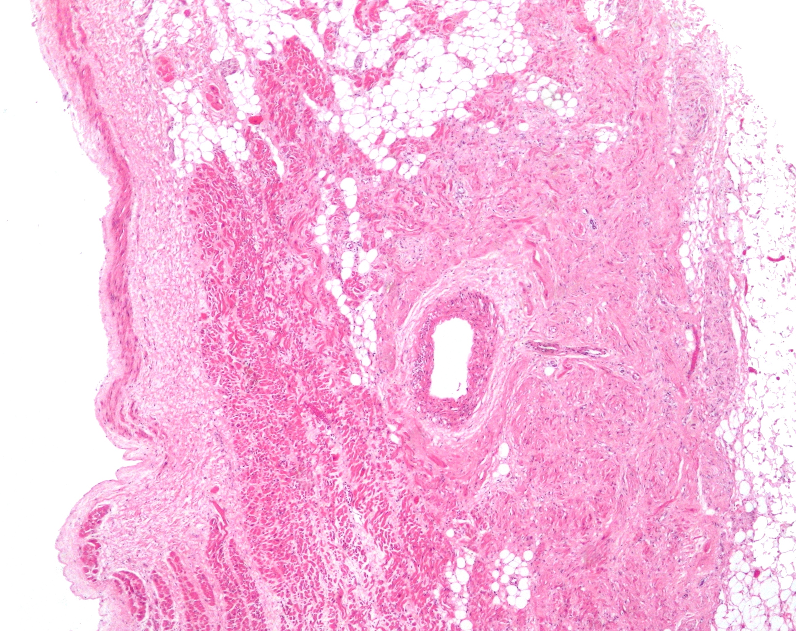
The sinoatrial node (also known as the sinuatrial node, SA node, sinus node or Keith–Flack node) is an oval shaped region of special
 The cells of the SA node are spread out within a mesh of
 This phase is also known as the pacemaker potential. Immediately following repolarization, when the membrane potential is very negative (it is hyperpolarised), the voltage slowly begins to increase. This is initially due to the closing of
Image:Reizleitungssystem 1.png, Heart; conduction system (SA node labeled 1)
Image:Gray501.png , Schematic representation of the atrioventricular bundle
Diagram at gru.net
* () * https://web.archive.org/web/20070929080346/http://www.healthyheart.nhs.uk/heart_works/heart03.shtml {{DEFAULTSORT:Sinoatrial Node Cardiac anatomy
cardiac muscle
Cardiac muscle (also called heart muscle or myocardium) is one of three types of vertebrate muscle tissues, the others being skeletal muscle and smooth muscle. It is an involuntary, striated muscle that constitutes the main tissue of the wall o ...
in the upper back wall of the right atrium
The atrium (; : atria) is one of the two upper chambers in the heart that receives blood from the circulatory system. The blood in the atria is pumped into the heart ventricles through the atrioventricular mitral and tricuspid heart valves.
...
made up of cells known as pacemaker cells. The sinus node is approximately 15 mm long, 3 mm wide, and 1 mm thick, located directly below and to the side of the superior vena cava
The superior vena cava (SVC) is the superior of the two venae cavae, the great venous trunks that return deoxygenated blood from the systemic circulation to the right atrium of the heart. It is a large-diameter (24 mm) short length vei ...
.
These cells produce an electrical impulse known as a cardiac action potential
Unlike the action potential in skeletal muscle cells, the cardiac action potential is not initiated by nervous activity. Instead, it arises from a group of specialized cells known as pacemaker cells, that have automatic action potential generati ...
that travels through the electrical conduction system of the heart
The cardiac conduction system (CCS, also called the electrical conduction system of the heart) transmits the Cardiac action potential, signals generated by the sinoatrial node – the heart's Cardiac pacemaker, pacemaker, to cause the heart musc ...
, causing it to contract
A contract is an agreement that specifies certain legally enforceable rights and obligations pertaining to two or more parties. A contract typically involves consent to transfer of goods, services, money, or promise to transfer any of thos ...
. In a healthy heart, the SA node continuously produces action potentials, setting the rhythm of the heart (sinus rhythm
A sinus rhythm is any cardiac rhythm in which depolarisation of the cardiac muscle begins at the sinus node. It is necessary, but not sufficient, for normal electrical activity within the heart. On the electrocardiogram (ECG), a sinus rhythm ...
), and so is known as the heart's natural pacemaker. The rate of action potentials produced (and therefore the heart rate
Heart rate is the frequency of the cardiac cycle, heartbeat measured by the number of contractions of the heart per minute (''beats per minute'', or bpm). The heart rate varies according to the body's Human body, physical needs, including the nee ...
) is influenced by the nerves that supply it.
Structure
The sinoatrial node is an oval-shaped structure that is approximately 15 mm long, 3 mm wide, and 1 mm thick, located directly below and to the side of thesuperior vena cava
The superior vena cava (SVC) is the superior of the two venae cavae, the great venous trunks that return deoxygenated blood from the systemic circulation to the right atrium of the heart. It is a large-diameter (24 mm) short length vei ...
. The size can vary but is usually between 10-30 mm long, 5–7 mm wide, and 1–2 mm deep.
Location
The SA node is located in the wall (epicardium
The pericardium (: pericardia), also called pericardial sac, is a double-walled sac containing the heart and the roots of the great vessels. It has two layers, an outer layer made of strong inelastic connective tissue (fibrous pericardium), a ...
) of the right atrium
The atrium (; : atria) is one of the two upper chambers in the heart that receives blood from the circulatory system. The blood in the atria is pumped into the heart ventricles through the atrioventricular mitral and tricuspid heart valves.
...
, laterally to the entrance of the superior vena cava
The superior vena cava (SVC) is the superior of the two venae cavae, the great venous trunks that return deoxygenated blood from the systemic circulation to the right atrium of the heart. It is a large-diameter (24 mm) short length vei ...
in a region called the sinus venarum
The sinus venarum (also known as the sinus of the vena cava, or sinus venarum cavarum) is the portion of the right atrium in the adult human heart where the inner surface of the right atrium is smooth, whereas the rest of the inner surface is rough ...
(hence ''sino-
The names of China include the many contemporary and historical designations given in various languages for the East Asian country known as in Standard Chinese, a form based on the Beijing dialect of Mandarin.
The English name "China" was bor ...
'' + ''atrial
The atrium (; : atria) is one of the two upper chambers in the heart that receives blood from the circulatory system. The blood in the atria is pumped into the heart ventricles through the atrioventricular mitral and tricuspid heart valves.
...
)''. It is positioned roughly between a groove called the crista terminalis
The crista terminalis (also known as the terminal crest, or crista terminalis of His) is a vertical ridge on the posterolateral inner surface of the adult right atrium extending between the superior vena cava, and the inferior vena cava. The cris ...
located on the internal surface of the heart
The heart is a muscular Organ (biology), organ found in humans and other animals. This organ pumps blood through the blood vessels. The heart and blood vessels together make the circulatory system. The pumped blood carries oxygen and nutrie ...
and the corresponding sulcus terminalis, on the external surface. These grooves run between the entrance of the superior vena cava
The superior vena cava (SVC) is the superior of the two venae cavae, the great venous trunks that return deoxygenated blood from the systemic circulation to the right atrium of the heart. It is a large-diameter (24 mm) short length vei ...
and the inferior vena cava
The inferior vena cava is a large vein that carries the deoxygenated blood from the lower and middle body into the right atrium of the heart. It is formed by the joining of the right and the left common iliac veins, usually at the level of the ...
.
Microanatomy
 The cells of the SA node are spread out within a mesh of
 connective tissue
Connective tissue is one of the four primary types of animal tissue, a group of cells that are similar in structure, along with epithelial tissue, muscle tissue, and nervous tissue. It develops mostly from the mesenchyme, derived from the mesod ...
, containing nerves, blood vessels
Blood vessels are the tubular structures of a circulatory system that transport blood throughout many animals’ bodies. Blood vessels transport blood cells, nutrients, and oxygen to most of the tissues of a body. They also take waste an ...
, collagen
Collagen () is the main structural protein in the extracellular matrix of the connective tissues of many animals. It is the most abundant protein in mammals, making up 25% to 35% of protein content. Amino acids are bound together to form a trip ...
and fat
In nutrition science, nutrition, biology, and chemistry, fat usually means any ester of fatty acids, or a mixture of such chemical compound, compounds, most commonly those that occur in living beings or in food.
The term often refers specif ...
. Immediately surrounding the SA node cells are paranodal cells. These cells have structures intermediate between that of the SA node cells and the rest of the atrium. The connective tissue, along with the paranodal cells, insulate the SA node from the rest of the atrium, preventing the electrical activity of the atrial cells from affecting the SA node cells. The SA node cells are smaller and paler than the surrounding atrial cells, with the average cell being around 8 micrometers in diameter and 20-30 micrometers in length (1 micrometer= 0.000001 meter). Unlike the atrial cells, SA node cells contain fewer mitochondria
A mitochondrion () is an organelle found in the cells of most eukaryotes, such as animals, plants and fungi. Mitochondria have a double membrane structure and use aerobic respiration to generate adenosine triphosphate (ATP), which is us ...
and myofibers, as well as a smaller sarcoplasmic reticulum. This means that the SA node cells are less equipped to contract compared to the atrial
The atrium (; : atria) is one of the two upper chambers in the heart that receives blood from the circulatory system. The blood in the atria is pumped into the heart ventricles through the atrioventricular mitral and tricuspid heart valves.
...
and ventricular cells.
Action potentials pass from one cardiac cell to the next through pores known as gap junctions. These gap junctions are made of proteins called connexin
Connexins (Cx)TC# 1.A.24, or gap junction proteins, are structurally related transmembrane proteins that assemble to form vertebrate gap junctions. An entirely different family of proteins, the innexins, forms gap junctions in invertebrates. Eac ...
s. There are fewer gap junctions within the SA node and they are smaller in size. This is again important in insulating the SA node from the surrounding atrial cells.
Blood supply
The sinoatrial node receives its blood supply from the sinoatrial nodal artery. This blood supply, however, can differ hugely between individuals. For example, in most humans, this is a singleartery
An artery () is a blood vessel in humans and most other animals that takes oxygenated blood away from the heart in the systemic circulation to one or more parts of the body. Exceptions that carry deoxygenated blood are the pulmonary arteries in ...
, although in some cases there have been either 2 or 3 sinoatrial node arteries supplying the SA node. Also, the SA node artery mainly originates as a branch of the right coronary artery
In the coronary circulation, blood supply of the heart, the right coronary artery (RCA) is an artery originating above the right cusp of the aortic valve, at the Aortic sinus, right aortic sinus in the heart. It travels down the right coronary su ...
; however in some individuals it has arisen from the circumflex artery, which is a branch of the left coronary artery. Finally, the SA node artery commonly passes behind the superior vena cava
The superior vena cava (SVC) is the superior of the two venae cavae, the great venous trunks that return deoxygenated blood from the systemic circulation to the right atrium of the heart. It is a large-diameter (24 mm) short length vei ...
, before reaching the SA node; however in some instances it passes in front. Despite these many differences, there doesn't appear to be any advantage to how many sinoatrial nodal arteries an individual has, or where they originate.
Venous drainage
There are no largeveins
Veins () are blood vessels in the circulatory system of humans and most other animals that carry blood towards the heart. Most veins carry deoxygenated blood from the tissues back to the heart; exceptions are those of the pulmonary and fetal c ...
that drain blood away from the SA node. Instead, smaller venule
A venule is a very small vein in the microcirculation that allows blood to return from the capillary beds to drain into the venous system via increasingly larger veins. Post-capillary venules are the smallest of the veins with a diameter of ...
s drain the blood directly into the right atrium
The atrium (; : atria) is one of the two upper chambers in the heart that receives blood from the circulatory system. The blood in the atria is pumped into the heart ventricles through the atrioventricular mitral and tricuspid heart valves.
...
.
Function
Pacemaking
The main role of a sinoatrial node cell is to initiate action potentials of the heart that can pass throughcardiac muscle cell
Cardiac muscle (also called heart muscle or myocardium) is one of three types of vertebrate muscle tissues, the others being skeletal muscle and smooth muscle. It is an involuntary, striated muscle that constitutes the main tissue of the Heart#Wa ...
s and cause contraction. An action potential is a rapid change in membrane potential
Membrane potential (also transmembrane potential or membrane voltage) is the difference in electric potential between the interior and the exterior of a biological cell. It equals the interior potential minus the exterior potential. This is th ...
, produced by the movement of charged atoms (ions
An ion () is an atom or molecule with a net electrical charge. The charge of an electron is considered to be negative by convention and this charge is equal and opposite to the charge of a proton, which is considered to be positive by convent ...
). In the absence of stimulation, non-pacemaker cells (including the ventricular and atrial cells) have a relatively constant membrane potential; this is known as a resting potential
The relatively static membrane potential of quiescent cells is called the resting membrane potential (or resting voltage), as opposed to the specific dynamic electrochemical phenomena called action potential and graded membrane potential. The re ...
. This resting phase (see cardiac action potential, phase 4) ends when an action potential reaches the cell. This produces a positive change in membrane potential, known as depolarization
In biology, depolarization or hypopolarization is a change within a cell (biology), cell, during which the cell undergoes a shift in electric charge distribution, resulting in less negative charge inside the cell compared to the outside. Depolar ...
, which is propagated throughout the heart and initiates muscle contraction
Muscle contraction is the activation of Tension (physics), tension-generating sites within muscle cells. In physiology, muscle contraction does not necessarily mean muscle shortening because muscle tension can be produced without changes in musc ...
. Pacemaker cells, however, do not have a resting potential. Instead, immediately after repolarization
In neuroscience, repolarization refers to the change in membrane potential that returns it to a negative value just after the depolarization phase of an action potential which has changed the membrane potential to a positive value. The repolarizat ...
, the membrane potential of these cells begins to depolarise again automatically, a phenomenon known as the pacemaker potential. Once the pacemaker potential reaches a set value, the threshold potential
In electrophysiology, the threshold potential is the critical level to which a membrane potential must be depolarized to initiate an action potential. In neuroscience, threshold potentials are necessary to regulate and propagate signaling in both ...
, it produces an action potential. Other cells within the heart (including the Purkinje fibers
The Purkinje fibers, named for Jan Evangelista Purkyně, ( ; ; Purkinje tissue or subendocardial branches) are located in the inner ventricular walls of the heart, just beneath the endocardium in a space called the subendocardium. The Purki ...
and atrioventricular node
The atrioventricular node (AV node, or Aschoff-Tawara node) electrically connects the heart's atria and ventricles to coordinate beating in the top of the heart; it is part of the electrical conduction system of the heart. The AV node lies at the ...
) can also initiate action potentials; however, they do so at a slower rate and therefore, if the SA node is functioning properly, its action potentials usually override those that would be produced by other tissues.
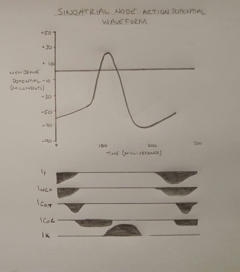
Outlined below are the 3 phases of a sinoatrial node action potential. In the cardiac action potential
Unlike the action potential in skeletal muscle cells, the cardiac action potential is not initiated by nervous activity. Instead, it arises from a group of specialized cells known as pacemaker cells, that have automatic action potential generati ...
, there are 5 phases (labelled 0-4), however pacemaker action potentials do not have an obvious phase 1 or 2.
Phase 4
 This phase is also known as the pacemaker potential. Immediately following repolarization, when the membrane potential is very negative (it is hyperpolarised), the voltage slowly begins to increase. This is initially due to the closing of
 potassium channel
Potassium channels are the most widely distributed type of ion channel found in virtually all organisms. They form potassium-selective pores that span cell membranes. Potassium channels are found in most cell types and control a wide variety of ...
s, which reduces the flow of potassium
Potassium is a chemical element; it has Symbol (chemistry), symbol K (from Neo-Latin ) and atomic number19. It is a silvery white metal that is soft enough to easily cut with a knife. Potassium metal reacts rapidly with atmospheric oxygen to ...
ions (Ik) out of the cell (see phase 2, below). Hyperpolarization also causes activation of hyperpolarisation-activated cyclic nucleotide–gated (HCN) channels. The activation of ion channels at very negative membrane potentials is unusual, therefore the flow of sodium (Na+) and some K+ through the activated HCN channel is referred to as a ''funny current
The pacemaker current (I''f'', or IK''f'', also called funny current) is an electric current in the heart that flows through the HCN channel or pacemaker channel. Such channels are important parts of the electrical conduction system of the heart an ...
'' (If). This funny current causes the membrane potential of the cell to gradually increase, as the positive charge (Na+ and K+) is flowing into the cell. Another mechanism involved in pacemaker potential is known as the calcium
Calcium is a chemical element; it has symbol Ca and atomic number 20. As an alkaline earth metal, calcium is a reactive metal that forms a dark oxide-nitride layer when exposed to air. Its physical and chemical properties are most similar to it ...
clock. This refers to the spontaneous release of calcium from the sarcoplasmic reticulum (a calcium store) into the cytoplasm, also known as calcium sparks. This increase in calcium within the cell then activates a sodium-calcium exchanger
The sodium-calcium exchanger (often denoted Na+/Ca2+ exchanger, exchange protein, or NCX) is an antiporter membrane protein that removes calcium from cells. It uses the energy that is stored in the electrochemical gradient of sodium (Na+) by ...
(NCX), which removes one Ca2+ from the cell, and exchanges it for 3 Na+ into the cell (therefore removing a charge of +2 from the cell, but allowing a charge of +3 to enter the cell) further increasing the membrane potential. Calcium later reenters the cell via SERCA and calcium channel
A calcium channel is an ion channel which shows selective permeability to calcium ions. It is sometimes synonymous with voltage-gated calcium channel, which are a type of calcium channel regulated by changes in membrane potential. Some calcium chan ...
s located on the cell membrane. The increase in membrane potential produced by these mechanisms, activates T-type calcium channels and then L-type calcium channels (which open very slowly). These channels allow a flow of Ca2+ into the cell, making the membrane potential even more positive.
Phase 0
This is the depolarization phase. When the membrane potential reaches the threshold potential (around -20 to -50 mV), the cell begins to rapidly depolarise (become more positive). This is mainly due to the flow of Ca2+ through L-type calcium channels, which are now fully open. During this stage, T-type calcium channels and HCN channels deactivate.
Phase 3
This phase is the repolarization phase. This occurs due to the inactivation of L-type calcium channels (preventing the movement of Ca2+ into the cell) and the activation of potassium channels, which allows the flow of K+ out of the cell, making the membrane potential more negative.
Nerve supply
Heart rate
Heart rate is the frequency of the cardiac cycle, heartbeat measured by the number of contractions of the heart per minute (''beats per minute'', or bpm). The heart rate varies according to the body's Human body, physical needs, including the nee ...
depends on the rate at which the sinoatrial node produces action potentials
An action potential (also known as a nerve impulse or "spike" when in a neuron) is a series of quick changes in voltage across a cell membrane. An action potential occurs when the membrane potential of a specific cell rapidly rises and falls. ...
. At rest, heart rate is between 60 and 100 beats per minute. This is a result of the activity of two sets of nerves, one acting to slow down action potential production (these are parasympathetic nerves) and the other acting to speed up action potential production ( sympathetic nerves).
Modulation of heart rate by ANS is carried by two types of channel: Kir and HCN (members of the CNG gated channels).
The sympathetic nerves begin in the thoracic
The thorax (: thoraces or thoraxes) or chest is a part of the anatomy of mammals and other tetrapod animals located between the neck and the abdomen.
In insects, crustaceans, and the extinct trilobites, the thorax is one of the three main ...
region of the spinal cord (in particular T1-T4). These nerves release a neurotransmitter called noradrenaline (NA). This binds to a receptor on the SA node membrane, called a beta-1adrenoceptor. Binding of NA to this receptor activates a G-protein (in particular a Gs-Protein, S for stimulatory) which initiates a series of reactions (known as the cAMP pathway) that results in the production of a molecule called cyclic adenosinemonophosphate (cAMP). This cAMP binds to the HCN channel (see above). Binding of cAMP to the HCN increases the flow of Na+ and K+ into the cell, speeding up the pacemaker potential, so producing action potentials at a quicker rate and increasing heart rate. An increase in heart rate is known as positive chronotropy.
The parasympathetic nerves supplying the SA node (in particular the Vagus nerves) originate in the brain
The brain is an organ (biology), organ that serves as the center of the nervous system in all vertebrate and most invertebrate animals. It consists of nervous tissue and is typically located in the head (cephalization), usually near organs for ...
. These nerves release a neurotransmitter called acetylcholine (ACh). ACh binds to a receptor called an M2 muscarinic receptor, located on the SA node membrane. Activation of this M2 receptor then activates a protein called a G-protein (in particular Gi protein, i for inhibitory). Activation of this G-protein blocks the cAMP pathway, reducing its effects, therefore inhibiting sympathetic activity and slowing action potential production. The G-protein also activates a potassium channel GIRK-1 and GIRK-4, which allows K+ to flow out of the cell, making the membrane potential more negative and slowing the pacemaker potential, therefore decreasing the rate of action potential production and therefore decreasing heart rate. A decrease in heart rate is known as negative chronotropy.
The first cell to produce the action potential in the SA node isn't always the same; this is known as pacemaker shift. In certain species of animals—for example, in dogs—a superior shift (i.e., the cell that produces the fastest action potential in the SA node is higher than previously) usually produces an increased heart rate whereas an inferior shift (i.e. the cell producing the fastest action potential within the SA node is further down than previously) produces a decreased heart rate.
Clinical significance
Sinus node dysfunction also known as ''sick sinus syndrome'' is a group of irregular heartbeat conditions caused by faulty electrical signals of the heart. When the heart's sinoatrial node is defective, the heart's rhythms become abnormal—typically too slow or exhibiting pauses in its function or a combination, and very rarely faster than normal. Blockage of the arterial blood supply to the SA node (most commonly due to amyocardial infarction
A myocardial infarction (MI), commonly known as a heart attack, occurs when Ischemia, blood flow decreases or stops in one of the coronary arteries of the heart, causing infarction (tissue death) to the heart muscle. The most common symptom ...
or progressive coronary artery disease
Coronary artery disease (CAD), also called coronary heart disease (CHD), or ischemic heart disease (IHD), is a type of cardiovascular disease, heart disease involving Ischemia, the reduction of blood flow to the cardiac muscle due to a build-up ...
) can therefore cause ischemia
Ischemia or ischaemia is a restriction in blood supply to any tissue, muscle group, or organ of the body, causing a shortage of oxygen that is needed for cellular metabolism (to keep tissue alive). Ischemia is generally caused by problems ...
and cell death in the SA node. This can disrupt the electrical pacemaker function of the SA node, and can result in sinus node dysfunction.
If the SA node does not function or the impulse generated in the SA node is blocked before it travels down the electrical conduction system, a group of cells further down the heart will become its pacemaker.
History
The sinoatrial node was first discovered by a young medical student, Martin Flack, in the heart of a mole, whilst his mentor, SirArthur Keith
Sir Arthur Keith FRS FRAI (5 February 1866 – 7 January 1955) was a British anatomist and anthropologist, and a proponent of scientific racism. He was a fellow and later the Hunterian Professor and conservator of the Hunterian Museum of the ...
, was on a bicycle ride with his wife. They made the discovery in a makeshift laboratory set up in a farmhouse in Kent
Kent is a Ceremonial counties of England, ceremonial county in South East England. It is bordered by Essex across the Thames Estuary to the north, the Strait of Dover to the south-east, East Sussex to the south-west, Surrey to the west, and Gr ...
, England
England is a Countries of the United Kingdom, country that is part of the United Kingdom. It is located on the island of Great Britain, of which it covers about 62%, and List of islands of England, more than 100 smaller adjacent islands. It ...
, called Mann's Place. Their discovery was published in 1907.
Additional images
See also
*Cardiac pacemaker
image:ConductionsystemoftheheartwithouttheHeart-en.svg, 350px, Image showing the cardiac pacemaker or SA node, the primary pacemaker within the electrical conduction system of the heart
The cardiac pacemaker is the heart's natural rhythm gener ...
* Cardiology
Cardiology () is the study of the heart. Cardiology is a branch of medicine that deals with disorders of the heart and the cardiovascular system. The field includes medical diagnosis and treatment of congenital heart defects, coronary artery di ...
* Heart block
* Sinus bradycardia
Sinus bradycardia is a sinus rhythm with a reduced rate of electrical discharge from the sinoatrial node, resulting in a bradycardia, a heart rate that is lower than the normal range (60–100 beats per minute for adult humans).
Signs and sympt ...
* Sinus tachycardia
Sinus tachycardia is a sinus rhythm of the heart, with an increased rate of electrical discharge from the sinoatrial node, resulting in a tachycardia, a heart rate that is higher than the upper limit of normal (90–100 beats per minute for adu ...
* Cardiothoracic Surgery
References
External links
* - "The conduction system of the heart."Diagram at gru.net
* () * https://web.archive.org/web/20070929080346/http://www.healthyheart.nhs.uk/heart_works/heart03.shtml {{DEFAULTSORT:Sinoatrial Node Cardiac anatomy