pyknosis on:
[Wikipedia]
[Google]
[Amazon]
Pyknosis, or karyopyknosis, is the irreversible condensation of
File:4_Bd_obs_4_680x512px.tif,
Pyknotic nuclei are often found in the

chromatin
Chromatin is a complex of DNA and protein found in eukaryote, eukaryotic cells. The primary function is to package long DNA molecules into more compact, denser structures. This prevents the strands from becoming tangled and also plays important r ...
in the nucleus of a cell undergoing necrosis
Necrosis () is a form of cell injury which results in the premature death of cells in living tissue by autolysis. The term "necrosis" came about in the mid-19th century and is commonly attributed to German pathologist Rudolf Virchow, who i ...
or apoptosis
Apoptosis (from ) is a form of programmed cell death that occurs in multicellular organisms and in some eukaryotic, single-celled microorganisms such as yeast. Biochemistry, Biochemical events lead to characteristic cell changes (Morphology (biol ...
. It is followed by karyorrhexis, or fragmentation of the nucleus.
Pyknosis (from Ancient Greek meaning "thick, closed or condensed") is also observed in the maturation of erythrocyte
Red blood cells (RBCs), referred to as erythrocytes (, with -''cyte'' translated as 'cell' in modern usage) in academia and medical publishing, also known as red cells, erythroid cells, and rarely haematids, are the most common type of blood ce ...
s (a red blood cell) and the neutrophil
Neutrophils are a type of phagocytic white blood cell and part of innate immunity. More specifically, they form the most abundant type of granulocytes and make up 40% to 70% of all white blood cells in humans. Their functions vary in differe ...
(a type of white blood cell). The maturing metarubricyte (a stage in RBC maturation) will condense its nucleus before expelling it to become a reticulocyte. The maturing neutrophil will condense its nucleus into several connected lobes that stay in the cell until the end of its cell life.
Micrograph
A micrograph is an image, captured photographically or digitally, taken through a microscope or similar device to show a magnify, magnified image of an object. This is opposed to a macrograph or photomacrograph, an image which is also taken ...
of an infarct in the biliary tract
The biliary tract (also biliary tree or biliary system) refers to the liver, gallbladder and bile ducts, and how they work together to make, store and secrete bile. Bile consists of water, electrolytes, bile acids, cholesterol, phospholipids an ...
, with pyknotic nuclei (arrows) (400x).
zona reticularis
The zona reticularis (sometimes, reticulate zone) is the innermost layer of the adrenal cortex, lying deep to the zona fasciculata and superficial to the adrenal medulla. The cells are arranged cords that project in different directions giving a n ...
of the adrenal gland. They are also found in the keratinocytes of the outermost layer in parakeratinised epithelium.
Overview of Pyknosis
Pyknosis, or the irreversible nuclear condensation (a nuclear morphology) in a cell (generally old vertebrateleukocyte
White blood cells (scientific name leukocytes), also called immune cells or immunocytes, are cells of the immune system that are involved in protecting the body against both infectious disease and foreign entities. White blood cells are genera ...
cells) is the result of a cell undergoing either apoptosis
Apoptosis (from ) is a form of programmed cell death that occurs in multicellular organisms and in some eukaryotic, single-celled microorganisms such as yeast. Biochemistry, Biochemical events lead to characteristic cell changes (Morphology (biol ...
or necrosis
Necrosis () is a form of cell injury which results in the premature death of cells in living tissue by autolysis. The term "necrosis" came about in the mid-19th century and is commonly attributed to German pathologist Rudolf Virchow, who i ...
. There are two types of pyknosis: nucleolytic pyknosis and anucleolytic pyknosis. Nucleolytic pyknosis occurs during apoptosis (a form of controlled/programmed cell death), while anucleolytic pyknosis occurs during necrosis. Necrosis is a form of regulated cell death due to toxins, infections, and other acute stressors. These stressors cause swelling/shape modification of cellular organelle
In cell biology, an organelle is a specialized subunit, usually within a cell (biology), cell, that has a specific function. The name ''organelle'' comes from the idea that these structures are parts of cells, as Organ (anatomy), organs are to th ...
s leading to the eventual loss of stability and integrity of the cell membrane
The cell membrane (also known as the plasma membrane or cytoplasmic membrane, and historically referred to as the plasmalemma) is a biological membrane that separates and protects the interior of a cell from the outside environment (the extr ...
.
In simpler terms, pyknosis is the process of nuclear shrinkage that may occur during both necrosis and apoptosis. Pyknosis is also characterized by eventual fragmentation ( karyorrhexis) of the dense nuclear chromatin
Chromatin is a complex of DNA and protein found in eukaryote, eukaryotic cells. The primary function is to package long DNA molecules into more compact, denser structures. This prevents the strands from becoming tangled and also plays important r ...
, resulting in dark, round, and dense nuclear fragments. Karyorrhexis is the fragmentation of a pyknotic cell’s nucleus and the cleavage and condensing of chromatin.
Pyknosis provides a distinction between apoptosis and necrosis
Apoptosis is characterized by nuclear condensation, shrinking of the cell, and blebbing of the nuclear and cell membrane, while necrosis is characterized by nuclear condensation, swelling of the cell, and breaks in the cell membrane. Both necrosis and apoptosis are regulated by a few of the same proteins: caspase-activated DNase (CAD), endonuclease G and DNase I. Pyknosis occurs in both an apoptotic and a necrotic cell. Pyknosis in an apoptotic cell is identified by nuclear condensation, chromatin fragmentation, and the formation of a few large clumps which are enveloped by apoptotic extracellular vesicles, which are to be released when the cell dies. Pyknosis in a necrotic cell is identified by nuclear condensation and fragmentation into small clumps that will be dissolved later in the process of the necrotic cell’s death. Consequently, pyknosis can be distinguished into two types, nucleolytic pyknosis (apoptotic cells) and anucleolytic pyknosis (necrotic cells).The types of pyknosis
Nucleolytic pyknosis
Nucleolytic pyknosis, which can also be referred to as apoptotic pyknosis, involves three main events. These are disrupting the nuclear membrane, the condensing of the chromatin, and lastly, nuclear cleavage/fragmentation. Throughout these events the cell shrinks in size and the cell membrane undergoes blebbing, which is the forming of membrane bulges across the exterior-facing surface of the cell membrane. During the first event (the disruption of the nuclear membrane), severalenzyme
An enzyme () is a protein that acts as a biological catalyst by accelerating chemical reactions. The molecules upon which enzymes may act are called substrate (chemistry), substrates, and the enzyme converts the substrates into different mol ...
s are used to cleave the proteins found in the nuclear membrane. These enzymes, caspase-3 and caspase-6, both target and cleave nuclear membrane proteins, including NUP153, LAP2, and B1 (proteins that are used for membrane structure and molecular transport). This cleavage, in turn, results in a disruption of the interior of the membrane, which is an initiating factor for chromatin condensation (the second event of nucleolytic pyknosis). This is due to the fact that caspase-3 cleaves Acinus, which has DNA
Deoxyribonucleic acid (; DNA) is a polymer composed of two polynucleotide chains that coil around each other to form a double helix. The polymer carries genetic instructions for the development, functioning, growth and reproduction of al ...
/RNA
Ribonucleic acid (RNA) is a polymeric molecule that is essential for most biological functions, either by performing the function itself (non-coding RNA) or by forming a template for the production of proteins (messenger RNA). RNA and deoxyrib ...
binding domains and ATPase
ATPases (, Adenosine 5'-TriPhosphatase, adenylpyrophosphatase, ATP monophosphatase, triphosphatase, ATP hydrolase, adenosine triphosphatase) are a class of enzymes that catalyze the decomposition of ATP into ADP and a free phosphate ion or ...
activity to initiate the condensation of chromatin.
Anucleolytic pyknosis
Anucleolytic pyknosis, which can also be referred to as necrotic pyknosis, involves the swelling of the cell, followed by the separation of the nuclear membrane from chromatin, the eventual collapse of both the nuclear membrane and chromatin, and finally the cell membrane ruptures (the cell dies). One protein that plays a significant role in necrotic pyknosis is the barrier-to-autointegration factor (BAF). The function of BAF is to facilitate the tethering of chromatin to the membrane of the nucleus, however in the case of necrosis, when BAF isphosphorylated
In biochemistry, phosphorylation is described as the "transfer of a phosphate group" from a donor to an acceptor. A common phosphorylating agent (phosphate donor) is ATP and a common family of acceptor are alcohols:
:
This equation can be writt ...
, it will initiate the dissociation between the nuclear membrane and the condensed chromatin. As a result, the nuclear membrane will collapse onto the condensed chromatin. Thus, the phosphorylation of BAF is a critical marker of necrotic pyknosis.
Significance of pyknosis
Pyknosis is a stage in the apoptotic or necrotic cell death pathways. It is an important stage that involves fragmentation and condensation of damaged DNA/chromatin. Without it, the apoptotic or necrotic cell death pathways would be interrupted. This disruption, in turn, may prompt the improper destruction or removal of a cell with damaged elements as well as other related issues. These issues include cell accumulation and uncontrolled cell growth, which results in the formation ofcancer
Cancer is a group of diseases involving Cell growth#Disorders, abnormal cell growth with the potential to Invasion (cancer), invade or Metastasis, spread to other parts of the body. These contrast with benign tumors, which do not spread. Po ...
ous and abnormal tissue masses known as tumors
A neoplasm () is a type of abnormal and excessive growth of tissue. The process that occurs to form or produce a neoplasm is called neoplasia. The growth of a neoplasm is uncoordinated with that of the normal surrounding tissue, and persists ...
. Therefore, being able to observe or identify when a cell is pyknotic (which may indicate that the cell is undergoing apoptosis or necrosis) and if it then successfully undergoes apoptosis or necrosis, may be crucial in determining if the cell will undergo uncontrolled cell growth and contribute to the formation of a tumor.
Techniques for detecting or observing pyknosis in cells
Various techniques are used to detect/observe pyknosis. These techniques also help to differentiate between apoptotic or necrotic cells. The techniques are identified and described as follows:Cellular staining
When stains and dyes are applied to locate pyknotic cells in a tissue sample, the cell becomes easily identifiable. The stains/dyes target the nuclear and blebbed fragments of a pyknotic cell, making them dark (light contrast) and more readily seen when the sample is placed under a light microscope. Fluorescence microscopy andflow cytometry
Flow cytometry (FC) is a technique used to detect and measure the physical and chemical characteristics of a population of cells or particles.
In this process, a sample containing cells or particles is suspended in a fluid and injected into the ...
also use staining ( fluorescent stains) to target the DNA/nuclear fragments of cells. The fluorescent staining creates a contrast between normal cell DNA and pyknotic cell DNA, because pyknotic cell nuclear material is condensed.

Gel electrophoresis
Gel electrophoresis is an electrophoresis method for separation and analysis of biomacromolecules (DNA, RNA, proteins, etc.) and their fragments, based on their size and charge through a gel. It is used in clinical chemistry to separate ...
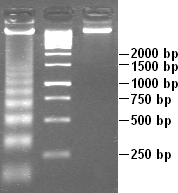
Gel electrophoresis is a standard technique that is frequently used to visualize DNA fragmentation (forming a ladder-like image on the gel), which is a characteristic of apoptosis and is associated with nuclear condensation (which characterizes pyknosis). Therefore, when referring to apoptosis, this technique is known as DNA laddering. Gel electrophoresis is also used to visualize the random DNA fragmentation of necrosis, which forms a smear on the gel.
Assays to detect DNA fragmentation or condensation
Various assays of DNA fragmentation or condensation include the APO single-stranded DNA ( ssDNA) assay which detects damaged DNA of cells undergoing apoptosis or necrosis, TUNEL assay which is used to locally find DNA strand breaks (DSBs), and ISEL. ISEL (in-situ labeling technique) is a labeling/tagging technique of apoptotic or necrotic cells. ISEL specifically targets unfragmented DNA that has condensed into anucleosome
A nucleosome is the basic structural unit of DNA packaging in eukaryotes. The structure of a nucleosome consists of a segment of DNA wound around eight histone, histone proteins and resembles thread wrapped around a bobbin, spool. The nucleosome ...
structure.
The APO ssDNA assay detects apoptotic cells by using an antibody
An antibody (Ab) or immunoglobulin (Ig) is a large, Y-shaped protein belonging to the immunoglobulin superfamily which is used by the immune system to identify and neutralize antigens such as pathogenic bacteria, bacteria and viruses, includin ...
that specifically binds to the ssDNA, which is accumulated during apoptosis as a result of DNA fragmentation. Therefore, the presence of ssDNA is an indicator of DNA damage in the apoptotic cell. For the assay process, cells are fixed (with e.g., formamide
Formamide is an amide derived from formic acid. It is a colorless liquid which is miscible with water and has an ammonia-like odor. It is chemical feedstock for the manufacture of sulfa drugs and other pharmaceuticals, herbicides and pesticides, ...
), and these cells then undergo incubation (at a predetermined temperature), which subjects the DNA to thermal denaturation and exposes the ssDNA. Next, the cells are incubated with an ssDNA-specific antibody along with a fluorescently labeled secondary antibody. The fluorescence amounts as a measure of apoptosis which can then be quantified using flow cytometry.
The TUNEL assay, otherwise known as the terminal deoxynucleotidyl transferase dUTP nick-end labeling assay, is a technique that measures DNA damage and breakage during apoptosis. During apoptosis, DNA fragmentation exposes numerous 3’OH ends, that are labeled with modified deoxy-uridine triphosphate (dUTP) by the TUNEL reaction. Then, this modified dUTP can be identified with specific fluorescent antibodies which can identify modified nucleotide
Nucleotides are Organic compound, organic molecules composed of a nitrogenous base, a pentose sugar and a phosphate. They serve as monomeric units of the nucleic acid polymers – deoxyribonucleic acid (DNA) and ribonucleic acid (RNA), both o ...
s or by using tagged nucleotides themselves. Flow cytometry can then be used to quantify fluorescence intensity, and thus provide a measure of apoptosis.
Detection methods of caspase activity
As mentioned above, caspase proteins, which areprotease
A protease (also called a peptidase, proteinase, or proteolytic enzyme) is an enzyme that catalysis, catalyzes proteolysis, breaking down proteins into smaller polypeptides or single amino acids, and spurring the formation of new protein products ...
enzymes, promote DNA condensation and fragmentation via the caspase (or proteolytic) cascade. These caspase proteins include, for example, caspase 9, caspase 6, caspase 7, and caspase 3. The caspase cascade directly activates caspase-activated DNase (CAD) which initiates DNA fragmentation into smaller pieces resulting in chromatin condensation. The biochemical techniques used to detect caspase activity include ELISA
The enzyme-linked immunosorbent assay (ELISA) (, ) is a commonly used analytical biochemistry assay, first described by Eva Engvall and Peter Perlmann in 1971. The assay is a solid-phase type of enzyme immunoassay (EIA) to detect the presence of ...
and fluorometric and colorimetric
Colorimetry is "the science and technology used to quantify and describe physically the human color perception".
It is similar to spectrophotometry, but is distinguished by its interest in reducing spectra to the physical correlates of color p ...
assays.
See also
*Apoptosis
Apoptosis (from ) is a form of programmed cell death that occurs in multicellular organisms and in some eukaryotic, single-celled microorganisms such as yeast. Biochemistry, Biochemical events lead to characteristic cell changes (Morphology (biol ...
* Necrosis
Necrosis () is a form of cell injury which results in the premature death of cells in living tissue by autolysis. The term "necrosis" came about in the mid-19th century and is commonly attributed to German pathologist Rudolf Virchow, who i ...
* Karyolysis
* Karyorrhexis
References
{{Pathology Cellular processes Programmed cell death Cellular senescence