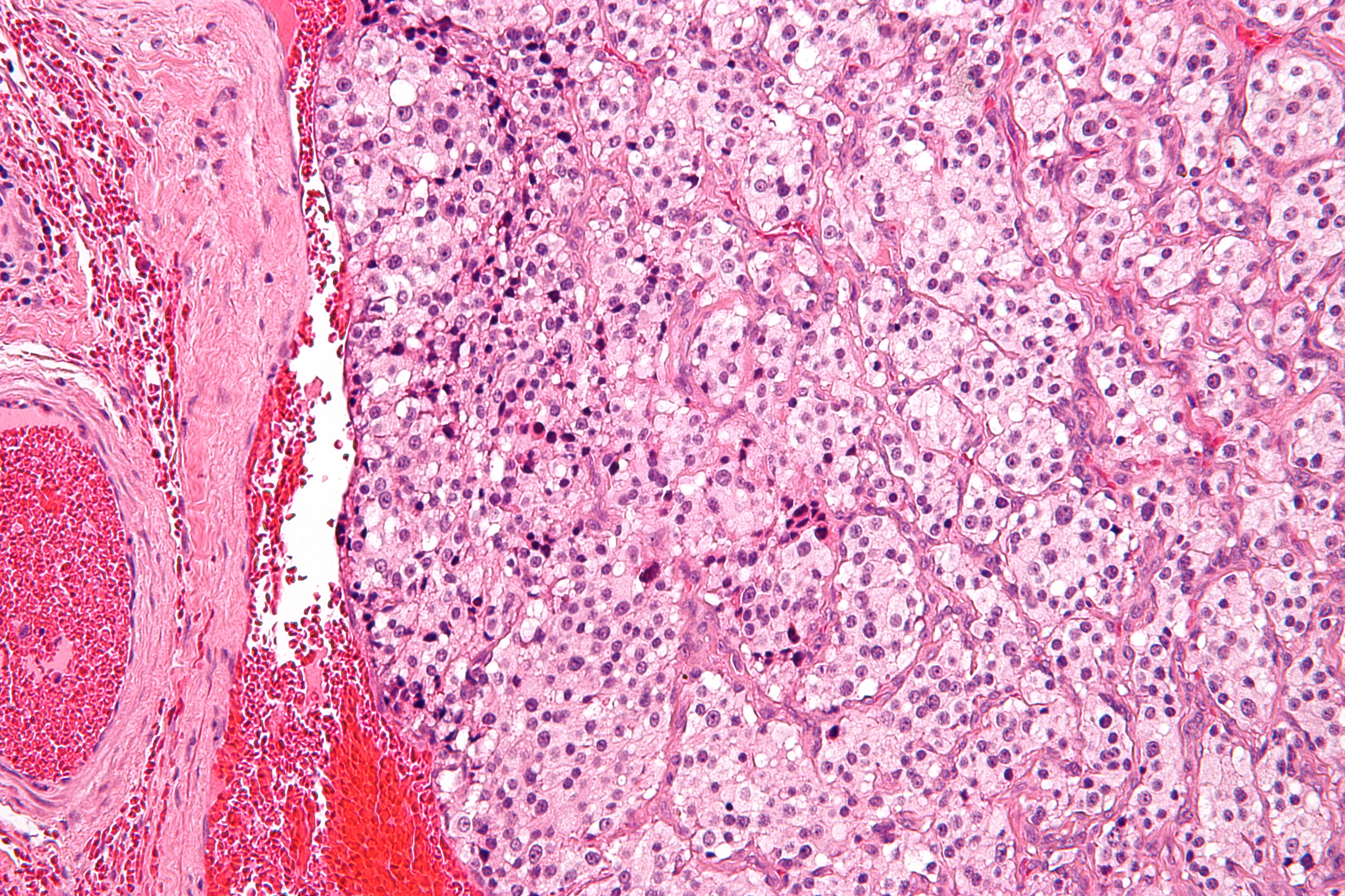
Carotid body on:
[Wikipedia]
[Google]
[Amazon]
The carotid body is a small cluster of peripheral chemoreceptor cells and supporting

sustentacular cell
A sustentacular cell is a type of cell primarily associated with structural support, they can be found in various tissues.
* Sustentacular cells of the olfactory epithelium (also called supporting cells or Sertoli cells) have been shown to be inv ...
s situated at the bifurcation of each common carotid artery
In anatomy, the left and right common carotid arteries (carotids) () are artery, arteries that supply the head and neck with oxygenated blood; they divide in the neck to form the external carotid artery, external and internal carotid artery, inte ...
in its tunica externa
The tunica externa (Neo-Latin "outer coat"), also known as the tunica adventitia (Neo-Latin "additional coat"), is the outermost tunica (biology), tunica (layer) of a blood vessel, surrounding the tunica media. It is mainly composed of collagen an ...
.
The carotid body detects changes in the composition of arterial blood
Arterial blood is the oxygenated blood in the circulatory system found in the pulmonary vein, the left chambers of the heart, and in the artery, arteries. It is bright red in color, while venous blood is dark red in color (but looks purple through ...
flowing through it, mainly the partial pressure of arterial oxygen, but also of carbon dioxide
Carbon dioxide is a chemical compound with the chemical formula . It is made up of molecules that each have one carbon atom covalent bond, covalently double bonded to two oxygen atoms. It is found in a gas state at room temperature and at norma ...
. It is also sensitive to changes in blood pH, and temperature
Temperature is a physical quantity that quantitatively expresses the attribute of hotness or coldness. Temperature is measurement, measured with a thermometer. It reflects the average kinetic energy of the vibrating and colliding atoms making ...
.
Structure
The carotid body is situated on the posterior aspect of the bifurcation of the common carotid artery. The carotid body is made up of two types of cells, called glomus cells: glomus type I cells are peripheral chemoreceptors, and glomus type II cells are sustentacular supportive cells. * Glomus type I cells are derived from theneural crest
The neural crest is a ridge-like structure that is formed transiently between the epidermal ectoderm and neural plate during vertebrate development. Neural crest cells originate from this structure through the epithelial-mesenchymal transition, ...
. They release a variety of neurotransmitter
A neurotransmitter is a signaling molecule secreted by a neuron to affect another cell across a Chemical synapse, synapse. The cell receiving the signal, or target cell, may be another neuron, but could also be a gland or muscle cell.
Neurotra ...
s, including acetylcholine
Acetylcholine (ACh) is an organic compound that functions in the brain and body of many types of animals (including humans) as a neurotransmitter. Its name is derived from its chemical structure: it is an ester of acetic acid and choline. Par ...
, ATP, and dopamine
Dopamine (DA, a contraction of 3,4-dihydroxyphenethylamine) is a neuromodulatory molecule that plays several important roles in cells. It is an organic chemical of the catecholamine and phenethylamine families. It is an amine synthesized ...
that trigger EPSPs in synapsed neurons leading to the respiratory center
The respiratory center is located in the medulla oblongata and pons, in the brainstem. The respiratory center is made up of three major respiratory groups of neurons, two in the medulla and one in the pons. In the medulla they are the dorsal ...
. They are innervated by axons of the glossopharyngeal nerve which collectively are called the carotid sinus nerve.
* Glomus type II cells resemble glial cells
Glia, also called glial cells (gliocytes) or neuroglia, are non-neuronal cells in the central nervous system (the brain and the spinal cord) and in the peripheral nervous system that do not produce electrical impulses. The neuroglia make up ...
, express the glial marker S100 and act as supporting cells.
Function
The carotid body functions as a sensor: it responds to a stimulus, primarily O2 partial pressure, which is detected by the type I (glomus) cells, and triggers anaction potential
An action potential (also known as a nerve impulse or "spike" when in a neuron) is a series of quick changes in voltage across a cell membrane. An action potential occurs when the membrane potential of a specific Cell (biology), cell rapidly ri ...
through the afferent fibers of the glossopharyngeal nerve
The glossopharyngeal nerve (), also known as the ninth cranial nerve, cranial nerve IX, or simply CN IX, is a cranial nerve that exits the brainstem from the sides of the upper Medulla oblongata, medulla, just anterior (closer to the nose) to t ...
, which relays the information to the central nervous system.
Stimulus
The carotid body peripheral chemoreceptors are primarily sensitive to decreases in the partial pressure of oxygen (PO2). This is in contrast to thecentral chemoreceptors
A central chemoreceptor is a chemoreceptor sensitive to the pH of its environment. Central chemoreceptors are located on the ventrolateral medulla oblongata, medullary surface in vicinity of the exit of CN IX and CN X in the central nervous syste ...
in the medulla oblongata
The medulla oblongata or simply medulla is a long stem-like structure which makes up the lower part of the brainstem. It is anterior and partially inferior to the cerebellum. It is a cone-shaped neuronal mass responsible for autonomic (involun ...
that are primarily sensitive to changes in pH and PCO2 (a decrease in pH and an increase in PCO2). The carotid body chemoreceptors are also sensitive to pH and PCO2, but only secondarily. More specifically, the sensitivity of carotid body chemoreceptors to decreased PO2 is greater when pH is decreased and PCO2 is increased.
Impulse rate for carotid bodies is particularly sensitive to changes in arterial PO2 in the range of 60 down to 30 mm Hg, a range in which hemoglobin saturation with oxygen decreases rapidly.
The output of the carotid bodies is low at an oxygen partial pressure
In a mixture of gases, each constituent gas has a partial pressure which is the notional pressure of that constituent gas as if it alone occupied the entire volume of the original mixture at the same temperature. The total pressure of an ideal g ...
above about 100mmHg (13,3 k Pa) (at normal physiological pH), but below 60mmHg the activity of the type I (glomus) cells increases rapidly due to a decrease in hemoglobin-oxygen saturation below 90%.
Detection
The mechanism for detecting reductions in PO2 has yet to be identified, there may be multiple mechanisms and could vary between species. Hypoxia detection has been shown to depend upon increasedhydrogen sulfide
Hydrogen sulfide is a chemical compound with the formula . It is a colorless chalcogen-hydride gas, and is toxic, corrosive, and flammable. Trace amounts in ambient atmosphere have a characteristic foul odor of rotten eggs. Swedish chemist ...
generation produced by cystathionine gamma-lyase as hypoxia detection is reduced in mice in which this enzyme is knocked out or pharmacologically inhibited. The process of detection involves the interaction of cystathionine gamma-lyase with hemeoxygenase-2 and the production of carbon monoxide
Carbon monoxide (chemical formula CO) is a poisonous, flammable gas that is colorless, odorless, tasteless, and slightly less dense than air. Carbon monoxide consists of one carbon atom and one oxygen atom connected by a triple bond. It is the si ...
.Peng Y-J, Nanduri J, Raghuraman G, Souvannakitti D, Gadalla M.M, Kumar GK, Snyder SH, Prabhakar NR. (2010). H2S mediates O2 sensing in the carotid body PNAS 107 (23) 10719-10724. Yet, some studies show that physiologic concentration of hydrogen sulfide may not be strong enough to trigger such responses.
Other theories suggest it may involve mitochondrial oxygen sensors and the haem-containing cytochromes that undergo reversible one-electron reduction during oxidative-phosphorylation. Haem reversibly binds O2 with an affinity similar to that of the carotid body, suggesting that haem containing proteins may have a role in O2, potentially this could be one of the complexes involved in oxidative-phosphorylation. This leads to increases in reactive oxygen species and rises in intracellular Ca2+. However, whether hypoxia leads to an increase or decrease in reactive oxygen species is unknown. The role of reactive oxygen species in hypoxia sensing is also under question.
The oxygen dependent enzyme haem-oxidase has also been put forward as a hypoxia sensor. In normoxia, haem-oxygenase generates carbon monoxide (CO), CO activates the large conductance calcium-activated potassium channel, BK. Falls in CO that occur as a consequence of hypoxia would lead to closure of this potassium channel and this would lead to membrane depolarisation and consequence activation of the carotid body. A role for the "energy sensor" AMP-activated protein kinase (AMPK) has also been proposed in hypoxia sensing. This enzyme is activated during times of net energy usage and metabolic stress, including hypoxia. AMPK has a number of targets and it appears that, in the carotid body, when AMPK is activated by hypoxia, it leads to downstream potassium channel closure of both O2-sentive TASK-like and BK channels
An increased PCO2 is detected because the CO2 diffuses into the cell, where it increases the concentration of carbonic acid
Carbonic acid is a chemical compound with the chemical formula . The molecule rapidly converts to water and carbon dioxide in the presence of water. However, in the absence of water, it is quite stable at room temperature. The interconversion ...
and thus protons
A proton is a stable subatomic particle, symbol , H+, or 1H+ with a positive electric charge of +1 ''e'' ( elementary charge). Its mass is slightly less than the mass of a neutron and approximately times the mass of an electron (the pro ...
. The precise mechanism of CO2 sensing is unknown, however it has been demonstrated that CO2 and low pH inhibit a TASK-like potassium conductance, reducing potassium current. This leads to depolarisation of the cell membrane which leads to Ca2+ entry, excitation of glomus cells and consequent neurotransmitter release.
Arterial acidosis
Acidosis is a biological process producing hydrogen ions and increasing their concentration in blood or body fluids. pH is the negative log of hydrogen ion concentration and so it is decreased by a process of acidosis.
Acidemia
The term ac ...
(either metabolic
Metabolism (, from ''metabolē'', "change") is the set of life-sustaining chemical reactions in organisms. The three main functions of metabolism are: the conversion of the energy in food to energy available to run cellular processes; the ...
or from altered PCO2) inhibits acid-base transporters (e.g. Na+-H+) which raise intracellular pH, and activates transporters (e.g. Cl−-HCO3−) which decrease it. Changes in proton concentration caused by acidosis (or the opposite from alkalosis
Alkalosis is the result of a process reducing hydrogen ion concentration of arterial blood plasma (alkalemia). In contrast to acidemia (serum pH 7.35 or lower), alkalemia occurs when the serum pH is higher than normal (7.45 or higher). Alkalosis ...
) inside the cell stimulates the same pathways involved in PCO2 sensing.
Another mechanism is through oxygen sensitive potassium channels. A drop in dissolved oxygen lead to closing of these channels which results in depolarization. This leads to release of the neurotransmitter dopamine in the glossopharyngeal and vagus afferente to the vasomotor area.
Action potential
The type I (glomus) cells in the carotid (and aortic bodies) are derived from neuroectoderm and are thus electrically excitable. A decrease in oxygen partial pressure, an increase in carbon dioxide partial pressure, and a decrease in arterial pH can all causedepolarization
In biology, depolarization or hypopolarization is a change within a cell (biology), cell, during which the cell undergoes a shift in electric charge distribution, resulting in less negative charge inside the cell compared to the outside. Depolar ...
of the cell membrane
The cell membrane (also known as the plasma membrane or cytoplasmic membrane, and historically referred to as the plasmalemma) is a biological membrane that separates and protects the interior of a cell from the outside environment (the extr ...
, and they affect this by blocking potassium
Potassium is a chemical element; it has Symbol (chemistry), symbol K (from Neo-Latin ) and atomic number19. It is a silvery white metal that is soft enough to easily cut with a knife. Potassium metal reacts rapidly with atmospheric oxygen to ...
currents. This reduction in the membrane potential
Membrane potential (also transmembrane potential or membrane voltage) is the difference in electric potential between the interior and the exterior of a biological cell. It equals the interior potential minus the exterior potential. This is th ...
opens voltage-gated calcium
Calcium is a chemical element; it has symbol Ca and atomic number 20. As an alkaline earth metal, calcium is a reactive metal that forms a dark oxide-nitride layer when exposed to air. Its physical and chemical properties are most similar to it ...
channels, which causes a rise in intracellular calcium concentration. This causes exocytosis
Exocytosis is a term for the active transport process that transports large molecules from cell to the extracellular area. Hormones, proteins and neurotransmitters are examples of large molecules that can be transported out of the cell. Exocytosis ...
of vesicles containing a variety of neurotransmitters
A neurotransmitter is a signaling molecule secreted by a neuron to affect another cell across a synapse. The cell receiving the signal, or target cell, may be another neuron, but could also be a gland or muscle cell.
Neurotransmitters are rele ...
, including acetylcholine
Acetylcholine (ACh) is an organic compound that functions in the brain and body of many types of animals (including humans) as a neurotransmitter. Its name is derived from its chemical structure: it is an ester of acetic acid and choline. Par ...
, noradrenaline
Norepinephrine (NE), also called noradrenaline (NA) or noradrenalin, is an organic chemical in the catecholamine family that functions in the brain and body as a hormone, neurotransmitter and neuromodulator. The name "noradrenaline" (from ...
, dopamine
Dopamine (DA, a contraction of 3,4-dihydroxyphenethylamine) is a neuromodulatory molecule that plays several important roles in cells. It is an organic chemical of the catecholamine and phenethylamine families. It is an amine synthesized ...
, adenosine
Adenosine (symbol A) is an organic compound that occurs widely in nature in the form of diverse derivatives. The molecule consists of an adenine attached to a ribose via a β-N9- glycosidic bond. Adenosine is one of the four nucleoside build ...
, ATP, substance P
Substance P (SP) is an undecapeptide (a peptide composed of a chain of 11 amino acid residues) and a type of neuropeptide, belonging to the tachykinin family of neuropeptides. It acts as a neurotransmitter and a neuromodulator. Substance P ...
, and met-enkephalin. These act on receptors on the afferent nerve fibres which lie in apposition to the glomus cell to cause an action potential.
Relay
The feedback from the carotid body is sent to the cardiorespiratory centers in themedulla oblongata
The medulla oblongata or simply medulla is a long stem-like structure which makes up the lower part of the brainstem. It is anterior and partially inferior to the cerebellum. It is a cone-shaped neuronal mass responsible for autonomic (involun ...
via the afferent branches of the glossopharyngeal nerve
The glossopharyngeal nerve (), also known as the ninth cranial nerve, cranial nerve IX, or simply CN IX, is a cranial nerve that exits the brainstem from the sides of the upper Medulla oblongata, medulla, just anterior (closer to the nose) to t ...
. (The efferent fibers of the aortic body
The aortic bodies are one of several small clusters of peripheral chemoreceptors located along the aortic arch. They are important in measuring partial pressures of oxygen and carbon dioxide in the blood, and blood pH.
Structure
The aortic b ...
chemoreceptors are relayed by the vagus nerve
The vagus nerve, also known as the tenth cranial nerve (CN X), plays a crucial role in the autonomic nervous system, which is responsible for regulating involuntary functions within the human body. This nerve carries both sensory and motor fibe ...
.) These centers, in turn, regulate breathing and blood pressure, with hypoxia causing an increase in ventilation.
Clinical significance

Paraganglioma
Aparaganglioma
A paraganglioma is a rare neuroendocrine tumour, neuroendocrine neoplasm that may develop at various body sites (including the head, neck, thorax and abdomen). When the same type of tumor is found in the adrenal gland, they are referred to as a p ...
is a tumor that may involve the carotid body and is usually benign
Malignancy () is the tendency of a medical condition to become progressively worse; the term is most familiar as a characterization of cancer.
A ''malignant'' tumor contrasts with a non-cancerous benign tumor, ''benign'' tumor in that a malig ...
. Rarely, a malignant neuroblastoma may originate from the carotid body.
See also
List of distinct cell types in the adult human body
The list of human cell types provides an enumeration and description of the various specialized cells found within the human body, highlighting their distinct functions, characteristics, and contributions to overall physiological processes. Cell ...
References
{{DEFAULTSORT:Carotid Body Angiology Respiration