|
Vascular Occlusion
Vascular occlusion is a blockage of a blood vessel, usually with a clot. It differs from thrombosis in that it can be used to describe any form of blockage, not just one formed by a clot. When it occurs in a major vein, it can, in some cases, cause deep vein thrombosis. The condition is also relatively common in the retina, and can cause partial or total loss of vision. An occlusion can often be diagnosed using Doppler sonography (a form of ultrasound). Some medical procedures, such as embolisation, involve occluding a blood vessel to treat a particular condition. This can be to reduce pressure on aneurysms (weakened blood vessels) or to restrict a haemorrhage. It can also be used to reduce blood supply to tumours or growths in the body, and therefore restrict their development. Occlusion can be carried out using a ligature; by implanting small coils which stimulate the formation of clots; or, particularly in the case of cerebral aneurysms, by clipping. See also * C ... [...More Info...] [...Related Items...] OR: [Wikipedia] [Google] [Baidu] |
Blood Clot
A thrombus (plural thrombi), colloquially called a blood clot, is the final product of the blood coagulation step in hemostasis. There are two components to a thrombus: aggregated platelets and red blood cells that form a plug, and a mesh of cross-linked fibrin protein. The substance making up a thrombus is sometimes called cruor. A thrombus is a healthy response to injury intended to stop and prevent further bleeding, but can be harmful in thrombosis, when a clot obstructs blood flow through healthy blood vessels in the circulatory system. In the microcirculation consisting of the very small and smallest blood vessels the capillaries, tiny thrombi known as microclots can obstruct the flow of blood in the capillaries. This can cause a number of problems particularly affecting the alveoli in the lungs of the respiratory system resulting from reduced oxygen supply. Microclots have been found to be a characteristic feature in severe cases of COVID-19, and in long COVID. Mural thr ... [...More Info...] [...Related Items...] OR: [Wikipedia] [Google] [Baidu] |
Ligature (medicine)
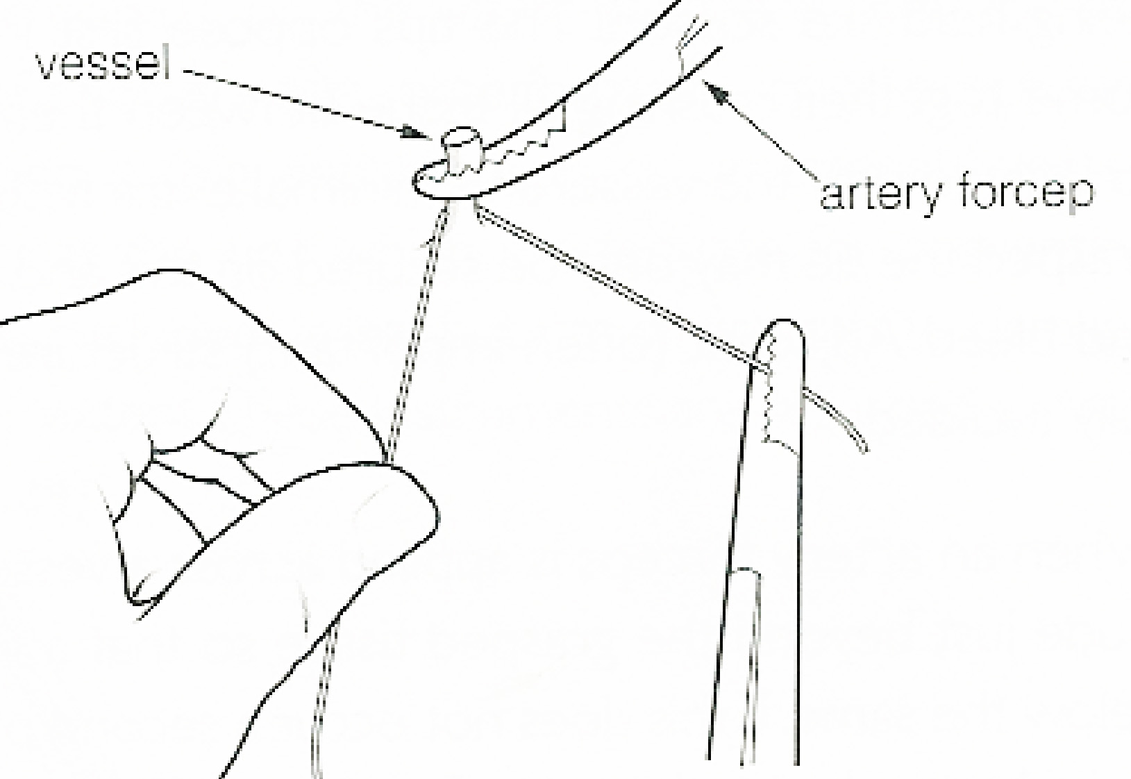
In surgery or medical procedure, a ligature consists of a piece of thread (suture) tied around an anatomical structure, usually a blood vessel or another hollow structure (e.g. urethra) to shut it off. History The principle of ligation is attributed to Hippocrates and Galen. In ancient Rome, ligatures were used to treat hemorrhoids. The concept of a ligature was reintroduced some 1,500 years later by Ambroise Paré, and finally it found its modern use in 1870–80, made popular by Jules-Émile Péan. Procedure With a blood vessel the surgeon will clamp the vessel perpendicular to the axis of the artery or vein with a hemostat, then secure it by ligating it; i.e. using a piece of suture around it before dividing the structure and releasing the hemostat. It is different from a tourniquet in that the tourniquet will not be secured by knots and it can therefore be released/tightened at will. Ligature is one of the remedies to treat skin tag, or acrochorda. It is done by tying st ... [...More Info...] [...Related Items...] OR: [Wikipedia] [Google] [Baidu] |
Branch Retinal Vein Occlusion
Branch retinal vein occlusion is a common retinal vascular disease of the elderly. It is caused by the occlusion of one of the branches of central retinal vein. Signs and symptoms Patients with branch retinal vein occlusion usually have a sudden onset of blurred vision or a central visual field defect. The eye examination findings of acute branch retinal vein occlusion include superficial hemorrhages, retinal edema, and often cotton-wool spots in a sector of retina drained by the affected vein. The obstructed vein is dilated and tortuous. The quadrant most commonly affected is the superotemporal (63%). Retinal neovascularization occurs in 20% of cases within the first 6–12 months of occlusion and depends on the area of retinal nonperfusion. Neovascularization is more likely to occur if more than five disc diameters of nonperfusion are present and vitreous hemorrhage can ensue. Risk factors Studies have identified the following abnormalities as risk factors for the development ... [...More Info...] [...Related Items...] OR: [Wikipedia] [Google] [Baidu] |
Branch Retinal Artery Occlusion
Branch retinal artery occlusion (BRAO) is a rare retinal vascular disorder in which one of the branches of the central retinal artery is obstructed. Presentation Abrupt painless loss of vision in the visual field corresponding to territory of the obstructed artery is the typical history of presentation. Patients can typically define the time and extent of visual loss precisely. Retinal whitening that corresponds to the area of ischemia is the most notable finding. In chronic phase the retinal whitening disappears. Risk factors Risk factors include: * Hypertension * Elevated lipid levels * cigarette smoking * Diabetes * Susac's syndrome - This is one of the key symptoms of the disease. Diagnosis Ancillary testing is not usually necessary to make the diagnosis. Fluorescein angiography reveals an abrupt diminution in dye at the site of the obstruction. Visual field testing can confirm the extent of visual loss. Treatment No proved treatment exists for branch retinal artery oc ... [...More Info...] [...Related Items...] OR: [Wikipedia] [Google] [Baidu] |
Central Retinal Vein Occlusion
Central retinal vein occlusion, also CRVO, is when the central retinal vein becomes occluded, usually through thrombosis. The central retinal vein is the venous equivalent of the central retinal artery and both may become occluded. Since the central retinal artery and vein are the sole source of blood supply and drainage for the retina, such occlusion can lead to severe damage to the retina and blindness, due to ischemia (restriction in blood supply) and edema (swelling). CRVO can cause ocular ischemic syndrome. Nonischemic CRVO is the milder form of the disease. It may progress to the more severe ischemic type. CRVO can also cause glaucoma. Diagnosis Despite the role of thrombosis in the development of CRVO, a systematic review found no increased prevalence of thrombophilia (an inherent propensity to thrombosis) in patients with retinal vascular occlusion. Treatment Treatment consists of Anti-VEGF drugs like Lucentis or intravitreal steroid implant (Ozurdex) and Pan-Retinal Lase ... [...More Info...] [...Related Items...] OR: [Wikipedia] [Google] [Baidu] |
Central Retinal Artery Occlusion
Central retinal artery occlusion (CRAO) is a disease of the eye where the flow of blood through the central retinal artery is blocked (occluded). There are several different causes of this occlusion; the most common is carotid artery atherosclerosis. Signs and symptoms Central retinal artery occlusion is characterized by painless, acute vision loss in one eye. Upon fundoscopic exam, one would expect to find: cherry-red spot (90%) (a morphologic description in which the normally red background of the choroid is sharply outlined by the swollen opaque retina in the central retina), retinal opacity in the posterior pole (58%), pallor (39%), retinal arterial attenuation (32%), and optic disk edema (22%). During later stages of onset, one may also find plaques, emboli, and optic atrophy. Diagnosis One diagnostic method for the confirmation of CRAO is Fluorescein angiography, it is used to examine the retinal artery filling time after the fluorescein dye is injected into the ... [...More Info...] [...Related Items...] OR: [Wikipedia] [Google] [Baidu] |
Clipping (medicine)
Clipping is a surgical procedure performed to treat an aneurysm. If the aneurysm is intracranial, a craniotomy is performed, and afterwards an Elgiloy (Phynox) or titanium Sugita clip is affixed around the aneurysm's neck. Surgical clipping was introduced by Walter Dandy of the Johns Hopkins Hospital in 1937. It consists of performing a craniotomy A craniotomy is a surgical operation in which a bone flap is temporarily removed from the skull to access the brain. Craniotomies are often critical operations, performed on patients who are suffering from brain lesions, such as tumors, blood clot ..., exposing the aneurysm, and closing the base of the aneurysm with a clip chosen specifically for the site. The surgical technique has been modified and improved over the years. Surgical clipping has a lower rate of aneurysm recurrence after treatment. Titanium Aneurysm Clips are being used to clip aneurysms and the procedure is known as aneurysm clipping. References Neurosurgery [...More Info...] [...Related Items...] OR: [Wikipedia] [Google] [Baidu] |
Cerebral Aneurysm
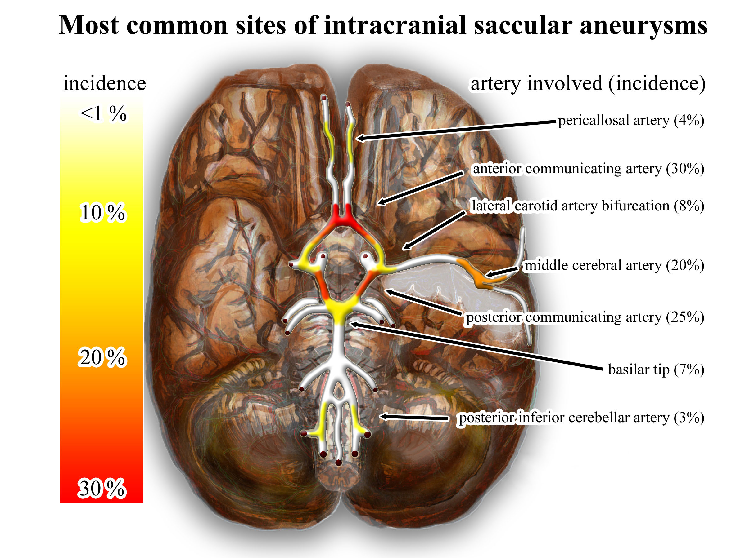
An intracranial aneurysm, also known as a brain aneurysm, is a cerebrovascular disorder in which weakness in the wall of a cerebral artery or vein causes a localized dilation or ballooning of the blood vessel. Aneurysms in the posterior circulation (basilar artery, vertebral arteries and posterior communicating artery) have a higher risk of rupture. Basilar artery aneurysms represent only 3–5% of all intracranial aneurysms but are the most common aneurysms in the posterior circulation. Classification Cerebral aneurysms are classified both by size and shape. Small aneurysms have a diameter of less than 15 mm. Larger aneurysms include those classified as large (15 to 25 mm), giant (25 to 50 mm), and super-giant (over 50 mm). Berry (saccular) aneurysms Saccular aneurysms, also known as berry aneurysms, appear as a round outpouching and are the most common form of cerebral aneurysm. Causes include connective tissue disorders, polycystic kidney disease, ar ... [...More Info...] [...Related Items...] OR: [Wikipedia] [Google] [Baidu] |
Haemorrhage
Bleeding, hemorrhage, haemorrhage or blood loss, is blood escaping from the circulatory system from damaged blood vessels. Bleeding can occur internally, or externally either through a natural opening such as the mouth, nose, ear, urethra, vagina or anus, or through a puncture in the skin. Hypovolemia is a massive decrease in blood volume, and death by excessive loss of blood is referred to as exsanguination. Typically, a healthy person can endure a loss of 10–15% of the total blood volume without serious medical difficulties (by comparison, blood donation typically takes 8–10% of the donor's blood volume). The stopping or controlling of bleeding is called hemostasis and is an important part of both first aid and surgery. Types * Upper head ** Intracranial hemorrhage – bleeding in the skull. ** Cerebral hemorrhage – a type of intracranial hemorrhage, bleeding within the brain tissue itself. ** Intracerebral hemorrhage – bleeding in the brain caused by the rupture ... [...More Info...] [...Related Items...] OR: [Wikipedia] [Google] [Baidu] |
Thrombosis
Thrombosis (from Ancient Greek "clotting") is the formation of a blood clot inside a blood vessel, obstructing the flow of blood through the circulatory system. When a blood vessel (a vein or an artery) is injured, the body uses platelets (thrombocytes) and fibrin to form a blood clot to prevent blood loss. Even when a blood vessel is not injured, blood clots may form in the body under certain conditions. A clot, or a piece of the clot, that breaks free and begins to travel around the body is known as an embolus. Thrombosis may occur in veins ( venous thrombosis) or in arteries ( arterial thrombosis). Venous thrombosis (sometimes called DVT, deep vein thrombosis) leads to a blood clot in the affected part of the body, while arterial thrombosis (and, rarely, severe venous thrombosis) affects the blood supply and leads to damage of the tissue supplied by that artery ( ischemia and necrosis). A piece of either an arterial or a venous thrombus can break off as an embolus, whic ... [...More Info...] [...Related Items...] OR: [Wikipedia] [Google] [Baidu] |
Aneurysm
An aneurysm is an outward bulging, likened to a bubble or balloon, caused by a localized, abnormal, weak spot on a blood vessel wall. Aneurysms may be a result of a hereditary condition or an acquired disease. Aneurysms can also be a nidus (starting point) for clot formation ( thrombosis) and embolization. As an aneurysm increases in size, the risk of rupture, which leads to uncontrolled bleeding, increases. Although they may occur in any blood vessel, particularly lethal examples include aneurysms of the Circle of Willis in the brain, aortic aneurysms affecting the thoracic aorta, and abdominal aortic aneurysms. Aneurysms can arise in the heart itself following a heart attack, including both ventricular and atrial septal aneurysms. There are congenital atrial septal aneurysms, a rare heart defect. Etymology The word is from Greek: ἀνεύρυσμα, aneurysma, "dilation", from ἀνευρύνειν, aneurynein, "to dilate". Classification Aneurysms are classified b ... [...More Info...] [...Related Items...] OR: [Wikipedia] [Google] [Baidu] |
Embolisation
Embolization refers to the passage and lodging of an embolus within the circulatory system, bloodstream. It may be of natural origin (pathological), in which word sense, sense it is also called embolism, for example a pulmonary embolism; or it may be artificially induced (therapy, therapeutic), as a hemostasis, hemostatic treatment for bleeding or as a treatment for some types of cancer by deliberately blocking blood vessels to starve the neoplasm, tumor cells. In the management of cancer, cancer management application, the embolus, besides blocking the blood supply to the tumor, also often includes an ingredient to attack the tumor chemically or with irradiation. When it bears a chemotherapy drug, the process is called chemoembolization. Transcatheter arterial chemoembolization (TACE) is the usual form. When the embolus bears a medicinal radiocompounds, radiopharmaceutical for unsealed source radiotherapy, the process is called radioembolization or selective internal radiatio ... [...More Info...] [...Related Items...] OR: [Wikipedia] [Google] [Baidu] |


.jpg)


