|
Voiding
Urination is the release of urine from the bladder through the urethra in placental mammals, or through the cloaca in other vertebrates. It is the urinary system's form of excretion. It is also known medically as micturition, voiding, uresis, or, rarely, emiction, and known colloquially by various names including peeing, weeing, pissing, and euphemistically number one. The process of urination is under voluntary control in healthy humans and other animals, but may occur as a reflex in infants, some elderly individuals, and those with neurological injury. It is normal for adult humans to urinate up to seven times during the day. In some animals, in addition to expelling waste material, urination can mark territory or express submissiveness. Physiologically, urination involves coordination between the central, autonomic, and somatic nervous systems. Brain centres that regulate urination include the pontine micturition center, periaqueductal gray, and the cerebral cortex. ... [...More Info...] [...Related Items...] OR: [Wikipedia] [Google] [Baidu] |
Manneken Pis
(; ) is a landmark bronze fountain sculpture in central Brussels, Belgium, depicting a puer mingens; a Nudity, naked little boy urinating into the fountain's basin. Though its existence is attested as early as the mid-15th century, ''Manneken Pis'' was redesigned by the Duchy of Brabant, Brabantine sculptor :fr:Jérôme Duquesnoy l'Ancien, Jérôme Duquesnoy the Elder and put in place in 1619. Its Petit Granit, blue stone niche in rocaille style dates from 1770. The statue has been repeatedly stolen or damaged throughout its history. Since 1965, a replica has been displayed on site, with the original stored in the Brussels City Museum. ''Manneken Pis'' is one of the best-known symbols of Brussels and Belgium, inspiring several legends, as well as numerous imitations and similar statues, both nationally and abroad. The figure is regularly dressed up and its wardrobe consists of around one thousand different costumes. Since 2017, they have been exhibited in a dedicated museum ... [...More Info...] [...Related Items...] OR: [Wikipedia] [Google] [Baidu] |
Somatic Nervous System
The somatic nervous system (SNS), also known as voluntary nervous system, is a part of the peripheral nervous system (PNS) that links brain and spinal cord to skeletal muscles under conscious control, as well as to sensory receptors in the skin. The other part complementary to the somatic nervous system is the autonomic nervous system (ANS). The somatic nervous system consists of nerves carrying afferent nerve fibers, which relay sensation from the body to the central nervous system (CNS), and nerves carrying efferent nerve fibers, which relay motor commands from the CNS to stimulate muscle contraction. Specialized nerve fiber ends called sensory receptors are responsible for detecting information both inside and outside the body. The ''a-'' of ''afferent'' and the ''e-'' of ''efferent'' correspond to the prefixes ''ad-'' (to, toward) and ''ex-'' (out of). Structure There are 43 segments of nerves in the human body. With each segment, there is a pair of sensory and motor n ... [...More Info...] [...Related Items...] OR: [Wikipedia] [Google] [Baidu] |
Sacrum
The sacrum (: sacra or sacrums), in human anatomy, is a triangular bone at the base of the spine that forms by the fusing of the sacral vertebrae (S1S5) between ages 18 and 30. The sacrum situates at the upper, back part of the pelvic cavity, between the two wings of the pelvis. It forms joints with four other bones. The two projections at the sides of the sacrum are called the alae (wings), and articulate with the ilium at the L-shaped sacroiliac joints. The upper part of the sacrum connects with the last lumbar vertebra (L5), and its lower part with the coccyx (tailbone) via the sacral and coccygeal cornua. The sacrum has three different surfaces which are shaped to accommodate surrounding pelvic structures. Overall, it is concave (curved upon itself). The base of the sacrum, the broadest and uppermost part, is tilted forward as the sacral promontory internally. The central part is curved outward toward the posterior, allowing greater room for the pelvic cavity. In a ... [...More Info...] [...Related Items...] OR: [Wikipedia] [Google] [Baidu] |
Parasympathetic
The parasympathetic nervous system (PSNS) is one of the three divisions of the autonomic nervous system, the others being the sympathetic nervous system and the enteric nervous system. The autonomic nervous system is responsible for regulating the body's unconscious actions. The parasympathetic system is responsible for stimulation of "rest-and-digest" or "feed-and-breed" activities that occur when the body is at rest, especially after eating, including sexual arousal, salivation, lacrimation (tears), urination, digestion, and defecation. Its action is described as being complementary to that of the sympathetic nervous system, which is responsible for stimulating activities associated with the fight-or-flight response. Nerve fibres of the parasympathetic nervous system arise from the central nervous system. Specific nerves include several cranial nerves, specifically the oculomotor nerve, facial nerve, glossopharyngeal nerve, and vagus nerve. Three spinal nerves in ... [...More Info...] [...Related Items...] OR: [Wikipedia] [Google] [Baidu] |
Spinal Cord
The spinal cord is a long, thin, tubular structure made up of nervous tissue that extends from the medulla oblongata in the lower brainstem to the lumbar region of the vertebral column (backbone) of vertebrate animals. The center of the spinal cord is hollow and contains a structure called the central canal, which contains cerebrospinal fluid. The spinal cord is also covered by meninges and enclosed by the neural arches. Together, the brain and spinal cord make up the central nervous system. In humans, the spinal cord is a continuation of the brainstem and anatomically begins at the occipital bone, passing out of the foramen magnum and then enters the spinal canal at the beginning of the cervical vertebrae. The spinal cord extends down to between the first and second lumbar vertebrae, where it tapers to become the cauda equina. The enclosing bony vertebral column protects the relatively shorter spinal cord. It is around long in adult men and around long in adult women. The diam ... [...More Info...] [...Related Items...] OR: [Wikipedia] [Google] [Baidu] |
Lumbar
In tetrapod anatomy, lumbar is an adjective that means of or pertaining to the abdominal segment of the torso, between the diaphragm (anatomy), diaphragm and the sacrum. Naming and location The lumbar region is sometimes referred to as the lower vertebral column, spine, or as an area of the back in its proximity. In human anatomy the five lumbar vertebrae (vertebrae in the lumbar region of the back) are the largest and strongest in the movable part of the spinal column, and can be distinguished by the absence of a foramen transversarium, foramen in the transverse process, and by the absence of facets on the sides of the body. In most mammals, the lumbar region of the spine curves outward. Description The actual spinal cord terminates between vertebrae one and two of this series, called L1 and L2. The central nervous system, nervous tissue that extends below this point are individual strands that collectively form the cauda equina. In between each lumbar vertebra a nerve root exi ... [...More Info...] [...Related Items...] OR: [Wikipedia] [Google] [Baidu] |
Sympathetic Nervous System
The sympathetic nervous system (SNS or SANS, sympathetic autonomic nervous system, to differentiate it from the somatic nervous system) is one of the three divisions of the autonomic nervous system, the others being the parasympathetic nervous system and the enteric nervous system. The enteric nervous system is sometimes considered part of the autonomic nervous system, and sometimes considered an independent system. The autonomic nervous system functions to regulate the body's unconscious actions. The sympathetic nervous system's primary process is to stimulate the body's fight or flight response. It is, however, constantly active at a basic level to maintain homeostasis. The sympathetic nervous system is described as being antagonistic to the parasympathetic nervous system. The latter stimulates the body to "feed and breed" and to (then) "rest-and-digest". The SNS has a major role in various physiological processes such as blood glucose levels, body temperature, cardiac output, ... [...More Info...] [...Related Items...] OR: [Wikipedia] [Google] [Baidu] |
Detrusor
The detrusor muscle, also detrusor urinae muscle, muscularis propria of the urinary bladder and (less precise) muscularis propria, is smooth muscle found in the wall of the urinary bladder, bladder. The detrusor muscle remains relaxed to allow the bladder to store urine, and contracts during urination to release urine. Related are the urethral sphincter muscles which envelop the urethra to control the flow of urine when they contract. Structure The fibers of the detrusor muscle arise from the Posterior (anatomy), posterior surface of the body of pubic bone, body of the pubis in both sexes (musculi pubovesicales), and in the male from the adjacent part of the prostate. These fibers pass, in a more or less wiktionary:longitudinal, longitudinal manner, up the Anatomical terms of location#Superior and inferior, inferior surface of the urinary bladder, bladder, over its Apex of urinary bladder, apex, and then descend along its fundus of the urinary bladder#Fundus, fundus to become atta ... [...More Info...] [...Related Items...] OR: [Wikipedia] [Google] [Baidu] |
Smooth Muscle
Smooth muscle is one of the three major types of vertebrate muscle tissue, the others being skeletal and cardiac muscle. It can also be found in invertebrates and is controlled by the autonomic nervous system. It is non- striated, so-called because it has no sarcomeres and therefore no striations (''bands'' or ''stripes''). It can be divided into two subgroups, ''single-unit'' and ''multi-unit'' smooth muscle. Within single-unit muscle, the whole bundle or sheet of smooth muscle cells contracts as a syncytium. Smooth muscle is found in the walls of hollow organs, including the stomach, intestines, bladder and uterus. In the walls of blood vessels, and lymph vessels, (excluding blood and lymph capillaries) it is known as vascular smooth muscle. There is smooth muscle in the tracts of the respiratory, urinary, and reproductive systems. In the eyes, the ciliary muscles, iris dilator muscle, and iris sphincter muscle are types of smooth muscles. The iris dilator and s ... [...More Info...] [...Related Items...] OR: [Wikipedia] [Google] [Baidu] |
Urinary Bladder
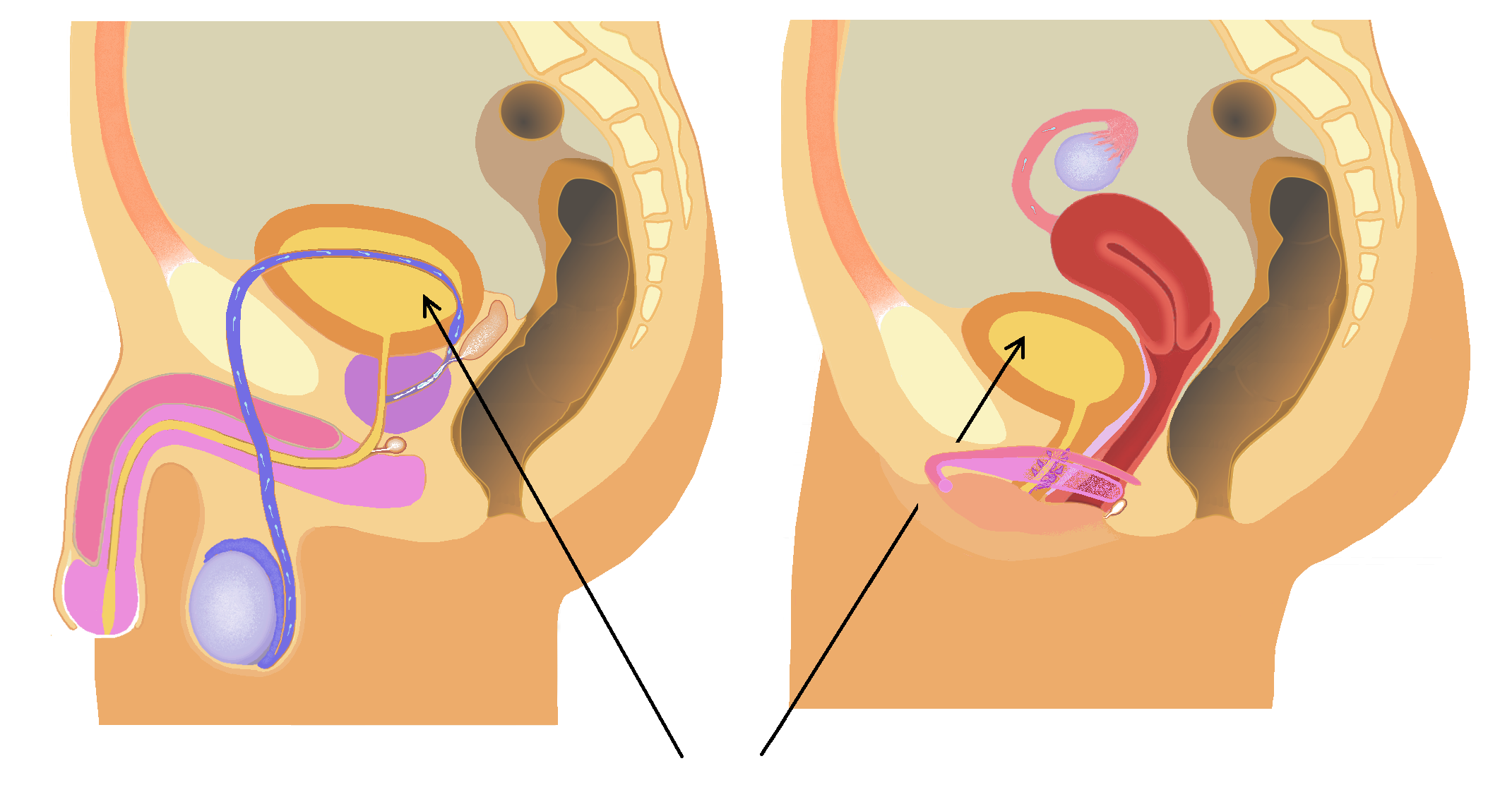
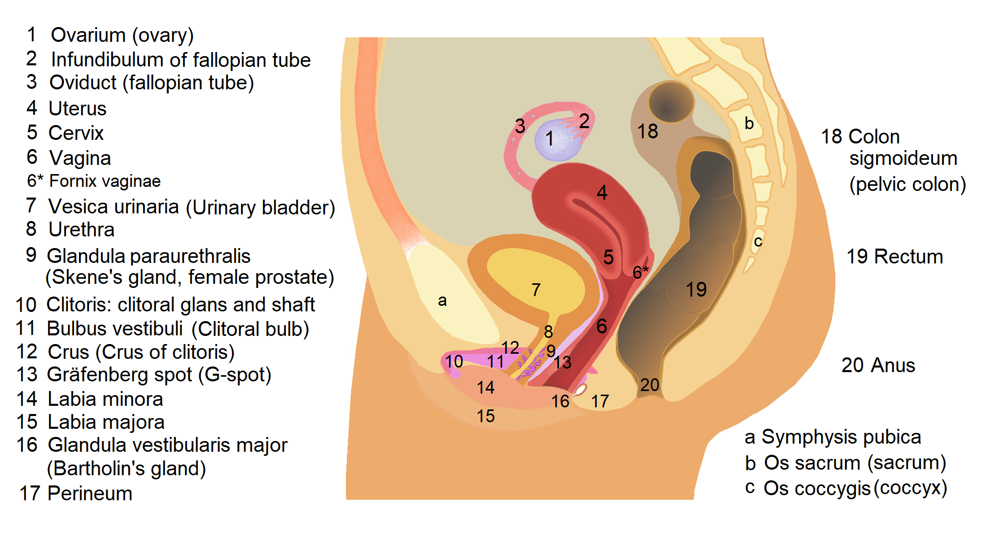
The bladder () is a hollow organ in humans and other vertebrates that stores urine from the Kidney (vertebrates), kidneys. In placental mammals, urine enters the bladder via the ureters and exits via the urethra during urination. In humans, the bladder is a distensible organ that sits on the pelvic floor. The typical adult human bladder will hold between 300 and (10 and ) before the urge to empty occurs, but can hold considerably more. The Latin phrase for "urinary bladder" is ''vesica urinaria'', and the term ''vesical'' or prefix ''vesico-'' appear in connection with associated structures such as vesical veins. The modern Latin word for "bladder" – ''cystis'' – appears in associated terms such as cystitis (inflammation of the bladder). Structure In humans, the bladder is a hollow muscular organ situated at the base of the pelvis. In gross anatomy, the bladder can be divided into a broad (base), a body, an apex, and a neck. The apex (also called the vertex) is directed ... [...More Info...] [...Related Items...] OR: [Wikipedia] [Google] [Baidu] |
Anatomical Plane
An anatomical plane is a hypothetical plane used to transect the body, in order to describe the location of structures or the direction of movements. In human anatomy and non-human anatomy, four principal planes are used: the median plane, sagittal plane, coronal plane, and transverse plane. * The median plane or midsagittal plane passes through the middle of the body, dividing it into left and right halves. * A para sagittal plane is any plane that runs parallel to the median plane, also dividing the body into left and right sections. * The dorsal plane divides the body into dorsal (towards the backbone) and ventral (towards the belly) parts. In human anatomy coronal plane is preferred, or sometimes the frontal plane, and the description may reference splitting the body into front and back parts, but this phrasing is not as clear for animals with a horizontal spine like quadrupeds or fish. * The transverse plane, also called the axial plane or horizontal plane, is perpendi ... [...More Info...] [...Related Items...] OR: [Wikipedia] [Google] [Baidu] |
External Urethral Orifice (male)
The urinary meatus (, ; : meati or meatuses), also known as the external urethral orifice, is the opening of the penis or vulva where urine exits the urethra during urination. It is also where semen exits during male ejaculation, and other fluids during female ejaculation. The meatus has varying degrees of sensitivity to touch. In human males The male external urethral orifice is the external opening of the urethra, normally located at the tip of the glans penis, at its junction with the frenular delta. It presents as a vertical slit, and continues longitudinally along the front aspect of the glans, which facilitates micturition. In some cases, the opening may be more rounded. This can occur naturally or may also occur as a side effect of excessive skin removal during circumcision. The meatus is a sensitive part of the male reproductive system. In human females The female external urethral orifice is where urine exits the urethra during urination. It is located about behin ... [...More Info...] [...Related Items...] OR: [Wikipedia] [Google] [Baidu] |


