|
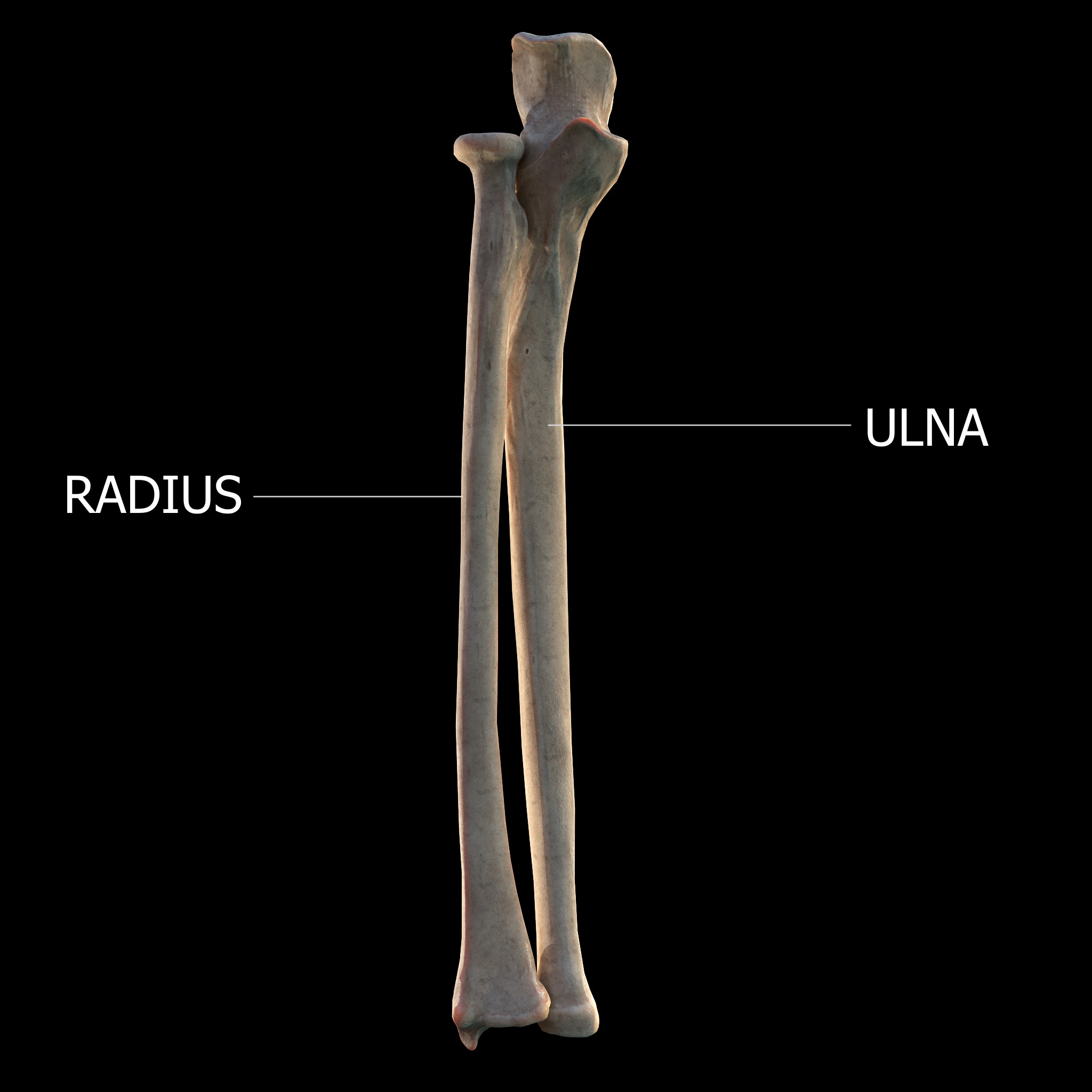
Ulna
The ulna or ulnar bone (: ulnae or ulnas) is a long bone in the forearm stretching from the elbow to the wrist. It is on the same side of the forearm as the little finger, running parallel to the Radius (bone), radius, the forearm's other long bone. Longer and thinner than the radius, the ulna is considered to be the smaller long bone of the lower arm. The corresponding bone in the Human leg#Structure, lower leg is the fibula. Structure The ulna is a long bone found in the forearm that stretches from the elbow to the wrist, and when in standard anatomical position, is found on the Medial (anatomy), medial side of the forearm. It is broader close to the elbow, and narrows as it approaches the wrist. Close to the elbow, the ulna has a bony Process (anatomy), process, the olecranon process, a hook-like structure that fits into the olecranon fossa of the humerus. This prevents hyperextension and forms a hinge joint with the trochlea of the humerus. There is also a radial notch for ... [...More Info...] [...Related Items...] OR: [Wikipedia] [Google] [Baidu] |
Elbow
The elbow is the region between the upper arm and the forearm that surrounds the elbow joint. The elbow includes prominent landmarks such as the olecranon, the cubital fossa (also called the chelidon, or the elbow pit), and the lateral and the medial epicondyles of the humerus. The elbow joint is a hinge joint between the arm and the forearm; more specifically between the humerus in the upper arm and the radius and ulna in the forearm which allows the forearm and hand to be moved towards and away from the body. The term ''elbow'' is specifically used for humans and other primates, and in other vertebrates it is not used. In those cases, forelimb plus joint is used. The name for the elbow in Latin is ''cubitus'', and so the word cubital is used in some elbow-related terms, as in ''cubital nodes'' for example. Structure Joint The elbow joint has three different portions surrounded by a common joint capsule. These are joints between the three bones of the elbow, the ... [...More Info...] [...Related Items...] OR: [Wikipedia] [Google] [Baidu] |
Forearm
The forearm is the region of the upper limb between the elbow and the wrist. The term forearm is used in anatomy to distinguish it from the arm, a word which is used to describe the entire appendage of the upper limb, but which in anatomy, technically, means only the region of the upper arm, whereas the lower "arm" is called the forearm. It is homologous to the region of the leg that lies between the knee and the ankle joints, the crus. The forearm contains two long bones, the radius and the ulna, forming the two radioulnar joints. The interosseous membrane connects these bones. Ultimately, the forearm is covered by skin, the anterior surface usually being less hairy than the posterior surface. The forearm contains many muscles, including the flexors and extensors of the wrist, flexors and extensors of the digits, a flexor of the elbow ( brachioradialis), and pronators and supinators that turn the hand to face down or upwards, respectively. In cross-section, the forearm can ... [...More Info...] [...Related Items...] OR: [Wikipedia] [Google] [Baidu] |
Radius (bone)
The radius or radial bone (: radii or radiuses) is one of the two large bones of the forearm, the other being the ulna. It extends from the Anatomical terms of location, lateral side of the Elbow-joint, elbow to the thumb side of the wrist and runs parallel to the ulna. The ulna is longer than the radius, but the radius is thicker. The radius is a long bone, Prism (geometry), prism-shaped and slightly curved longitudinally. The radius is part of two joint (anatomy), joints: the elbow and the wrist. At the elbow, it joins with the capitulum of the humerus, and in a separate region, with the ulna at the radial notch. At the wrist, the radius forms a joint with the ulna bone. The corresponding bone in the human leg, lower leg is the tibia. Structure The long narrow medullary cavity is enclosed in a strong wall of compact bone. It is thickest along the interosseous border and thinnest at the extremities, same over the cup-shaped articular surface (fovea) of the head. The tra ... [...More Info...] [...Related Items...] OR: [Wikipedia] [Google] [Baidu] |
Wrist
In human anatomy, the wrist is variously defined as (1) the carpus or carpal bones, the complex of eight bones forming the proximal skeletal segment of the hand; "The wrist contains eight bones, roughly aligned in two rows, known as the carpal bones." (2) the wrist joint or radiocarpal joint, the joint between the radius and the carpus and; (3) the anatomical region surrounding the carpus including the distal parts of the bones of the forearm and the proximal parts of the metacarpus or five metacarpal bones and the series of joints between these bones, thus referred to as ''wrist joints''. "With the large number of bones composing the wrist (ulna, radius, eight carpas, and five metacarpals), it makes sense that there are many, many joints that make up the structure known as the wrist." This region also includes the carpal tunnel, the anatomical snuff box, bracelet lines, the flexor retinaculum, and the extensor retinaculum. As a consequence of these various definitions, f ... [...More Info...] [...Related Items...] OR: [Wikipedia] [Google] [Baidu] |
Flexor Carpi Ulnaris
The flexor carpi ulnaris (FCU) is a skeletal muscle, muscle of the forearm that flexion, flexes and Adduction, adducts at the wrist joint. Structure Origin The flexor carpi ulnaris has two heads; a humeral head and ulnar head. The humeral head originates from the medial epicondyle of the humerus via the common flexor tendon. The ulnar head originates from the medial margin of the olecranon of the ulna and the upper two-thirds of the dorsal border of the ulna by an aponeurosis. Between the two heads passes the ulnar nerve and ulnar artery. Insertion The flexor carpi ulnaris inserts onto the pisiform bone, pisiform, hook of the hamate (via the pisohamate ligament) and the anterior surface of the base of the fifth metacarpal bone, fifth metacarpal (via the pisometacarpal ligament). Action The flexor carpi ulnaris flexes and adducts at the Wrist, wrist joint. Innervation The flexor carpi ulnaris is innervated by the ulnar nerve. The corresponding spinal nerves are Cervical spinal ... [...More Info...] [...Related Items...] OR: [Wikipedia] [Google] [Baidu] |
Ulnar Styloid Process
The styloid process of the ulna is a bony prominence found at distal end of the ulna in the forearm. Structure The styloid process of the ulna projects from the medial and back part of the ulna. It descends a little lower than the head. The head is separated from the styloid process by a depression for the attachment of the apex of the triangular articular disk, and behind, by a shallow groove for the tendon of the extensor carpi ulnaris muscle. The styloid process of the ulna varies from 2 to 6 mm in length. Function The rounded end of the styloid process of the ulna connects to the ulnar collateral ligament of the wrist. The radioulnar ligaments also attaches to the base of the styloid process of the ulna. Clinical significance Fractures of the styloid process of the ulna seldom require treatment when they occur in association with a distal radius fracture. The major exception is when the joint between these bones, the distal radioulnar joint (or DRUJ), is unstable ... [...More Info...] [...Related Items...] OR: [Wikipedia] [Google] [Baidu] |
Coronoid Process Of The Ulna
The coronoid process of the ulna is a triangular process projecting forward from the anterior proximal portion of the ulna. Structure Its ''base'' is continuous with the body of the bone, and of considerable strength. Its ''apex'' is pointed, slightly curved upward, and in flexion of the forearm is received into the coronoid fossa of the humerus. Its ''upper surface'' is smooth, convex, and forms the lower part of the semilunar notch. Its ''antero-inferior'' surface is concave, and marked by a rough impression for the insertion of the brachialis muscle. At the junction of this surface with the front of the body is a rough eminence, the tuberosity of the ulna, which gives insertion to a part of the brachialis; to the lateral border of this tuberosity the oblique cord is attached. Its ''lateral surface'' presents a narrow, oblong, articular depression, the radial notch. Its ''medial surface'', by its prominent, free margin, serves for the attachment of part of the ulnar ... [...More Info...] [...Related Items...] OR: [Wikipedia] [Google] [Baidu] |
Ulnar Collateral Ligament Of Elbow Joint
The ulnar collateral ligament (UCL) or internal lateral ligament is a thick triangular ligament at the medial aspect of the elbow uniting the distal aspect of the humerus to the proximal aspect of the ulna. Structure It consists of two portions, an anterior and posterior united by a thinner intermediate portion. Note that this ligament is also referred to as the medial collateral ligament and should not be confused with the lateral ulnar collateral ligament (LUCL). The ''anterior portion'', directed obliquely forward, is attached, above, by its apex, to the front part of the medial epicondyle of the humerus; and, below, by its broad base to the medial margin of the coronoid process of the ulna. The ''posterior portion'', also of triangular form, is attached, above, by its apex, to the lower and back part of the medial epicondyle; below, to the medial margin of the olecranon. Between these two bands a few intermediate fibers descend from the medial epicondyle to blend with ... [...More Info...] [...Related Items...] OR: [Wikipedia] [Google] [Baidu] |
Olecranon Process
The olecranon (, ), is a large, thick, curved bony process on the proximal, posterior end of the ulna. It forms the protruding part of the elbow and is opposite to the cubital fossa or elbow pit (trochlear notch). The olecranon serves as a lever for the extensor muscles that straighten the elbow joint. Structure The olecranon is situated at the proximal end of the ulna, one of the two bones in the forearm. When the hand faces forward (supination) the olecranon faces towards the back (posteriorly). It is bent forward at the summit so as to present a prominent lip which is received into the olecranon fossa of the humerus during extension of the forearm. Its base is contracted where it joins the body and the narrowest part of the upper end of the ulna. Its posterior surface, directed backward, is triangular, smooth, subcutaneous, and covered by a bursa. Its superior surface is of quadrilateral form, marked behind by a rough impression for the insertion of the triceps brachii; an ... [...More Info...] [...Related Items...] OR: [Wikipedia] [Google] [Baidu] |
Humerus
The humerus (; : humeri) is a long bone in the arm that runs from the shoulder to the elbow. It connects the scapula and the two bones of the lower arm, the radius (bone), radius and ulna, and consists of three sections. The humeral upper extremity of humerus, upper extremity consists of a rounded head, a narrow neck, and two short processes (tubercles, sometimes called tuberosities). The body of humerus, body is cylindrical in its upper portion, and more prism (geometry), prismatic below. The lower extremity of humerus, lower extremity consists of 2 epicondyles, 2 processes (trochlea of the humerus, trochlea and capitulum of the humerus, capitulum), and 3 fossae (radial fossa, coronoid fossa, and olecranon fossa). As well as its true anatomical neck, the constriction below the greater and lesser tubercles of the humerus is referred to as its Surgical neck of the humerus, surgical neck due to its tendency to fracture, thus often becoming the focus of surgeons. Etymology The word ... [...More Info...] [...Related Items...] OR: [Wikipedia] [Google] [Baidu] |
Olecranon
The olecranon (, ), is a large, thick, curved bony process on the proximal, posterior end of the ulna. It forms the protruding part of the elbow and is opposite to the cubital fossa or elbow pit (trochlear notch). The olecranon serves as a lever for the extensor muscles that straighten the elbow joint. Structure The olecranon is situated at the proximal end of the ulna, one of the two bones in the forearm. When the hand faces forward ( supination) the olecranon faces towards the back (posteriorly). It is bent forward at the summit so as to present a prominent lip which is received into the olecranon fossa of the humerus during extension of the forearm. Its base is contracted where it joins the body and the narrowest part of the upper end of the ulna. Its posterior surface, directed backward, is triangular, smooth, subcutaneous, and covered by a bursa. Its superior surface is of quadrilateral form, marked behind by a rough impression for the insertion of the triceps brachi ... [...More Info...] [...Related Items...] OR: [Wikipedia] [Google] [Baidu] |
Flexor Digitorum Profundus
The flexor digitorum profundus or flexor digitorum communis profundus is a muscle in the forearm of humans that flexes the fingers (also known as digits). It is considered an Muscles of the hand#Extrinsic, extrinsic hand muscle because it acts on the hand while its muscle belly is located in the forearm. Together the Flexor pollicis longus muscle, flexor pollicis longus, Pronator quadratus muscle, pronator quadratus, and flexor digitorum profundus form the deep layer of ventral forearm muscles.Platzer 2004, p 162 The muscle is named . Structure Flexor digitorum profundus originates in the upper 3/4 of the anterior and medial surfaces of the ulna, interosseous membrane and deep fascia of the forearm. The muscle fans out into four tendons (one to each of the second to fifth fingers) to the palmar base of the distal phalanges, distal phalanx. Along with the flexor digitorum superficialis, it has long tendons that run down the arm and through the carpal tunnel and attach to the p ... [...More Info...] [...Related Items...] OR: [Wikipedia] [Google] [Baidu] |


