|
Troponin I
Troponin I is a cardiac and skeletal muscle protein family. It is a part of the troponin protein complex, where it binds to actin in thin myofilaments to hold the actin-tropomyosin complex in place. Troponin I prevents myosin from binding to actin in relaxed muscle. When calcium binds to the troponin C, it causes conformational changes which lead to dislocation of troponin I. Afterwards, tropomyosin leaves the binding site for myosin on actin leading to contraction of muscle. The letter ''I'' is given due to its inhibitory character. It is a useful marker in the laboratory diagnosis of heart attack. It occurs in different plasma concentration but the same circumstances as troponin T - either test can be performed for confirmation of cardiac muscle damage and laboratories usually offer one test or the other. Three paralogs with unique tissue-specific expression patterns are expressed in humans, listed below with their locations and OMIM accessions: * Slow-twitch skeletal muscl ... [...More Info...] [...Related Items...] OR: [Wikipedia] [Google] [Baidu] |
Troponin
image:Troponin Ribbon Diagram.png, 400px, Ribbon representation of the human cardiac troponin core complex (52 kDa core) in the calcium-saturated form. Blue = troponin C; green = troponin I; magenta = troponin T.; ; rendered with PyMOL Troponin, or the troponin complex, is a complex of three regulatory proteins (troponin C, troponin I, and troponin T) that are integral to muscle contraction in skeletal muscle and cardiac muscle, but not smooth muscle. Measurements of cardiac-specific troponins I and T are extensively used as diagnostic and prognostic indicators in the management of myocardial infarction and acute coronary syndrome. Blood troponin levels may be used as a diagnostic marker for stroke or other myocardial injury that is ongoing, although the sensitivity of this measurement is low. Function Troponin is attached to the protein tropomyosin and lies within the groove between actin filaments in muscle tissue. In a relaxed muscle, tropomyosin blocks the attachment site fo ... [...More Info...] [...Related Items...] OR: [Wikipedia] [Google] [Baidu] |
P21-activated Kinases
p21 activated kinases (PAKs) are members of a family of enzymes. They serve as targets for the small GTP binding proteins CDC42 and Rac and have been implicated in a wide range of biological activities. Members include: * PAK1, regulating cell motility and morphology * PAK2, possibly playing a role in apoptosis * PAK3, possibly for dendritic development and for the rapid cytoskeletal reorganization in dendritic spines associated with synaptic plasticity * PAK4, a mediator of filopodia formation * PAK5, a mediator of filopodia formation * PAK6 Serine/threonine-protein kinase PAK 6 is an enzyme that in humans is encoded by the ''PAK6'' gene. PAK6 belongs to the Group II PAK family members. Like other family members, PAK6 is also evolutionary conserved across species. Discovery The PAK ..., involved in cytoskeleton rearrangement References External linksPAKInfo with links in thCell Migration Gateway Human proteins {{Protein-stub ... [...More Info...] [...Related Items...] OR: [Wikipedia] [Google] [Baidu] |
Troponin C
Troponin C is a protein which is part of the troponin complex. It contains four calcium-binding EF hands, although different isoforms may have fewer than four functional calcium-binding subdomains. It is a component of thin filaments, along with actin and tropomyosin. It contains an N lobe and a C lobe. The C lobe serves a structural purpose and binds to the N domain of troponin I (TnI). The C lobe can bind either Ca2+ or Mg2+. The N lobe, which binds only Ca2+, is the regulatory lobe and binds to the C domain of troponin I after calcium binding. Isoforms The tissue specific subtypes are: * Slow troponin C, TNNC1 (3p21.1 ) * Fast troponin C, TNNC2 (20q12-q13.11, ) Mutations Point mutations can occur in troponin C inducing alterations to Ca2+ and Mg2+ binding and protein structure, leading to abnormalities in muscle contraction. In cardiac muscle, they are related to dilated cardiomyopathy (DCM) and hypertrophic cardiomyopathy (HCM). These known point mutations are: ... [...More Info...] [...Related Items...] OR: [Wikipedia] [Google] [Baidu] |
Troponin T
Troponin T (shortened TnT or TropT) is a part of the troponin complex, which are proteins integral to the contraction of skeletal and heart muscles. They are expressed in skeletal and cardiac myocytes. Troponin T binds to tropomyosin and helps position it on actin, and together with the rest of the troponin complex, modulates contraction of striated muscle. The cardiac subtype of troponin T is especially useful in the laboratory diagnosis of heart attack because it is released into the blood-stream when damage to heart muscle occurs. It was discovered by the German physician Hugo A. Katus at the University of Heidelberg, who also developed the troponin T assay. Subtypes * Slow skeletal troponin T1, TNNT1 (19q13.4, ) * Cardiac troponin T2, TNNT2 (1q32, ) * Fast skeletal troponin T3, TNNT3 (11p15.5, ) Reference values The 99th percentile cutoff for cardiac troponin T (cTnT) is 0.01 ng/mL. The reference range for the high sensitivity troponin T is a normal 52 ng/L. Backgro ... [...More Info...] [...Related Items...] OR: [Wikipedia] [Google] [Baidu] |
Pulmonary Embolus
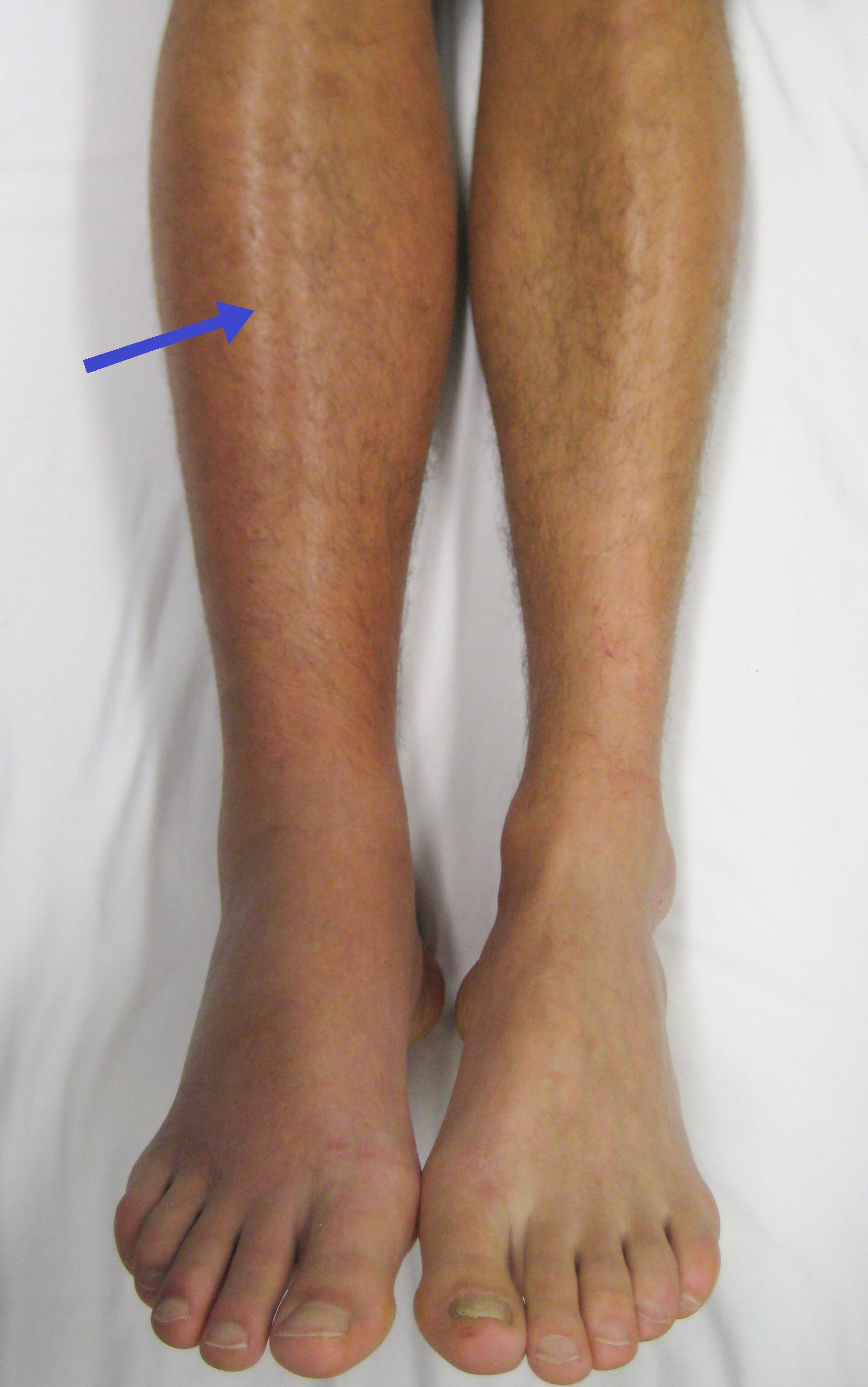
Pulmonary embolism (PE) is a blockage of an artery in the lungs by a substance that has moved from elsewhere in the body through the bloodstream (embolism). Symptoms of a PE may include shortness of breath, chest pain particularly upon breathing in, and coughing up blood. Symptoms of a blood clot in the leg may also be present, such as a red, warm, swollen, and painful leg. Signs of a PE include low blood oxygen levels, rapid breathing, rapid heart rate, and sometimes a mild fever. Severe cases can lead to passing out, abnormally low blood pressure, obstructive shock, and sudden death. PE usually results from a blood clot in the leg that travels to the lung. The risk of blood clots is increased by advanced age, cancer, prolonged bed rest and immobilization, smoking, stroke, long-haul travel over 4 hours, certain genetic conditions, estrogen-based medication, pregnancy, obesity, trauma or bone fracture, and after some types of surgery. A small proportion of cases ... [...More Info...] [...Related Items...] OR: [Wikipedia] [Google] [Baidu] |
Subarachnoid Haemorrhage
Subarachnoid hemorrhage (SAH) is bleeding into the subarachnoid space—the area between the arachnoid membrane and the pia mater surrounding the brain. Symptoms may include a severe headache of rapid onset, vomiting, decreased level of consciousness, fever, and sometimes seizures. Neck stiffness or neck pain are also relatively common. In about a quarter of people a small bleed with resolving symptoms occurs within a month of a larger bleed. SAH may occur as a result of a head injury or spontaneously, usually from a ruptured cerebral aneurysm. Risk factors for spontaneous cases include high blood pressure, smoking, family history, alcoholism, and cocaine use. Generally, the diagnosis can be determined by a CT scan of the head if done within six hours of symptom onset. Occasionally, a lumbar puncture is also required. After confirmation further tests are usually performed to determine the underlying cause. Treatment is by prompt neurosurgery or endovascular coiling. M ... [...More Info...] [...Related Items...] OR: [Wikipedia] [Google] [Baidu] |
Heart Failure
Heart failure (HF), also known as congestive heart failure (CHF), is a syndrome, a group of signs and symptoms caused by an impairment of the heart's blood pumping function. Symptoms typically include shortness of breath, excessive fatigue, and leg swelling. The shortness of breath may occur with exertion or while lying down, and may wake people up during the night. Chest pain, including angina, is not usually caused by heart failure, but may occur if the heart failure was caused by a heart attack. The severity of the heart failure is measured by the severity of symptoms during exercise. Other conditions that may have symptoms similar to heart failure include obesity, kidney failure, liver disease, anemia, and thyroid disease. Common causes of heart failure include coronary artery disease, heart attack, high blood pressure, atrial fibrillation, valvular heart disease, excessive alcohol consumption, infection, and cardiomyopathy. These cause heart failure by alterin ... [...More Info...] [...Related Items...] OR: [Wikipedia] [Google] [Baidu] |
Chronic Kidney Failure
Chronic kidney disease (CKD) is a type of kidney disease in which a gradual loss of kidney function occurs over a period of months to years. Initially generally no symptoms are seen, but later symptoms may include leg swelling, feeling tired, vomiting, loss of appetite, and confusion. Complications can relate to hormonal dysfunction of the kidneys and include (in chronological order) high blood pressure (often related to activation of the renin–angiotensin system system), bone disease, and anemia. Additionally CKD patients have markedly increased cardiovascular complications with increased risks of death and hospitalization. Causes of chronic kidney disease include diabetes, high blood pressure, glomerulonephritis, and polycystic kidney disease. Risk factors include a family history of chronic kidney disease. Diagnosis is by blood tests to measure the estimated glomerular filtration rate (eGFR), and a urine test to measure albumin. Ultrasound or kidney biopsy may be perfor ... [...More Info...] [...Related Items...] OR: [Wikipedia] [Google] [Baidu] |
Isoenzyme
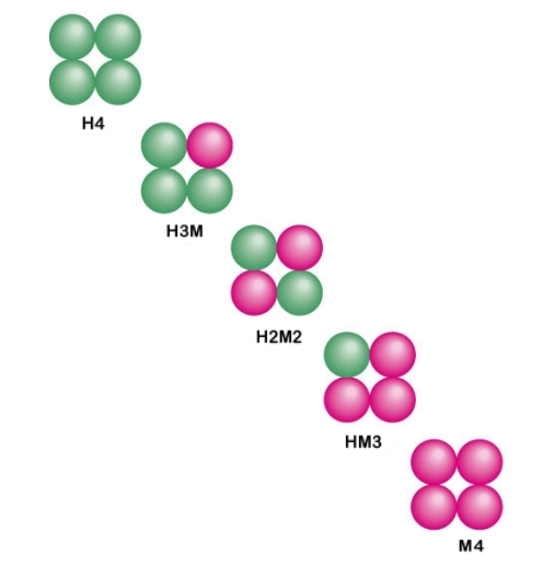
In biochemistry, isozymes (also known as isoenzymes or more generally as multiple forms of enzymes) are enzymes that differ in amino acid sequence but catalyze the same chemical reaction. Isozymes usually have different kinetic parameters (e.g. different ''K''M values), or are regulated differently. They permit the fine-tuning of metabolism to meet the particular needs of a given tissue or developmental stage. In many cases, isozymes are encoded by homologous genes that have diverged over time. Strictly speaking, enzymes with different amino acid sequences that catalyse the same reaction are isozymes if encoded by different genes, or allozymes if encoded by different alleles of the same gene; the two terms are often used interchangeably. Introduction Isozymes were first described by R. L. Hunter and Clement Markert (1957) who defined them as ''different variants of the same enzyme having identical functions and present in the same individual''. This definition encompasses (1) ... [...More Info...] [...Related Items...] OR: [Wikipedia] [Google] [Baidu] |
Lactate Dehydrogenase
Lactate dehydrogenase (LDH or LD) is an enzyme found in nearly all living cells. LDH catalyzes the conversion of lactate to pyruvate and back, as it converts NAD+ to NADH and back. A dehydrogenase is an enzyme that transfers a hydride from one molecule to another. LDH exists in four distinct enzyme classes. This article is specifically about the NAD(P)-dependent L-lactate dehydrogenase. Other LDHs act on D-lactate and/or are dependent on cytochrome c: D-lactate dehydrogenase (cytochrome) and L-lactate dehydrogenase (cytochrome). LDH is expressed extensively in body tissues, such as blood cells and heart muscle. Because it is released during tissue damage, it is a marker of common injuries and disease such as heart failure. Reaction Lactate dehydrogenase catalyzes the interconversion of pyruvate and lactate with concomitant interconversion of NADH and NAD+. It converts pyruvate, the final product of glycolysis, to lactate when oxygen is absent or in short supply, a ... [...More Info...] [...Related Items...] OR: [Wikipedia] [Google] [Baidu] |
Myoglobin
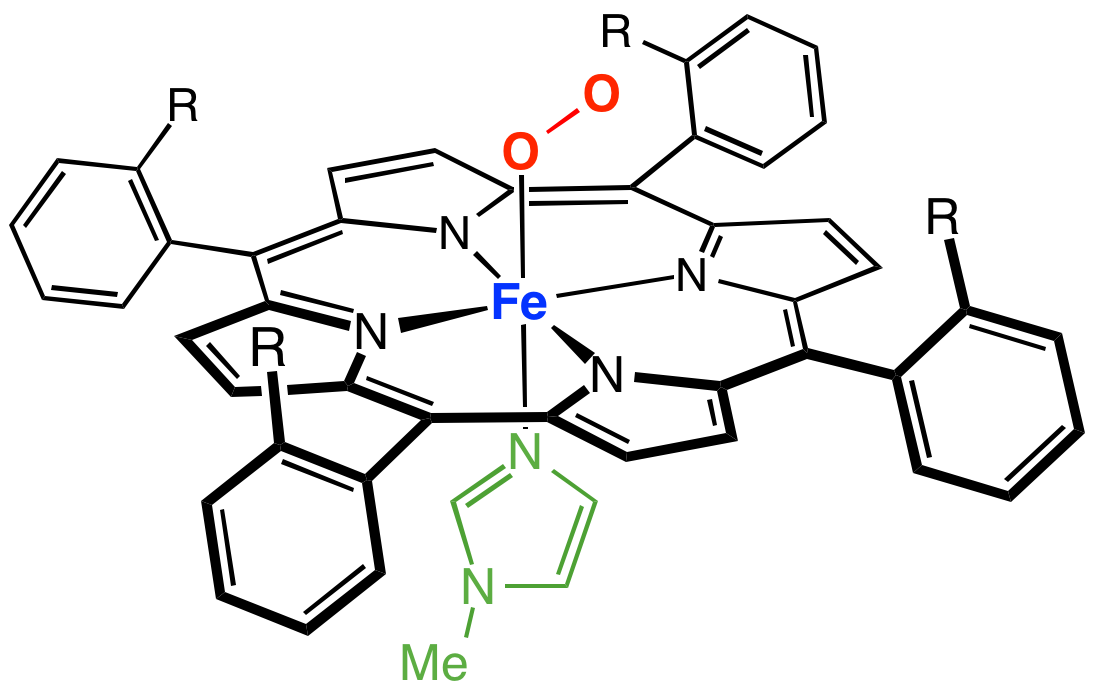
Myoglobin (symbol Mb or MB) is an iron- and oxygen-binding protein found in the cardiac and skeletal muscle tissue of vertebrates in general and in almost all mammals. Myoglobin is distantly related to hemoglobin. Compared to hemoglobin, myoglobin has a higher affinity for oxygen and does not have cooperative binding with oxygen like hemoglobin does. In humans, myoglobin is only found in the bloodstream after muscle injury. (Google books link is the 2008 edition) High concentrations of myoglobin in muscle cells allow organisms to hold their breath for a longer period of time. Diving mammals such as whales and seals have muscles with particularly high abundance of myoglobin. Myoglobin is found in Type I muscle, Type II A, and Type II B; although many texts consider myoglobin not to be found in smooth muscle, this has proved erroneous: there is also myoglobin in smooth muscle cells. Myoglobin was the first protein to have its three-dimensional structure revealed by X-ray cryst ... [...More Info...] [...Related Items...] OR: [Wikipedia] [Google] [Baidu] |
Creatine Kinase
Creatine kinase (CK), also known as creatine phosphokinase (CPK) or phosphocreatine kinase, is an enzyme () expressed by various tissues and cell types. CK catalyses the conversion of creatine and uses adenosine triphosphate (ATP) to create phosphocreatine (PCr) and adenosine diphosphate (ADP). This CK enzyme reaction is reversible and thus ATP can be generated from PCr and ADP. In tissues and cells that consume ATP rapidly, especially skeletal muscle, but also brain, photoreceptor cells of the retina, hair cells of the inner ear, spermatozoa and smooth muscle, PCr serves as an energy reservoir for the rapid buffering and regeneration of ATP ''in situ'', as well as for intracellular energy transport by the PCr shuttle or circuit. Thus creatine kinase is an important enzyme in such tissues. Clinically, creatine kinase is assayed in blood tests as a marker of damage of CK-rich tissue such as in myocardial infarction (heart attack), rhabdomyolysis (severe muscle breakdown), muscul ... [...More Info...] [...Related Items...] OR: [Wikipedia] [Google] [Baidu] |


