|
Triceps Brachii
The triceps, or triceps brachii (Latin for "three-headed muscle of the arm"), is a large muscle on the back of the upper limb of many vertebrates. It consists of 3 parts: the medial, lateral, and long head. It is the muscle principally responsible for extension of the elbow joint (straightening of the arm). Structure The long head arises from the infraglenoid tubercle of the scapula. It extends distally anterior to the teres minor and posterior to the teres major. The medial head arises proximally in the humerus, just inferior to the groove of the radial nerve; from the dorsal (back) surface of the humerus; from the medial intermuscular septum; and its distal part also arises from the lateral intermuscular septum. The medial head is mostly covered by the lateral and long heads, and is only visible distally on the humerus. The lateral head arises from the dorsal surface of the humerus, lateral and proximal to the groove of the radial nerve, from the greater tuber ... [...More Info...] [...Related Items...] OR: [Wikipedia] [Google] [Baidu] |
Infraglenoid Tubercle Of Scapula
The infraglenoid tubercle is the part of the scapula from which the long head of the triceps brachii muscle originates. The infraglenoid tubercle is a tubercle located on the lateral part of the scapula, inferior to (below) the glenoid cavity. The name infraglenoid tubercle refers to its location below the glenoid cavity. Function The infraglenoid tubercle is the origin of the long head of the triceps brachii muscle. It helps to stabilise the muscle origin. Additional images File:Infraglenoid tubercle of left scapula - animation.gif, Left scapula. Infraglenoid tubercle shown in red. File:Infraglenoid tubercle of scapula - animation01.gif, Animation. Infraglenoid tubercle shown in red. File:Infraglenoid tubercle of left scapula01.png, Lateral view of left scapula. Infraglenoid tubercle shown in red. File:Scapula ant numbered.png, Anterior surface of left scapula. Infraglenoid tubercle is "11" File:Infraglenoid tubercle of left scapula03.png, Anterior surface of left scapul ... [...More Info...] [...Related Items...] OR: [Wikipedia] [Google] [Baidu] |
Infraglenoid Tubercle
The infraglenoid tubercle is the part of the scapula from which the long head of the triceps brachii muscle originates. The infraglenoid tubercle is a tubercle located on the lateral part of the scapula, inferior to (below) the glenoid cavity. The name infraglenoid tubercle refers to its location below the glenoid cavity. Function The infraglenoid tubercle is the origin of the long head of the triceps brachii muscle The triceps, or triceps brachii (Latin for "three-headed muscle of the arm"), is a large muscle on the back of the upper limb of many vertebrates. It consists of 3 parts: the medial, lateral, and long head. It is the muscle principally respon .... It helps to stabilise the muscle origin. Additional images File:Infraglenoid tubercle of left scapula - animation.gif, Left scapula. Infraglenoid tubercle shown in red. File:Infraglenoid tubercle of scapula - animation01.gif, Animation. Infraglenoid tubercle shown in red. File:Infraglenoid tubercle of left scap ... [...More Info...] [...Related Items...] OR: [Wikipedia] [Google] [Baidu] |
Synovial Bursa
Synovial () may refer to: * Synovial fluid * Synovial joint * Synovial membrane The synovial membrane (also known as the synovial stratum, synovium or stratum synoviale) is a specialized connective tissue that lines the inner surface of capsules of synovial joints and tendon sheath. It makes direct contact with the fibrous ... * Synovial bursa {{disambiguation ... [...More Info...] [...Related Items...] OR: [Wikipedia] [Google] [Baidu] |
Olecranon
The olecranon (, ), is a large, thick, curved bony eminence of the ulna, a long bone in the forearm that projects behind the elbow. It forms the most pointed portion of the elbow and is opposite to the cubital fossa or elbow pit. The olecranon serves as a lever for the extensor muscles that straighten the elbow joint. Structure The olecranon is situated at the proximal end of the ulna, one of the two bones in the forearm. When the hand faces forward (supination) the olecranon faces towards the back (posteriorly). It is bent forward at the summit so as to present a prominent lip which is received into the olecranon fossa of the humerus during extension of the forearm. Its base is contracted where it joins the body and the narrowest part of the upper end of the ulna. Its posterior surface, directed backward, is triangular, smooth, subcutaneous, and covered by a bursa. Its superior surface is of quadrilateral form, marked behind by a rough impression for the insertion of the Tr ... [...More Info...] [...Related Items...] OR: [Wikipedia] [Google] [Baidu] |
Skeletal Striated Muscle
Skeletal muscles (commonly referred to as muscles) are organs of the vertebrate muscular system and typically are attached by tendons to bones of a skeleton. The muscle cells of skeletal muscles are much longer than in the other types of muscle tissue, and are often known as muscle fibers. The muscle tissue of a skeletal muscle is striated – having a striped appearance due to the arrangement of the sarcomeres. Skeletal muscles are voluntary muscles under the control of the somatic nervous system. The other types of muscle are cardiac muscle which is also striated and smooth muscle which is non-striated; both of these types of muscle tissue are classified as involuntary, or, under the control of the autonomic nervous system. A skeletal muscle contains multiple fascicles – bundles of muscle fibers. Each individual fiber, and each muscle is surrounded by a type of connective tissue layer of fascia. Muscle fibers are formed from the fusion of developmental myoblasts in a pro ... [...More Info...] [...Related Items...] OR: [Wikipedia] [Google] [Baidu] |
Greater Tubercle
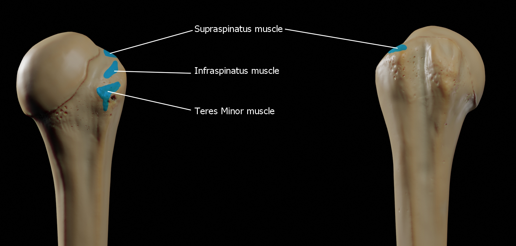
The greater tubercle of the humerus is the outward part the upper end of that bone, adjacent to the large rounded prominence of the humerus head. It provides attachment points for the supraspinatus, infraspinatus, and teres minor muscles, three of the four muscles of the rotator cuff, a muscle group that stabilizes the shoulder joint. In doing so the tubercle acts as a location for the transfer of forces from the rotator cuff muscles to the humerus. Structure The upper surface of the greater tubercle is rounded, and marked by three flat impressions: * the highest ("superior facet") gives insertion to the supraspinatus muscle. * the middle ("middle facet") gives insertion to the infraspinatus muscle. * the lowest ("inferior facet"), and the body of the bone for about 2.5 cm, gives insertion to the teres minor muscle. The lateral surface of the greater tubercle is convex, rough, and continuous with the lateral surface of the body of the humerus. It can be described a ... [...More Info...] [...Related Items...] OR: [Wikipedia] [Google] [Baidu] |
Lateral Intermuscular Septum Of Arm
The fascial compartments of arm refers to the specific anatomical term of the compartments within the upper segment of the upper limb (the arm) of the body. The upper limb is divided into two segments, the arm and the forearm. Each of these segments is further divided into two compartments which are formed by deep fascia – tough connective tissue septa (walls). Each compartment encloses specific muscles and nerves. The compartments of the arm are the anterior compartment of the arm and the posterior compartment of the arm, divided by the lateral and the medial intermuscular septa. The compartments of the forearm are the anterior compartment of the forearm and posterior compartment of the forearm. Intermuscular septa The lateral intermuscular septum extends from the lower part of the crest of the greater tubercle of the humerus, along the lateral supracondylar ridge, to the lateral epicondyle; it is blended with the tendon of the deltoid muscle, gives attachment to the ... [...More Info...] [...Related Items...] OR: [Wikipedia] [Google] [Baidu] |
Medial Intermuscular Septum Of Arm
The fascial compartments of arm refers to the specific anatomical term of the compartments within the upper segment of the upper limb (the arm) of the body. The upper limb is divided into two segments, the arm and the forearm. Each of these segments is further divided into two compartments which are formed by deep fascia – tough connective tissue septa (walls). Each compartment encloses specific muscles and nerves. The compartments of the arm are the anterior compartment of the arm and the posterior compartment of the arm, divided by the lateral and the medial intermuscular septa. The compartments of the forearm are the anterior compartment of the forearm and posterior compartment of the forearm. Intermuscular septa The lateral intermuscular septum extends from the lower part of the crest of the greater tubercle of the humerus, along the lateral supracondylar ridge, to the lateral epicondyle; it is blended with the tendon of the deltoid muscle, gives attachment to the ... [...More Info...] [...Related Items...] OR: [Wikipedia] [Google] [Baidu] |
Radial Sulcus
The radial groove (also known as the musculospiral groove, radial sulcus, or spiral groove) is a broad but shallow oblique depression for the radial nerve and deep brachial artery. It is located on the center of the lateral border of the humerus bone. Although it provides protection to the radial nerve, it is often involved in compressions on the nerve (due to external pressure due to surgery) that can cause radial nerve palsy. See also * Intertubercular groove * Triceps brachii muscle The triceps, or triceps brachii (Latin for "three-headed muscle of the arm"), is a large muscle on the back of the upper limb of many vertebrates. It consists of 3 parts: the medial, lateral, and long head. It is the muscle principally responsibl ... Additional images File:Gray413_color.png, Cross-section through the middle of upper arm. File:Gray525.png, The brachial artery. File:Gray818.png, The suprascapular, axillary, and radial nerves. References Bibliography Humerus ... [...More Info...] [...Related Items...] OR: [Wikipedia] [Google] [Baidu] |
Humerus
The humerus (; ) is a long bone in the arm that runs from the shoulder to the elbow. It connects the scapula and the two bones of the lower arm, the radius and ulna, and consists of three sections. The humeral upper extremity consists of a rounded head, a narrow neck, and two short processes (tubercles, sometimes called tuberosities). The body is cylindrical in its upper portion, and more prismatic below. The lower extremity consists of 2 epicondyles, 2 processes ( trochlea & capitulum), and 3 fossae ( radial fossa, coronoid fossa, and olecranon fossa). As well as its true anatomical neck, the constriction below the greater and lesser tubercles of the humerus is referred to as its surgical neck due to its tendency to fracture, thus often becoming the focus of surgeons. Etymology The word "humerus" is derived from la, humerus, umerus meaning upper arm, shoulder, and is linguistically related to Gothic ''ams'' shoulder and Greek ''ōmos''. Structure Upper extremity The ... [...More Info...] [...Related Items...] OR: [Wikipedia] [Google] [Baidu] |
Thieme Medical Publishers
Thieme Medical Publishers is a German medical and science publisher in the Thieme Publishing Group. It produces professional journals, textbooks, atlases, monographs and reference books in both German and English covering a variety of medical specialties, including neurosurgery, orthopaedics, endocrinology, urology, radiology, anatomy, chemistry, otolaryngology, ophthalmology, audiology and speech-language pathology, complementary and alternative medicine. Thieme has more than 1,000 employees and maintains offices in seven cities worldwide, including New York City, Beijing, Delhi, Stuttgart, and three other cities in Germany. History Georg Thieme Verlag was founded in 1886 in Leipzig, Germany, by Georg Thieme when he was 26 years old. Thieme remains privately held and family-owned. The company received some early success in 1896 by publishing Wilhelm Röntgen's famous picture of his wife's hand in what is still one of Thieme's and Germany's oldest journals, the '' Deut ... [...More Info...] [...Related Items...] OR: [Wikipedia] [Google] [Baidu] |


