|
Trigone Of Urinary Bladder
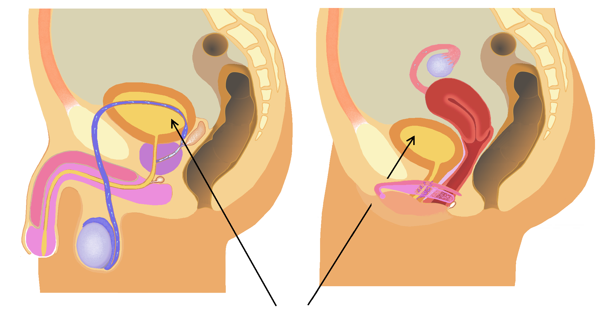
The trigone of urinary bladder (also known as the vesical trigone) is a smooth triangular region of the urinary bladder formed by the two ureteric orifices and the internal urethral orifice. Between the ureteric openings, there is a fold of mucous membrane called the ''interureteric crest'' or ''Mercier bar''. The trigone lies between the crest or ridge, and the neck of the bladder. The area is very sensitive to expansion and once stretched to a certain degree, stretch receptors in the urinary bladder signal the brain of its need to empty. The signals become stronger as the bladder continues to fill. Embryologically, the trigone of the bladder is derived from the caudal end of mesonephric ducts, which is of intermediate mesodermal origin (the rest of the bladder is endodermal). In the female the mesonephric ducts regress, causing the trigone to be less prominent, but still present. Clinical significance The trigone can become irritated in a condition known as trigonitis Tr ... [...More Info...] [...Related Items...] OR: [Wikipedia] [Google] [Baidu] |
Urinary Bladder
The bladder () is a hollow organ in humans and other vertebrates that stores urine from the Kidney (vertebrates), kidneys. In placental mammals, urine enters the bladder via the ureters and exits via the urethra during urination. In humans, the bladder is a distensible organ that sits on the pelvic floor. The typical adult human bladder will hold between 300 and (10 and ) before the urge to empty occurs, but can hold considerably more. The Latin phrase for "urinary bladder" is ''vesica urinaria'', and the term ''vesical'' or prefix ''vesico-'' appear in connection with associated structures such as vesical veins. The modern Latin word for "bladder" – ''cystis'' – appears in associated terms such as cystitis (inflammation of the bladder). Structure In humans, the bladder is a hollow muscular organ situated at the base of the pelvis. In gross anatomy, the bladder can be divided into a broad (base), a body, an apex, and a neck. The apex (also called the vertex) is directed ... [...More Info...] [...Related Items...] OR: [Wikipedia] [Google] [Baidu] |
Ureteric Orifice
The ureters are tubes composed of smooth muscle that transport urine from the kidneys to the urinary bladder. In an adult human, the ureters typically measure 20 to 30 centimeters in length and about 3 to 4 millimeters in diameter. They are lined with urothelial cells, a form of transitional epithelium, and feature an extra layer of smooth muscle in the lower third to aid in peristalsis. The ureters can be affected by a number of diseases, including urinary tract infections and kidney stone. is when a ureter is narrowed, due to for example chronic inflammation. Congenital abnormalities that affect the ureters can include the development of two ureters on the same side or abnormally placed ureters. Additionally, reflux of urine from the bladder back up the ureters is a condition commonly seen in children. The ureters have been identified for at least two thousand years, with the word "ureter" stemming from the stem relating to urinating and seen in written records since at leas ... [...More Info...] [...Related Items...] OR: [Wikipedia] [Google] [Baidu] |
Internal Urethral Orifice
The internal urethral orifice is the opening of the urinary bladder into the urethra. Anatomy It is usually somewhat crescent-shaped. Relations It is formed by the neck of the urinary bladder. It opens at the apex/inferior angle of the trigone of the bladder, some 2-3 cm anteromedial to either ureteral orifice. The mucous membrane immediately posterior to it presents a slight elevation in males - the uvula vesicae - caused by the middle lobe of the prostate The prostate is an male accessory gland, accessory gland of the male reproductive system and a muscle-driven mechanical switch between urination and ejaculation. It is found in all male mammals. It differs between species anatomically, chemica .... See also * Internal sphincter muscle of urethra References External links * - "The Male Pelvis: The Urethra" Urinary system Urethra {{genitourinary-stub ... [...More Info...] [...Related Items...] OR: [Wikipedia] [Google] [Baidu] |
Bladder
The bladder () is a hollow organ in humans and other vertebrates that stores urine from the kidneys. In placental mammals, urine enters the bladder via the ureters and exits via the urethra during urination. In humans, the bladder is a distensible organ that sits on the pelvic floor. The typical adult human bladder will hold between 300 and (10 and ) before the urge to empty occurs, but can hold considerably more. The Latin phrase for "urinary bladder" is ''vesica urinaria'', and the term ''vesical'' or prefix ''vesico-'' appear in connection with associated structures such as vesical veins. The modern Latin word for "bladder" – ''cystis'' – appears in associated terms such as cystitis (inflammation of the bladder). Structure In humans, the bladder is a hollow muscular organ situated at the base of the pelvis. In gross anatomy, the bladder can be divided into a broad (base), a body, an apex, and a neck. The apex (also called the vertex) is directed forward toward th ... [...More Info...] [...Related Items...] OR: [Wikipedia] [Google] [Baidu] |
Brain
The brain is an organ (biology), organ that serves as the center of the nervous system in all vertebrate and most invertebrate animals. It consists of nervous tissue and is typically located in the head (cephalization), usually near organs for special senses such as visual perception, vision, hearing, and olfaction. Being the most specialized organ, it is responsible for receiving information from the sensory nervous system, processing that information (thought, cognition, and intelligence) and the coordination of motor control (muscle activity and endocrine system). While invertebrate brains arise from paired segmental ganglia (each of which is only responsible for the respective segmentation (biology), body segment) of the ventral nerve cord, vertebrate brains develop axially from the midline dorsal nerve cord as a brain vesicle, vesicular enlargement at the rostral (anatomical term), rostral end of the neural tube, with centralized control over all body segments. All vertebr ... [...More Info...] [...Related Items...] OR: [Wikipedia] [Google] [Baidu] |
Mesonephric Ducts
The mesonephric duct, also known as the Wolffian duct, archinephric duct, Leydig's duct or nephric duct, is a paired organ that develops in the early stages of embryonic development in humans and other mammals. It is an important structure that plays a critical role in the formation of male reproductive organs. The duct is named after Caspar Friedrich Wolff, a German physiologist and embryologist who first described it in 1759. During embryonic development, the mesonephric ducts form as a part of the urogenital system. Structure The mesonephric duct connects the primitive kidney, the ''mesonephros'', to the cloaca. It also serves as the primordium for male urogenital structures including the epididymides, vasa deferentia, and seminal vesicles. Development In both males and females, the mesonephric ducts develop into the trigone of urinary bladder, a part of the bladder wall, but the sexes differentiate in other ways during development of the urinary and reproductive organs. ... [...More Info...] [...Related Items...] OR: [Wikipedia] [Google] [Baidu] |
Intermediate Mesoderm
Intermediate mesoderm or intermediate mesenchyme is a narrow section of the mesoderm (one of the three primary germ layers) located between the paraxial mesoderm and the lateral plate of the developing embryo. The intermediate mesoderm develops into vital parts of the urogenital system (kidneys, gonads and respective tracts). Early formation Factors regulating the formation of the intermediate mesoderm are not fully understood. It is believed that bone morphogenic proteins, or BMPs, specify regions of growth along the dorsal-ventral axis of the mesoderm and plays a central role in formation of the intermediate mesoderm. Vg1/wikt:node, Nodal signalling is an identified regulator of intermediate mesoderm formation acting through BMP signalling. Excess Vg1/Nodal signalling during early gastrulation stages results in expansion of the intermediate mesoderm at the expense of the adjacent paraxial mesoderm, whereas inhibition of Vg1/Nodal signalling represses intermediate mesoderm formati ... [...More Info...] [...Related Items...] OR: [Wikipedia] [Google] [Baidu] |
Endoderm
Endoderm is the innermost of the three primary germ layers in the very early embryo. The other two layers are the ectoderm (outside layer) and mesoderm (middle layer). Cells migrating inward along the archenteron form the inner layer of the gastrula, which develops into the endoderm. The endoderm consists at first of flattened cells, which subsequently become columnar. It forms the epithelial lining of multiple systems. In plant biology, endoderm corresponds to the innermost part of the cortex ( bark) in young shoots and young roots often consisting of a single cell layer. As the plant becomes older, more endoderm will lignify. Production The following chart shows the tissues produced by the endoderm. The embryonic endoderm develops into the interior linings of two tubes in the body, the digestive and respiratory tube. Liver and pancreas cells are believed to derive from a common precursor. In humans, the endoderm can differentiate into distinguishable organs after 5 w ... [...More Info...] [...Related Items...] OR: [Wikipedia] [Google] [Baidu] |
Trigonitis
Trigonitis is a condition of inflammation of the trigone region of the bladder. It is more common in women. The cause of trigonitis is not known, and there is no solid treatment. Electrocautery is sometimes used, but is generally unreliable as a treatment, and typically does not have quick results. Several drugs, such as muscle relaxants including urinary antispasmodics, antibiotics, and antiseptics have varied and unreliable results. Other forms of treatment include urethrotomy, cryosurgery, and neurostimulation Neurostimulation is the purposeful modulation of the nervous system's activity using invasive (e.g. microelectrodes) or Non-invasive procedure, non-invasive means (e.g. transcranial magnetic stimulation, transcranial electric stimulation such as .... References External links Urinary bladder disorders Inflammations {{genitourinary-disease-stub ... [...More Info...] [...Related Items...] OR: [Wikipedia] [Google] [Baidu] |


