|
Transthoracic Echocardiogram
A transthoracic echocardiogram (TTE) is the most common type of Echocardiography, echocardiogram, which is a still or moving image of the internal parts of the heart using ultrasound. In this case, the probe (or ultrasonic transducer) is placed on the Thorax, chest or abdomen of the subject to get various views of the heart. It is used as a non-invasive assessment of the overall health of the heart, including a patient's heart valves and degree of heart muscle contraction (an indicator of the ejection fraction). The images are displayed on a monitor for real-time viewing and then recorded. Often abbreviated "TTE", it can be easily confused with transesophageal echocardiography which is abbreviated "TEE". Pronunciation of "TTE" and "TEE" are similar, and full use of "transthoracic" or "transesophageal" can minimize any verbal miscommunication. Details A TTE is a clinical tool to evaluate the structure and function of the heart. All four chambers and all four valves can be assessed ... [...More Info...] [...Related Items...] OR: [Wikipedia] [Google] [Baidu] [Amazon] |
Echocardiography
Echocardiography, also known as cardiac ultrasound, is the use of ultrasound to examine the heart. It is a type of medical imaging, using standard ultrasound or Doppler ultrasound. The visual image formed using this technique is called an echocardiogram, a cardiac echo, or simply an echo. Echocardiography is routinely used in the diagnosis, management, and follow-up of patients with any suspected or known heart diseases. It is one of the most widely used diagnostic imaging modalities in cardiology. It can provide a wealth of helpful information, including the size and shape of the heart (internal chamber size quantification), pumping capacity, location and extent of any tissue damage, and assessment of valves. An echocardiogram can also give physicians other estimates of heart function, such as a calculation of the cardiac output, ejection fraction, and diastolic function (how well the heart relaxes). Echocardiography is an important tool in assessing wall motion abnorma ... [...More Info...] [...Related Items...] OR: [Wikipedia] [Google] [Baidu] [Amazon] |
Arteriovenous Malformation
An arteriovenous malformation (AVM) is an abnormal connection between arteries and veins, bypassing the capillary system. Usually congenital, this vascular anomaly is widely known because of its occurrence in the central nervous system (usually as a cerebral AVM), but can appear anywhere in the body. The symptoms of AVMs can range from none at all to intense pain or bleeding, and they can lead to other serious medical problems. Signs and symptoms Symptoms of AVMs vary according to their location. Most neurological AVMs produce few to no symptoms. Often the malformation is discovered as part of an autopsy or during treatment of an unrelated disorder (an " incidental finding"); in rare cases, its expansion or a micro-bleed from an AVM in the brain can cause epilepsy, neurological deficit, or pain. The most general symptoms of a cerebral AVM include headaches and epileptic seizures, with more specific symptoms that normally depend on its location and the individual, in ... [...More Info...] [...Related Items...] OR: [Wikipedia] [Google] [Baidu] [Amazon] |
Focused Assessment With Sonography For Trauma
Focused assessment with sonography in trauma (commonly abbreviated as FAST) is a rapid bedside ultrasound examination performed by surgeons, emergency physicians, and paramedics as a screening test for blood around the heart (pericardial effusion) or abdominal organs ( hemoperitoneum) after trauma. There is also the extended FAST (eFAST) which includes some additional ultrasound views to assess for pneumothorax. It may be useful prior to conducting more accurate tests such as CT in a stable trauma patient. The four classic areas that are examined for free fluid are the perihepatic space (including Morison's pouch or the hepatorenal recess), peri splenic space, pericardium, and the pelvis. With this technique it is possible to identify the presence of moderate to large amounts of intraperitoneal or pericardial free fluid. In the context of traumatic injury, this fluid will usually be due to bleeding. FAST is poor at detecting smaller amounts of free fluid. Indications Reas ... [...More Info...] [...Related Items...] OR: [Wikipedia] [Google] [Baidu] [Amazon] |
Cardiac Tamponade
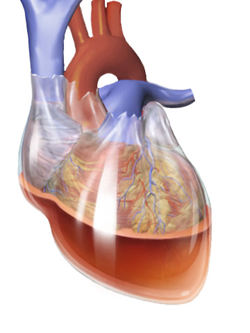
Cardiac tamponade, also known as pericardial tamponade (), is a compression of the heart due to pericardial effusion (the build-up of pericardial fluid in the pericardium, sac around the heart). Onset may be rapid or gradual. Symptoms typically include those of obstructive shock including shortness of breath, weakness, lightheadedness, and cough. Other symptoms may relate to the underlying cause. Common causes of cardiac tamponade include cancer, kidney failure, chest trauma, myocardial infarction, and pericarditis. Other causes include connective tissue disease, connective tissues diseases, hypothyroidism, aortic rupture, autoimmune disease, and complications of cardiac surgery. In Africa, tuberculosis is a relatively common cause. Diagnosis may be suspected based on hypotension, low blood pressure, jugular venous distension, or quiet heart sounds (together known as Beck's triad (cardiology), Beck's triad). A pericardial rub may be present in cases due to inflammation. The dia ... [...More Info...] [...Related Items...] OR: [Wikipedia] [Google] [Baidu] [Amazon] |
Echocardiogram In US Navy
Echocardiography, also known as cardiac ultrasound, is the use of ultrasound to examine the heart. It is a type of medical imaging, using standard ultrasound or Doppler ultrasound. The visual image formed using this technique is called an echocardiogram, a cardiac echo, or simply an echo. Echocardiography is routinely used in the diagnosis, management, and follow-up of patients with any suspected or known heart diseases. It is one of the most widely used diagnostic imaging modalities in cardiology. It can provide a wealth of helpful information, including the size and shape of the heart (internal chamber size quantification), pumping capacity, location and extent of any tissue damage, and assessment of valves. An echocardiogram can also give physicians other estimates of heart function, such as a calculation of the cardiac output, ejection fraction, and diastolic function (how well the heart relaxes). Echocardiography is an important tool in assessing wall motion abnormality in ... [...More Info...] [...Related Items...] OR: [Wikipedia] [Google] [Baidu] [Amazon] |
Hypoxemia
Hypoxemia (also spelled hypoxaemia) is an abnormally low level of oxygen in the blood. More specifically, it is oxygen deficiency in arterial blood. Hypoxemia is usually caused by pulmonary disease. Sometimes the concentration of oxygen in the air is decreased leading to hypoxemia. Definition ''Hypoxemia'' refers to the low level of oxygen in arterial blood. Tissue hypoxia refers to low levels of oxygen in the tissues of the body and the term ''hypoxia'' is a general term for low levels of oxygen. Hypoxemia is usually caused by pulmonary disease whereas tissue oxygenation requires additionally adequate circulation of blood and perfusion of tissue to meet metabolic demands. Hypoxemia is usually defined in terms of reduced partial pressure of oxygen (mm Hg) in arterial blood, but also in terms of reduced content of oxygen (ml oxygen per dl blood) or percentage saturation of hemoglobin (the oxygen-binding protein within red blood cells) with oxygen, which is either found singly o ... [...More Info...] [...Related Items...] OR: [Wikipedia] [Google] [Baidu] [Amazon] |
Endocarditis
Endocarditis is an inflammation of the inner layer of the heart, the endocardium. It usually involves the heart valves. Other structures that may be involved include the interventricular septum, the chordae tendineae, the mural endocardium, or the surfaces of intracardiac devices. Endocarditis is characterized by lesions, known as '' vegetations'', which are masses of platelets, fibrin, microcolonies of microorganisms, and scant inflammatory cells. In the subacute form of infective endocarditis, a vegetation may also include a center of granulomatous tissue, which may fibrose or calcify. There are several ways to classify endocarditis. The simplest classification is based on cause: either ''infective'' or ''non-infective'', depending on whether a microorganism is the source of the inflammation or not. Regardless, the diagnosis of endocarditis is based on clinical features, investigations such as an echocardiogram, and blood cultures demonstrating the presence of endocar ... [...More Info...] [...Related Items...] OR: [Wikipedia] [Google] [Baidu] [Amazon] |
Congenital Heart Disease
A congenital heart defect (CHD), also known as a congenital heart anomaly, congenital cardiovascular malformation, and congenital heart disease, is a defect in the structure of the heart or great vessels that is present at birth. A congenital heart defect is classed as a cardiovascular disease. Signs and symptoms depend on the specific type of defect. Symptoms can vary from none to life-threatening. When present, symptoms are variable and may include rapid breathing, bluish skin (cyanosis), poor weight gain, and feeling tired. CHD does not cause chest pain. Most congenital heart defects are not associated with other diseases. A complication of CHD is heart failure. Congenital heart defects are the most common birth defect. In 2015, they were present in 48.9 million people globally. They affect between 4 and 75 per 1,000 live births, depending upon how they are diagnosed. In about 6 to 19 per 1,000 they cause a moderate to severe degree of problems. Congenital heart defects are th ... [...More Info...] [...Related Items...] OR: [Wikipedia] [Google] [Baidu] [Amazon] |
Chemotherapy
Chemotherapy (often abbreviated chemo, sometimes CTX and CTx) is the type of cancer treatment that uses one or more anti-cancer drugs (list of chemotherapeutic agents, chemotherapeutic agents or alkylating agents) in a standard chemotherapy regimen, regimen. Chemotherapy may be given with a cure, curative intent (which almost always involves combinations of drugs), or it may aim only to prolong life or to Palliative care, reduce symptoms (Palliative care, palliative chemotherapy). Chemotherapy is one of the major categories of the medical discipline specifically devoted to pharmacotherapy for cancer, which is called ''oncology#Specialties, medical oncology''. The term ''chemotherapy'' now means the non-specific use of intracellular poisons to inhibit mitosis (cell division) or to induce DNA damage (naturally occurring), DNA damage (so that DNA repair can augment chemotherapy). This meaning excludes the more-selective agents that block extracellular signals (signal transduction) ... [...More Info...] [...Related Items...] OR: [Wikipedia] [Google] [Baidu] [Amazon] |
Heart Murmur
Heart murmurs are unique heart sounds produced when blood flows across a heart valve or blood vessel. This occurs when turbulent blood flow creates a sound loud enough to hear with a stethoscope. The sound differs from normal heart sounds by their characteristics. For example, heart murmurs may have a distinct pitch, duration and timing. The major way health care providers examine the heart on physical exam is heart auscultation; another clinical technique is palpation, which can detect by touch when such turbulence causes the vibrations called cardiac thrill. A murmur is a sign found during the cardiac exam. Murmurs are of various types and are important in the detection of cardiac and valvular pathologies (i.e. can be a sign of heart diseases or defects). There are two types of murmur. A functional murmur is a benign heart murmur that is primarily due to physiologic conditions outside the heart. The other type of heart murmur is due to a structural defect in the heart itsel ... [...More Info...] [...Related Items...] OR: [Wikipedia] [Google] [Baidu] [Amazon] |
Heart Failure
Heart failure (HF), also known as congestive heart failure (CHF), is a syndrome caused by an impairment in the heart's ability to Cardiac cycle, fill with and pump blood. Although symptoms vary based on which side of the heart is affected, HF typically presents with shortness of breath, Fatigue (medical), excessive fatigue, and bilateral peripheral edema, leg swelling. The severity of the heart failure is mainly decided based on ejection fraction and also measured by the severity of symptoms. Other conditions that have symptoms similar to heart failure include obesity, kidney failure, liver disease, anemia, and thyroid disease. Common causes of heart failure include coronary artery disease, heart attack, hypertension, high blood pressure, atrial fibrillation, valvular heart disease, alcohol use disorder, excessive alcohol consumption, infection, and cardiomyopathy. These cause heart failure by altering the structure or the function of the heart or in some cases both. There are ... [...More Info...] [...Related Items...] OR: [Wikipedia] [Google] [Baidu] [Amazon] |
Shortness Of Breath
Shortness of breath (SOB), known as dyspnea (in AmE) or dyspnoea (in BrE), is an uncomfortable feeling of not being able to breathe well enough. The American Thoracic Society defines it as "a subjective experience of breathing discomfort that consists of qualitatively distinct sensations that vary in intensity", and recommends evaluating dyspnea by assessing the intensity of its distinct sensations, the degree of distress and discomfort involved, and its burden or impact on the patient's activities of daily living. Distinct sensations include effort/work to breathe, chest tightness or pain, and "air hunger" (the feeling of not enough oxygen). The tripod position is often assumed to be a sign. Dyspnea is a normal symptom of heavy physical exertion but becomes disease, pathological if it occurs in unexpected situations, when resting or during light exertion. In 85% of cases it is due to asthma, pneumonia, cardiac ischemia, reflux/LPR, cardiac ischemia, COVID-19, interstitial lung di ... [...More Info...] [...Related Items...] OR: [Wikipedia] [Google] [Baidu] [Amazon] |


