|
Thenar Eminence
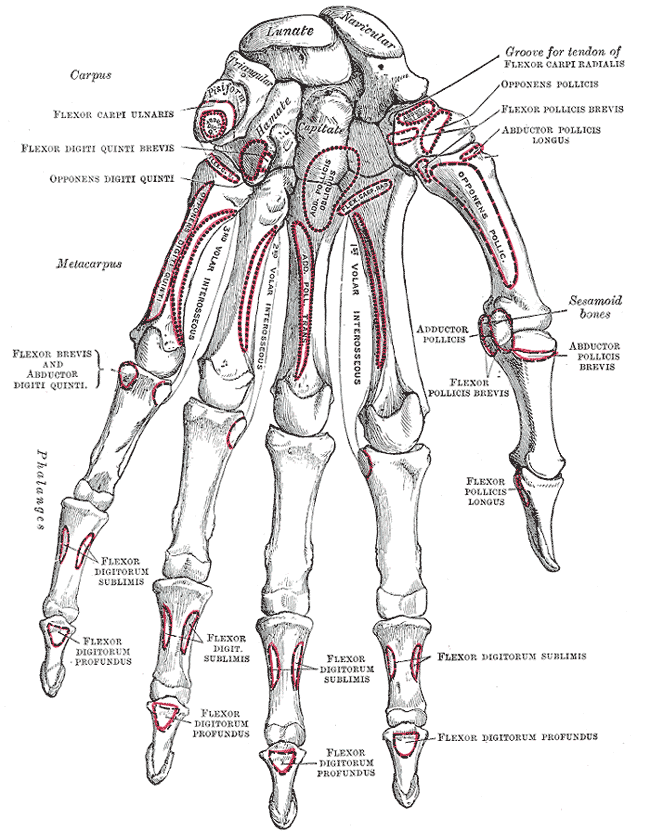
The thenar eminence is the mound formed at the base of the thumb on the palm of the hand by the intrinsic group of muscles of the thumb. The skin overlying this region is the area stimulated when trying to elicit a palmomental reflex. The word thenar comes . Structure The following three muscles are considered part of the thenar eminence: * Abductor pollicis brevis abducts the thumb. This muscle is the most superficial of the thenar group. * Flexor pollicis brevis, which lies next to the abductor, will flex the thumb, curling it up in the palm. (The flexor pollicis longus, which is inserted into the distal phalanx of the thumb, is not considered part of the thenar eminence.) * Opponens pollicis lies deep to abductor pollicis brevis. As its name suggests it opposes the thumb, bringing it against the fingers. This is a very important movement, as most of human hand dexterity comes from this action. Another muscle that controls movement of the thumb is adductor pollicis ... [...More Info...] [...Related Items...] OR: [Wikipedia] [Google] [Baidu] |
Wrist
In human anatomy, the wrist is variously defined as (1) the carpus or carpal bones, the complex of eight bones forming the proximal skeletal segment of the hand; "The wrist contains eight bones, roughly aligned in two rows, known as the carpal bones." (2) the wrist joint or radiocarpal joint, the joint between the radius and the carpus and; (3) the anatomical region surrounding the carpus including the distal parts of the bones of the forearm and the proximal parts of the metacarpus or five metacarpal bones and the series of joints between these bones, thus referred to as ''wrist joints''. "With the large number of bones composing the wrist (ulna, radius, eight carpas, and five metacarpals), it makes sense that there are many, many joints that make up the structure known as the wrist." This region also includes the carpal tunnel, the anatomical snuff box, bracelet lines, the flexor retinaculum, and the extensor retinaculum. As a consequence of these various definitions, f ... [...More Info...] [...Related Items...] OR: [Wikipedia] [Google] [Baidu] |
Adductor Pollicis
In human anatomy, the adductor pollicis muscle is a muscle in the hand that functions to Adduction, adduct the thumb. It has two heads: transverse and oblique. It is a fleshy, flat, triangular, and fan-shaped muscle deep in the Thenar eminence, thenar compartment beneath the long flexor tendons and the Lumbricals of the hand, lumbrical muscles at the center of the palm. It overlies the Metacarpus, metacarpal bones and the Palmar interossei muscles, interosseous muscles. Structure Oblique head The oblique head (Latin: ''adductor obliquus pollicis'') arises by several slips from the capitate bone, the bases of the second and third metacarpus, metacarpals, the intercarpal ligaments, and the sheath of the tendon of the flexor carpi radialis. Gray's Anatomy 1918. (See infobox) From this origin the greater number of fibers pass obliquely downward and converge to a tendon, which, uniting with the tendons of the medial portion of the flexor pollicis brevis and the transverse head of ... [...More Info...] [...Related Items...] OR: [Wikipedia] [Google] [Baidu] |
Lumbricals Of The Hand
The lumbricals are intrinsic muscles of the hand that flexion, flex the metacarpophalangeal joints, and extension (kinesiology), extend the Interphalangeal articulations of hand, interphalangeal joints. p. 97 The lumbrical muscle of the foot, lumbrical muscles of the foot also have a similar action, though they are of less clinical concern. Structure The lumbricals are four, small, worm-like muscles on each hand. These muscles are unusual in that they do not attach to bone. Instead, they attach proximally to the tendons of flexor digitorum profundus, and distally to the extensor expansions. The first and second lumbricals are Anatomical terms of muscle, unipennate, while the third and fourth lumbricals are Anatomical terms of muscle, bipennate. Nerve supply The first and second lumbricals (the most radial two) are Nerve, innervated by the median nerve. The third and fourth lumbricals (most ulnar two) are innervated by the deep branch of ulnar nerve. This is the usual inne ... [...More Info...] [...Related Items...] OR: [Wikipedia] [Google] [Baidu] |
Hypothenar Eminence
The hypothenar muscles are a group of three muscles of the palm that control the motion of the little finger. The three muscles are: * Abductor digiti minimi * Flexor digiti minimi brevis * Opponens digiti minimi Structure The muscles of hypothenar eminence are from medial to lateral: * Opponens digiti minimi * Flexor digiti minimi brevis * Abductor digiti minimi The intrinsic muscles of hand can be remembered using the mnemonic, "A OF A OF A" for, Abductor pollicis brevis, Opponens pollicis, Flexor pollicis brevis (the three thenar muscles), Adductor pollicis, and the three hypothenar muscles, Opponens digiti minimi, Flexor digiti minimi brevis, Abductor digiti minimi. Clinical significance "Hypothenar atrophy" is associated with the lesion of the ulnar nerve, which supplies the three hypothenar muscles. Hypothenar hammer syndrome is a vascular occlusion of this region. See also * Thenar eminence * Palmaris brevis Palmaris brevis muscle is a thin, quadrilateral muscle, ... [...More Info...] [...Related Items...] OR: [Wikipedia] [Google] [Baidu] |
Brachial Plexus
The brachial plexus is a network of nerves (nerve plexus) formed by the anterior rami of the lower four Spinal nerve#Cervical nerves, cervical nerves and first Spinal nerve#Thoracic nerves, thoracic nerve (cervical spinal nerve 5, C5, Cervical spinal nerve 6, C6, cervical spinal nerve 7, C7, cervical spinal nerve 8, C8, and thoracic spinal nerve 1, T1). This plexus extends from the spinal cord, through the cervicoaxillary canal in the neck, over the first rib, and into the axilla, armpit, it supplies Afferent nerve fiber, afferent and efferent nerve fibers to the chest, shoulder, arm, forearm, and hand. Structure The brachial plexus is divided into five ''roots'', three ''trunks'', six ''divisions'' (three anterior and three posterior), three ''cords'', and five ''branches''. There are five "terminal" branches and numerous other "pre-terminal" or "collateral" branches, such as the subscapular nerve, the thoracodorsal nerve, and the long thoracic nerve, that leave the plexus at vari ... [...More Info...] [...Related Items...] OR: [Wikipedia] [Google] [Baidu] |
Gray's Anatomy
''Gray's Anatomy'' is a reference book of human anatomy written by Henry Gray, illustrated by Henry Vandyke Carter and first published in London in 1858. It has had multiple revised editions, and the current edition, the 42nd (October 2020), remains a standard reference, often considered "the doctors' bible". Earlier editions were called ''Anatomy: Descriptive and Surgical'', ''Anatomy of the Human Body'' and ''Gray's Anatomy: Descriptive and Applied'', but the book's name is commonly shortened to, and later editions are titled, ''Gray's Anatomy''. The book is widely regarded as an extremely influential work on the subject. Publication history Origins The English anatomist Henry Gray was born in 1827. He studied the development of the endocrine glands and spleen and in 1853 was appointed Lecturer on Anatomy at St George's Hospital Medical School in London. In 1855, he approached his colleague Henry Vandyke Carter with his idea to produce an inexpensive and access ... [...More Info...] [...Related Items...] OR: [Wikipedia] [Google] [Baidu] |
Thoracic Spinal Nerve 1
The thoracic spinal nerve 1 (T1) is a spinal nerve A spinal nerve is a mixed nerve, which carries Motor neuron, motor, Sensory neuron, sensory, and Autonomic nervous system, autonomic signals between the spinal cord and the body. In the human body there are 31 pairs of spinal nerves, one on each s ... of the thoracic segment. Nervous System -- Groups of Nerves It originates from the spinal column from below the thoracic vertebra 1 (T1). Additional images References Spinal nerves {{neuroanatomy-stub ...[...More Info...] [...Related Items...] OR: [Wikipedia] [Google] [Baidu] |
Cervical Spinal Nerve 8
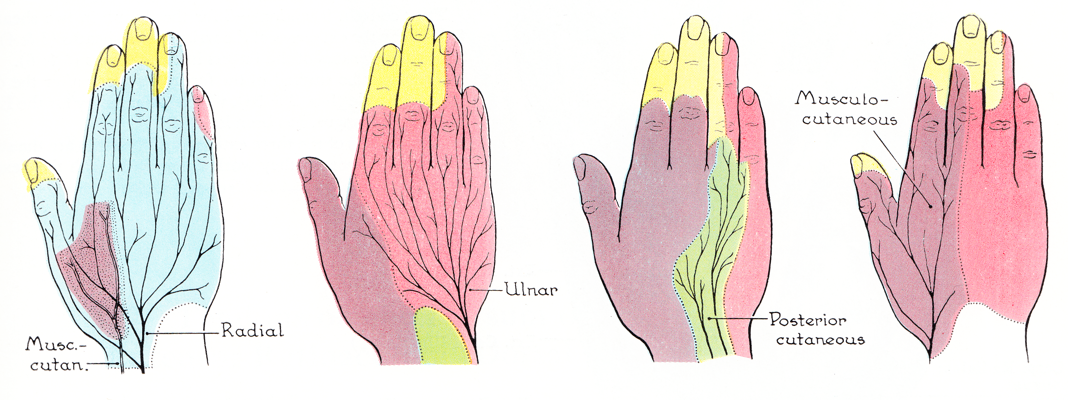
The cervical spinal nerve 8 (C8) is a spinal nerve of the cervical segment. It originates from the spinal column from below the cervical vertebra 7 (C7). Innervation The C8 nerve forms part of the radial and ulnar nerves via the brachial plexus, and therefore has motor and sensory function in the upper limb. Sensory The C8 nerve receives sensory afferents from the C8 dermatome. This consists of all the skin on the little finger, and continuing up slightly past the wrist on the palmar and dorsal aspects of the hand and forearm.Drake et al. Gray's Anatomy for Students. Second Edition (2010). Clinically, a test of the pad of the little finger is often used to assess C8 integrity.Aland et al. University of Queensland School of Medicine Clinical Skills Handbook 2010 Motor The C8 nerve contributes to the motor innervation of many of the muscles in the trunk and upper limb. Its primary function is the flexion of the fingers, and this is used as the clinical test for C8 inte ... [...More Info...] [...Related Items...] OR: [Wikipedia] [Google] [Baidu] |
Deep Branch Of Ulnar Nerve
The deep branch of the ulnar nerve is a terminal, primarily motor branch of the ulnar nerve. It is accompanied by the deep palmar branch of ulnar artery. Structure It passes between the abductor digiti minimi and the flexor digiti minimi brevis. It then perforates the opponens digiti minimi and follows the course of the deep palmar arch beneath the flexor tendons. As the deep ulnar nerve passes across the palm, it lies in a fibrous tunnel formed between the hook of the hamate and the pisiform ( Guyon's canal). Function At its origin it innervates the hypothenar muscles. As it crosses the deep part of the hand, it innervates all the interosseous muscles and the third and fourth lumbricals. It ends by innervating the adductor pollicis and the medial (deep) head of the flexor pollicis brevis The flexor pollicis brevis is a muscle in the hand that flexes the thumb. It is one of three thenar muscles. It has both a superficial part and a deep part. Origin and insertion The ... [...More Info...] [...Related Items...] OR: [Wikipedia] [Google] [Baidu] |
Median Nerve
The median nerve is a nerve in humans and other animals in the upper limb. It is one of the five main nerves originating from the brachial plexus. The median nerve originates from the lateral and medial cords of the brachial plexus, and has contributions from ventral roots of C6-C7 (lateral cord) and C8 and T1 (medial cord). The median nerve is the only nerve that passes through the carpal tunnel. Carpal tunnel syndrome is the disability that results from the median nerve being pressed in the carpal tunnel. Structure The median nerve arises from the branches from lateral and medial cords of the brachial plexus, courses through the anterior part of arm, forearm, and hand, and terminates by supplying the muscles of the hand. Arm After receiving inputs from both the lateral and medial cords of the brachial plexus, the median nerve enters the arm from the axilla at the inferior margin of the teres major muscle. It then passes vertically down and courses lateral to the brac ... [...More Info...] [...Related Items...] OR: [Wikipedia] [Google] [Baidu] |


